
Everted filter device

a filter device and eversion technology, applied in the field of medical devices, can solve the problems of pain and amputation, decreased renal function, myocardial infarction, etc., and achieve the effects of small volume, small profile, and increased interior volume of the filter

Active Publication Date: 2005-12-22
TYCO HEALTHCARE GRP LP
View PDF25 Cites 263 Cited by
  • Summary
  • Abstract
  • Description
  • Claims
  • Application Information

AI Technical Summary

Benefits of technology

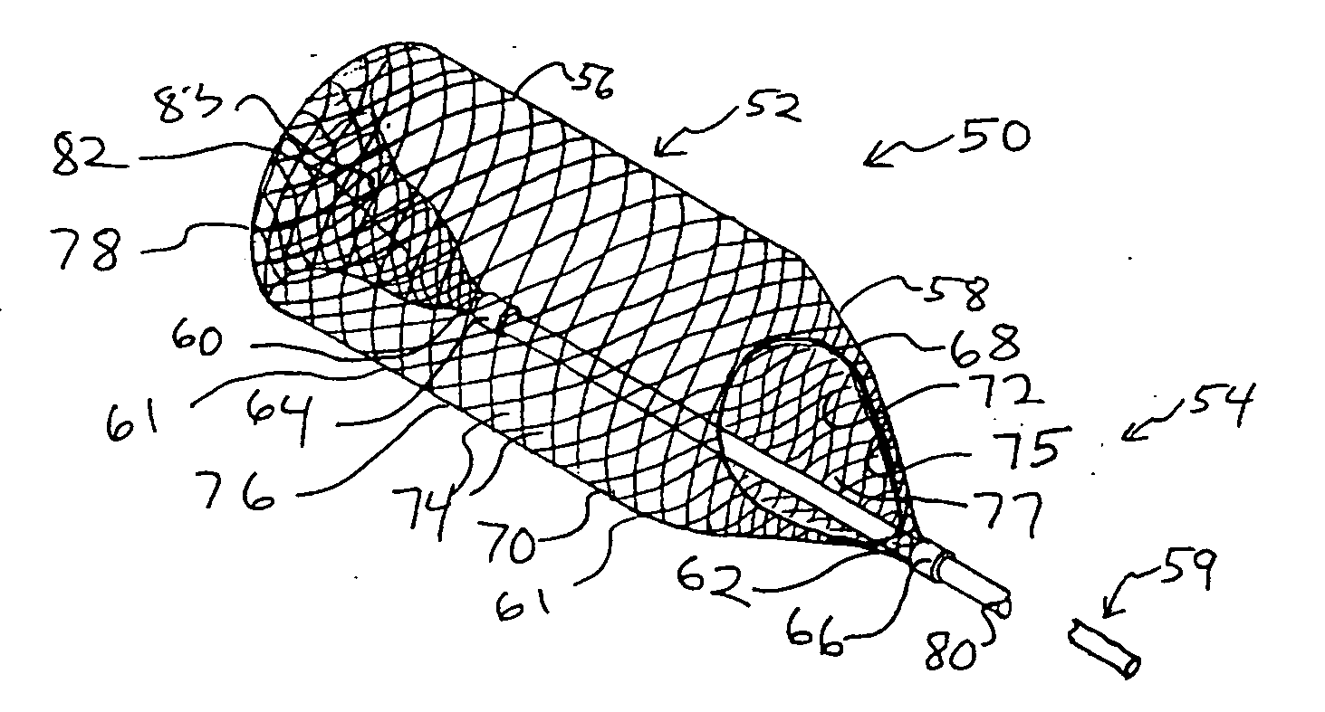
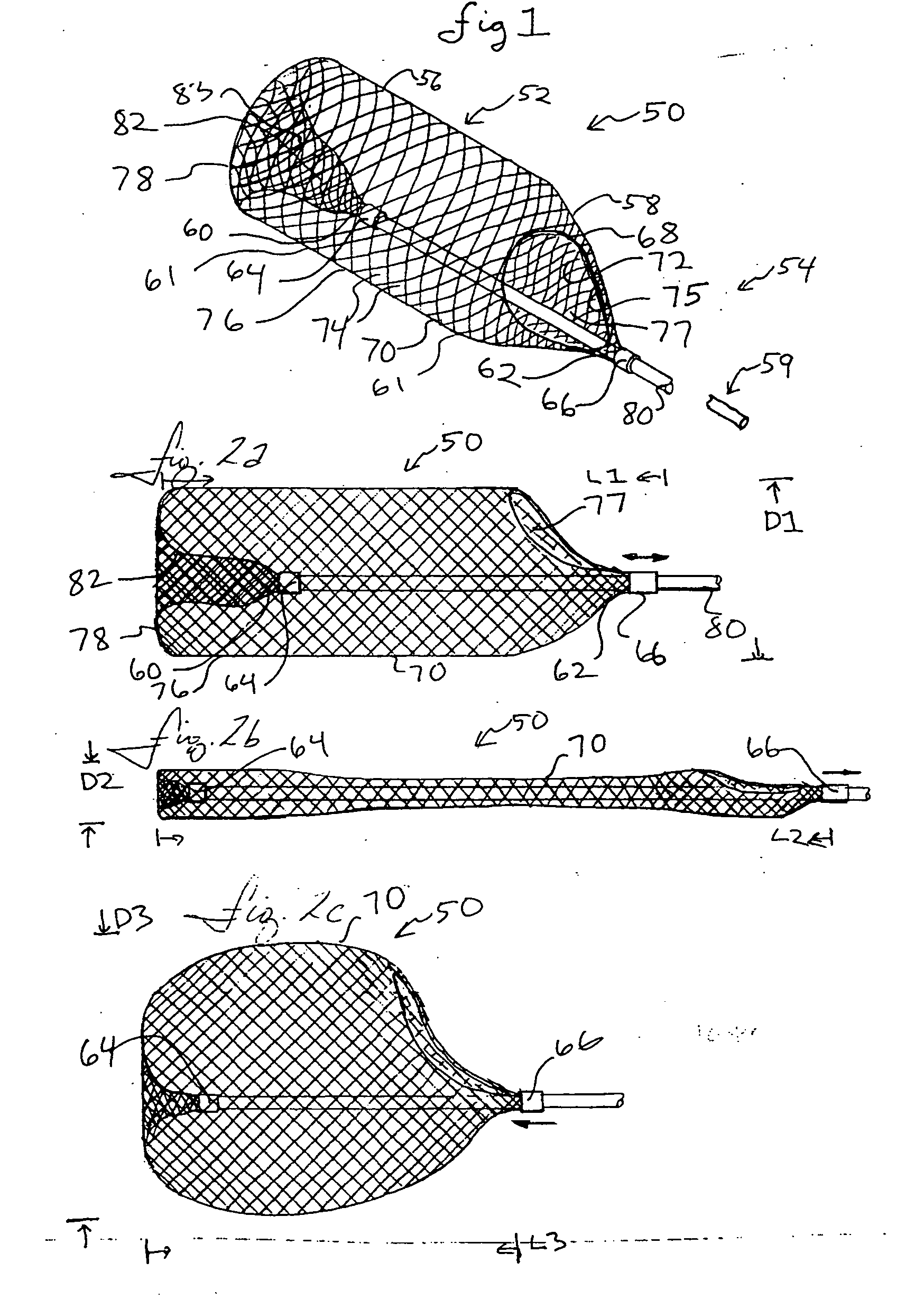
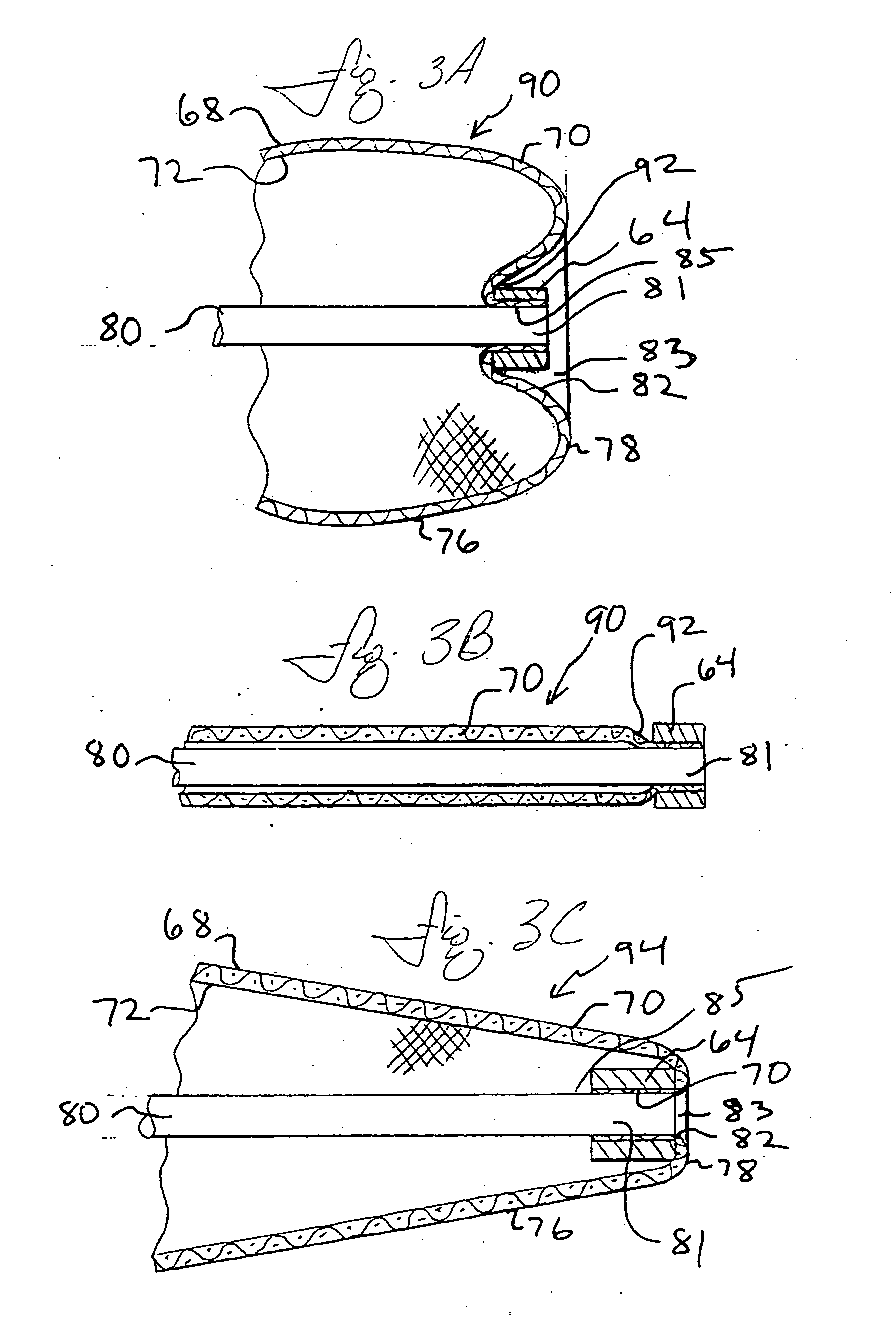
[0010] The everted filters can include a tubular filter body having a large proximal opening and a closed distal end. The filter body can extend distally from a first end region near the filter body proximal opening, further extending over an intermediate, non-everted region, continuing distally and everting over a distal-most region. The everting causes the filter body exterior to converge inwardly and proximally to a closed, filter body second end region. The proximally extending and inwardly converging exterior surface of the filter body defines a distal, everted cavity bounded distally by the distal-most extent of the everting filter. The distal everted cavity has exterior cavity walls which extend proximally back towards the proximal opening. The degree of eversion of the filter body may be changed through controlling the relative positions of the filter body first and second end regions. When the filter body diameter is held constant, proximally withdrawing the everted second end region toward the first end region increases the degree of eversion, increasing the distal everted cavity volume and length and increasing the length of filter body exterior surface within the distal, everted cavity.
[0018] The everted filters also provide a filter device able to significantly increase the interior volume of the filter after positioning the filter in a vessel region. An everted filter can be advanced to a vessel site in a compressed state, having a small volume, small profile, and a high strand density due to a small inter-strand distance and pore size. In place, the filter can be both elongated and radially expanded to decrease the strand density and increase the pore size and filter interior volume.

Problems solved by technology

While some stenoses remain in place once dilated, others are more brittle, and may partially crack and fragment, allowing the fragments to flow downstream where they may block more distal and smaller coronary vessels, possibly causing myocardial infarction, from that site.
Consequences of embolization include stroke, diminished renal function, and impairment of peripheral circulation possibly leading to pain and amputation.
With time, the grafts can become occluded with grumous.
The grumous can present an even more difficult material to remove than thrombus, as the material is very friable, and likely to break into smaller fragments during the removal procedure.
However, aspiration cannot remove large particles because they won't fit through the aspiration lumen.
During the occlusion, the lack of fluid flow can be deleterious.
In coronary applications, the lack of perfusing blood flow can cause angina.
In both coronaries and carotids it is not possible to predict who will suffer from angina or seizure due to vessel occlusion.
While the replacement is occurring, there is a period of no embolic protection for the patient and a risk of dislodging emboli during filter manipulation.
If the filter is left in place, the vessel will be occluded during the remainder of the procedure and the patient can suffer the consequences of occlusion described earlier.
Both choices are less than optimal.
Multiple filters may be required, one for each vessel branch, which is cumbersome and may not be done well, if attempted at all.
Further, the use of multiple filters may not be compatible with other needed equipment such as angioplasty balloons and / or stents.
However, occlusive devices compromise fluoroscopic imaging due to the lack of flow during radiopaque dye injection.

Method used


Image

  • Everted filter device
  • Everted filter device
  • Everted filter device

Examples


Embodiment Construction

[0061] The following detailed description should be read with reference to the drawings, in which like elements in different drawings are numbered identically. The drawings, which are not necessarily to scale, depict selected embodiments and are not intended to limit the scope of the invention. Several forms of invention have been shown and described, and other forms will now be apparent to those skilled in art. It will be understood that embodiments shown in drawings and described above are merely for illustrative purposes, and are not intended to limit scope of the invention as defined in the claims which follow.

[0062]FIG. 1 illustrates the distal portion of an everting filter 50. Everting filter 50 may be viewed as having a distal portion 52 and an intermediate portion 54. Distal portion 52 may be further divided into a distal portion distal region 56 and a distal portion proximal region 58. The device can extend proximally to a proximal portion 59.

[0063] Everting filter 50 inc...


PUM


Abstract

Everting filter devices and methods for using the devices, including using the devices as intra-vascular filters to filter thrombus, emboli, and plaque fragments from blood vessels. The filter devices include a filter body nominally tubular in shape and having a large proximal opening. The filter body can extend from a proximal first end region distally over the non-everted exterior surface of the filter, further extending distally to a distal-most region, then converging inwardly and extending proximally toward the filter second end region, forming a distal everted cavity. The degree of eversion of the filter can be controlled by varying the distance between the filter first end region near the proximal opening and the closed second end region. Bringing the filter first and second end regions closer together can bring filter material previously on the non-everted filter exterior to occupy the distal-most region. The everting process can also bring filter material previously in the distal-most position further into the distal everted cavity. The filter devices can be used to remove filtrate from body vessels, with the filtrate eventually occluding the distal-most region. The filter can then be further everted, bringing fresh, unoccluded filter material into place to provide additional filter capacity. Some everting filters have the capability of switching between occluding and filtering modes of operation, thereby allowing a treating physician to postpone the decision to use filtering or occluding devices until well after insertion of the device into the patient's body.

Description

FIELD OF THE INVENTION [0001] The present invention is related generally to medical devices. The present invention includes intravascular filter devices. BACKGROUND OF THE INVENTION [0002] Coronary vessels, partially occluded by plaque, may become totally occluded by a thrombus or blood clot causing myocardial infarction, angina and other conditions. A number of medical procedures have been developed to allow for the removal of plaque from vessel walls or to clear a channel through the thrombus or clot to restore blood flow and minimize the risk of myocardial infarction. Carotid, renal, peripheral, and other blood vessels can also be blocked and require treatment. For example, atherectomy or thrombectomy devices can be used to remove atheroma or thrombus. Alternatively, in percutaneous transluminal coronary angioplasty (PTCA), a guide wire or guide catheter is inserted into the femoral artery of a patient near the groin, advanced through the artery, over the aorta, and into a corona...

Claims


Application Information

Patent Timeline
IPC IPC(8): A61F2/01
CPCA61F2/013A61F2230/0069A61F2230/0006A61F2002/018A61F2230/008A61F2/0105
Inventor BERRADA, MARWANEKUSLEIKA, RICHARDANDERSON, KENT
Owner TYCO HEALTHCARE GRP LP
Learn More