
Method and apparatus for tissue expansion

a tissue and tissue technology, applied in the field of biological tissue expansion, can solve the problems of poor skin healing, large defects are often left open to heal, burn victims often suffer many painful surgeries, etc., and achieve the effect of reducing the presence or likelihood of noticeable holes or scars and improving outcomes

Inactive Publication Date: 2011-10-13
THE GENERAL HOSPITAL CORP
View PDF12 Cites 94 Cited by
  • Summary
  • Abstract
  • Description
  • Claims
  • Application Information

AI Technical Summary

Benefits of technology

[0010]Embodiments of the present disclosure are directed to method and apparatus for facilitating an expansion of a portion of tissue, while reducing a presence or likelihood of noticeable holes or scars after healing of the expanded tissue. The exemplary method and apparatus can be applied in situ for releasing local skin tension, to cover damaged or removed regions of tissue using adjacent healthy tissue, and other tissue expansion applications, or ex vivo for expansion of split-thickness and / or full thickness grafts while providing an improved outcome.
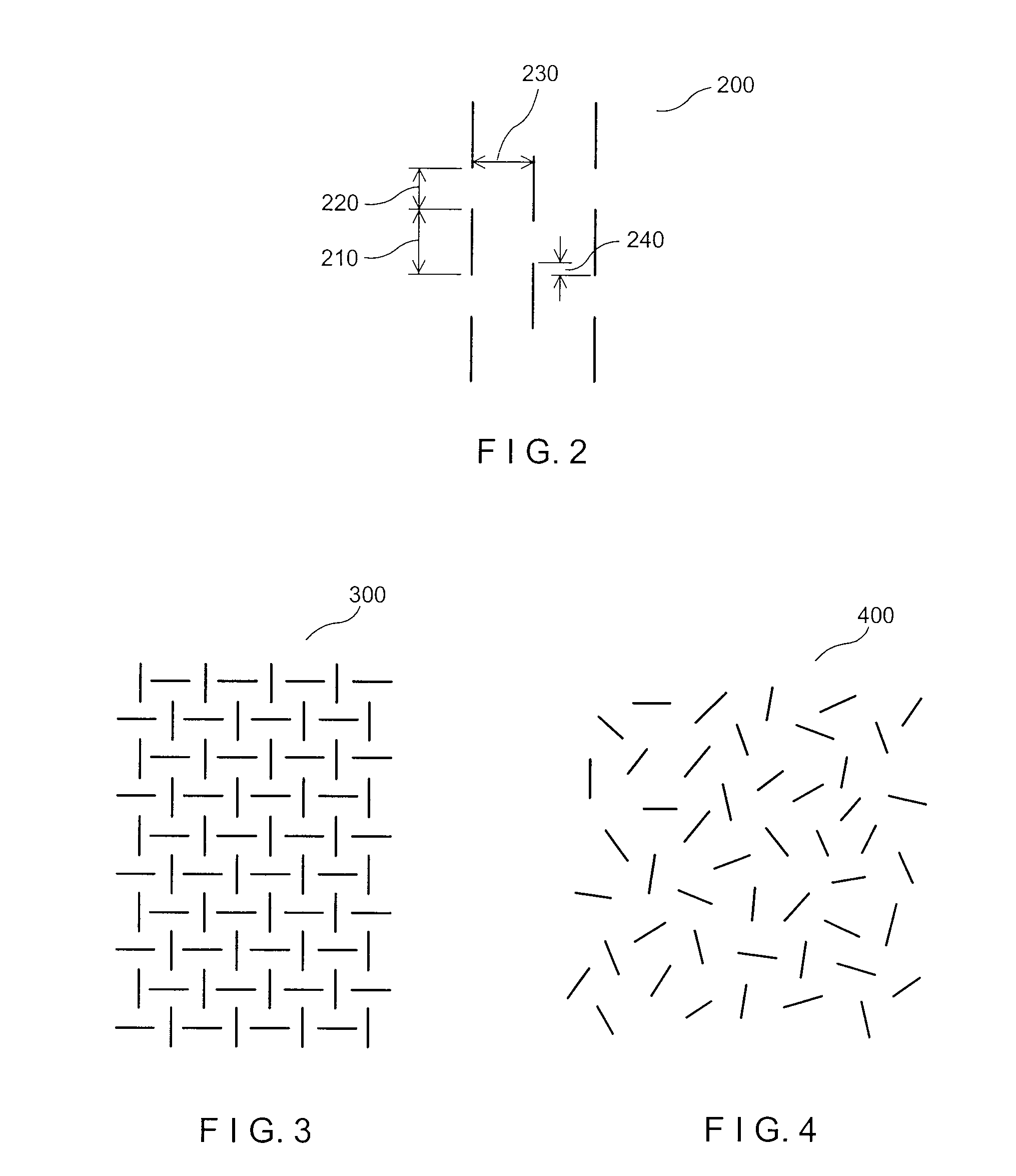
[0011]According to one exemplary embodiment of the present disclosure, a method can be provided for facilitating tissue expansion that includes a formation of a plurality of micro-slits in the tissue to be stretched. The length of extension of the micro-slits can be less than about 2 mm, or less than about 1.5 mm, or less than about 1 mm, or even less than about 0.8 mm. The length of each micro-slit can be greater than about 0.1 mm to allow sufficient expansion of the tissue. The width of the gap formed by a micro-slit when the tissue is expanded can be less than about 1 mm, or preferably less than about 0.5 mm.
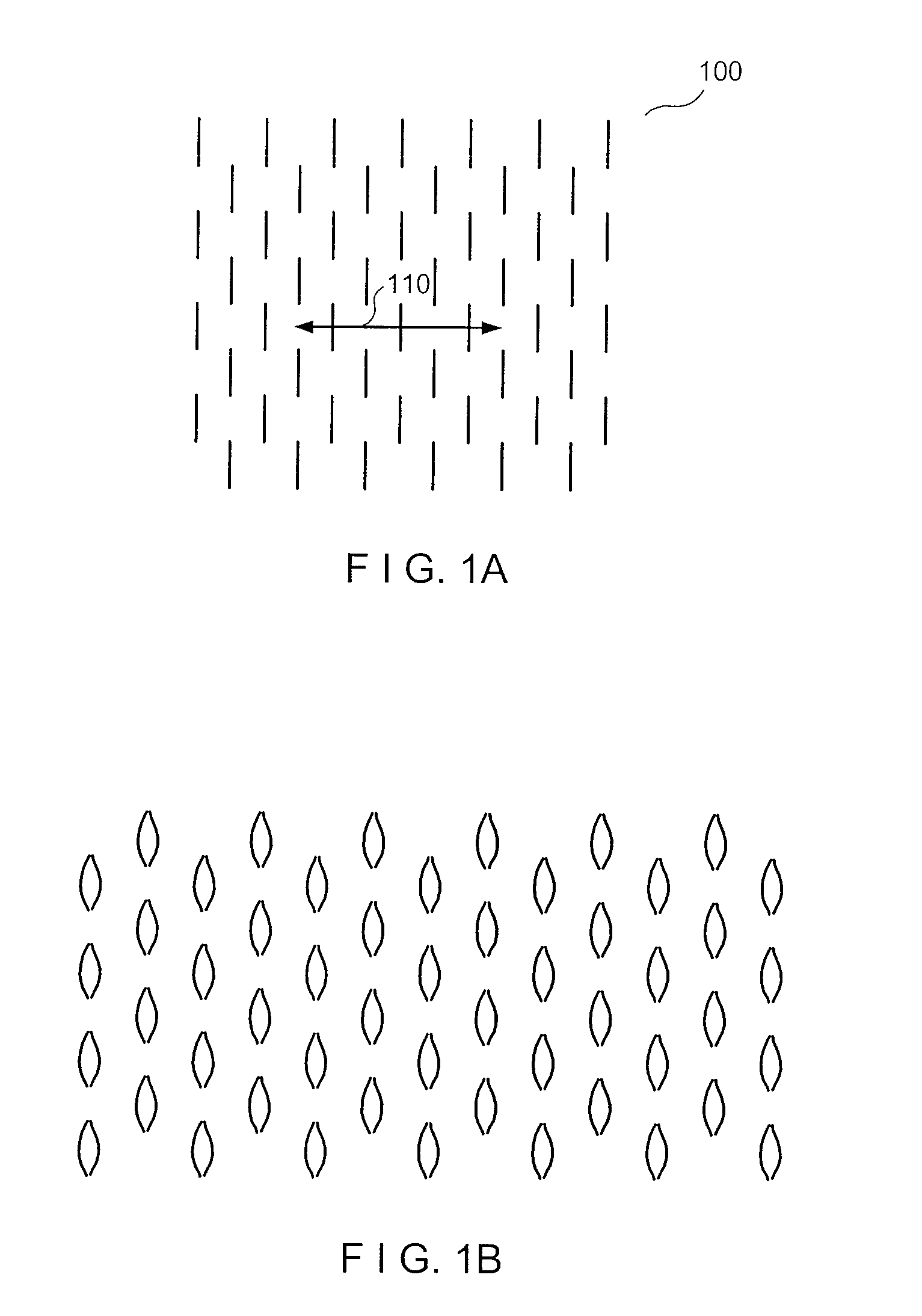
[0012]Such micro-slits may extend through an entire depth of the tissue (e.g., in a full-thickness graft or a split-thickness graft), or to a particular depth such as, e.g., the depth of the dermis (e.g., for in situ meshing and stretching). A force can then be applied to the tissue to expand it, which can widen the micro-slits to form lens-shaped or rounded gaps therefrom. The small dimensions of the micro-slits—and the gaps formed therefrom after stretching—can facilitate rapid regrowth and filling of these gaps, which can be facilitated by adjacent functional tissue. A shallower depth can also be formed, e.g., a depth of about 0.6 mm in skin tissue, or about one-third (⅓) of the skin thickness, which can assist in avoiding formation of scars after the tissue is stretched and allowed to heal. Accordingly, the use of such micro-slits can reduce healing times and result in an aesthetically pleasing appearance of the stretched tissue.

Problems solved by technology

For example, burn victims often suffer through many painful surgeries performed to relieve the tension and contraction of hypertrophic scars.
After excision of skin cancer on the forehead and scalp, large defects are often left to heal openly because the surrounding skin cannot be moved enough to close the wound.
On the legs, skin healing can be poor and the skin is relatively inelastic, such that surgical wounds and ulcers on the legs are difficult to close.
Scalp reduction surgery can be limited by the rigidity of scalp skin.
Meshing may not be ideal for expanding full-thickness grafts because, for example, (a) the lens-shaped holes left by gross meshing and expansion of full-thickness skin can be even more disfiguring than in split-thickness grafts because they are much deeper, and may look like an array of acne scars after healing, and (b) the large, full-thickness holes can take weeks to heal because cells from the surrounding dermis have to build new fibrotic tissue to fill in the substantial volume of each hole.
Similarly, meshing of flaps may not be appropriate because skin incisions within a flap can sever some of the blood supply to the flap, thus impairing the viability of the flap tissue.
For these reasons, conventional tissue meshing may generally not be suitable for expansion of skin grafts or flaps during surgery, and not for relief of tension on scars, nor to modify the scar tissue itself.

Method used


Image

  • Method and apparatus for tissue expansion
  • Method and apparatus for tissue expansion
  • Method and apparatus for tissue expansion

Examples


example

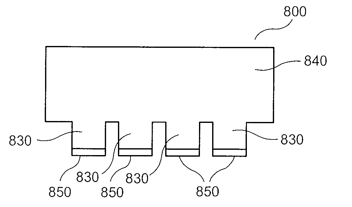
[0111]Images of an exemplary blade in accordance with embodiments of the present disclosure are shown in FIGS. 15A and 15B. The exemplary blade illustrated in FIG. 15A can be similar to the exemplary blade 800 illustrated in FIG. 8A. A close-up view of the extensions provided on the blade 800, each with a cutting edge at a distal end thereof, is shown in FIG. 15B.

[0112]Images of the exemplary apparatus in accordance with certain exemplary embodiments of the present disclosure are shown in FIGS. 16A-16D. The exemplary apparatus shown in FIG. 16A can include a handle attached to plurality of blades, where each blade is similar to the blade shown in FIG. 15A. FIGS. 16B and 16C are close-up images of the stack of blades having spacers between them, similar to the stack of the blades 900 shown in FIG. 9. FIG. 16D provides an image of the array of cutting edges provided by the exemplary apparatus, which can be used to form a pattern of micro-slits that is similar, e.g., to the pattern of ...


PUM


Abstract

Exemplary embodiments of the present disclosure provide method and apparatus for facilitating stretching of a bio-logical tissue by forming a plurality of micro-slits in the tissue. Each micro-slit can be less than about 2 mm or less than about 1.5 mm long, or even less than about 1 mm, such that small gaps that can heal quickly can be formed when the tissue is stretched. The micro-slits can be formed using a plurality of cutting arrangements or an ablative laser. The micro-slits can be formed in various patterns, including staggered rows, circular or spiral patterns, or random patterns.

Description

CROSS REFERENCE TO RELATED APPLICATION[0001]The present application claims priority from U.S. Provisional Patent Application Ser. No. 61 / 041,591 filed Apr. 1, 2008, the disclosure of which is incorporated herein by reference in its entirety.FIELD OF THE DISCLOSURE[0002]The present disclosure relates to an expansion of biological tissue, and in particular to exemplary embodiments of method and apparatus which can provide such expansion.BACKGROUND INFORMATION[0003]Expansion of connective tissue, especially skin, fascia, cartilage and tendon, is often desirable for cosmetic or functional purposes. Achieving tissue expansion without damaging, impairing, or aesthetically harming the tissue is a common concern in reconstruction after surgery, trauma, and for various medical and congenital disorders.[0004]Tissue expansion can be used for tension relief of contracted surgical, traumatic, and burn scars. For example, burn victims often suffer through many painful surgeries performed to relie...

Claims


Application Information

Patent Timeline
Patent Type & Authority Applications(United States)
IPC IPC(8): A61B18/20A61B17/32
CPCA61B17/32093A61B19/24A61B18/20A61B17/322A61B90/02A61F2/0059A61F2/105
Inventor ANDERSON, RICHARD ROXWILLEY, ANDREAFARINELLI, WILLIAM A.
Owner THE GENERAL HOSPITAL CORP
Learn More