Generation and isolation of antigen-specific T cells
a technology of antigen-specific t cells and t cells, which is applied in the field of generating, isolating, and expanding antigen-specific t cells, can solve the problems of reducing the number of cancer cells, requiring labor intensive and expensive cloning, and affecting the survival rate of t cells during long-term culture. it can reduce the presence of cancer cells
- Summary
- Abstract
- Description
- Claims
- Application Information
AI Technical Summary
Benefits of technology
Problems solved by technology
Method used
Image
Examples
example 1
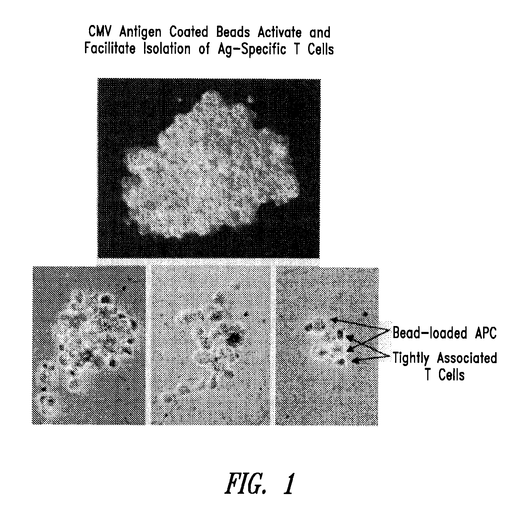
CMV Antigen Coated Beads Activate and Facilitate Isolation of Antigen-Specific T Cells
[0141]In this experiment, cytomegalovirus (CMV)-coated beads were used to activate and isolate antigen-specific T cells.
[0142]CMV lysate prepared using standard techniques was mixed at room temperature for 1-2 hours with Dynabead M-450 while rotating. Beads were then washed once, and added to PBMC. Within hours, the beads were phagocytosed in the APC. Within 72 hours, CMVpp65-HLA-A2 tetramers detected CD25-high (activated) T cell specific for CMV pp65. Magnetic selection of the bead-loaded APC with the associated antigen-specific T cells was carried out at day 5, thereby enriching for CMV-specific T cells. As shown in FIG. 1, following magnetic separation, CMV-specific T cells were still tightly associated with bead-loaded APC. It should be noted that magnetic separation can be carried out anywhere from about day 1 to about day 10.
example 2
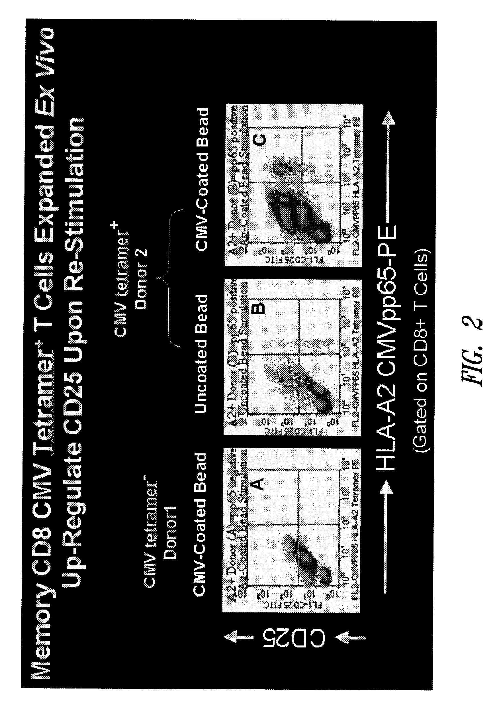
Memory CD8 CMV Tetramer+ T Cells Expanded Ex Vivo Up-Regulate CD25 Upon Re-Stimulation
[0143]In this example, antigen-coated beads were used to activate CMV-specific CD8+ T cells ex vivo.
[0144]PBMC from CMV pp65 tetramer-positive and tetramer-negative donors were stimulated with paramagnetic Dynal M-450 beads coated with CMV lysate. As controls, CMV pp65 tetramer-negative PBMC were cultured with CMV-lysate coated beads (FIG. 2, panel A), CMV pp65 tetramer-positive PBMC were cultured with “naked” beads (no CMV antigen) (FIG. 2, panel B). CMV pp65 tetramer-positive PBMC were cultured with CMV-lysate coated beads (FIG. 2, panel C). Following stimulation, activation of CMV-specific T cells was measured on Day 10 by CMV pp65 HLA-A2 tetramer stain and CD25 expression as an indicator of activation. As shown in FIG. 2, up-regulation of CD25 was observed in memory CD8 CMV tetramer+ T cells expanded ex vivo using antigen-coated beads.
[0145]Antigen-coated beads can be used to activate and stimu...
example 3
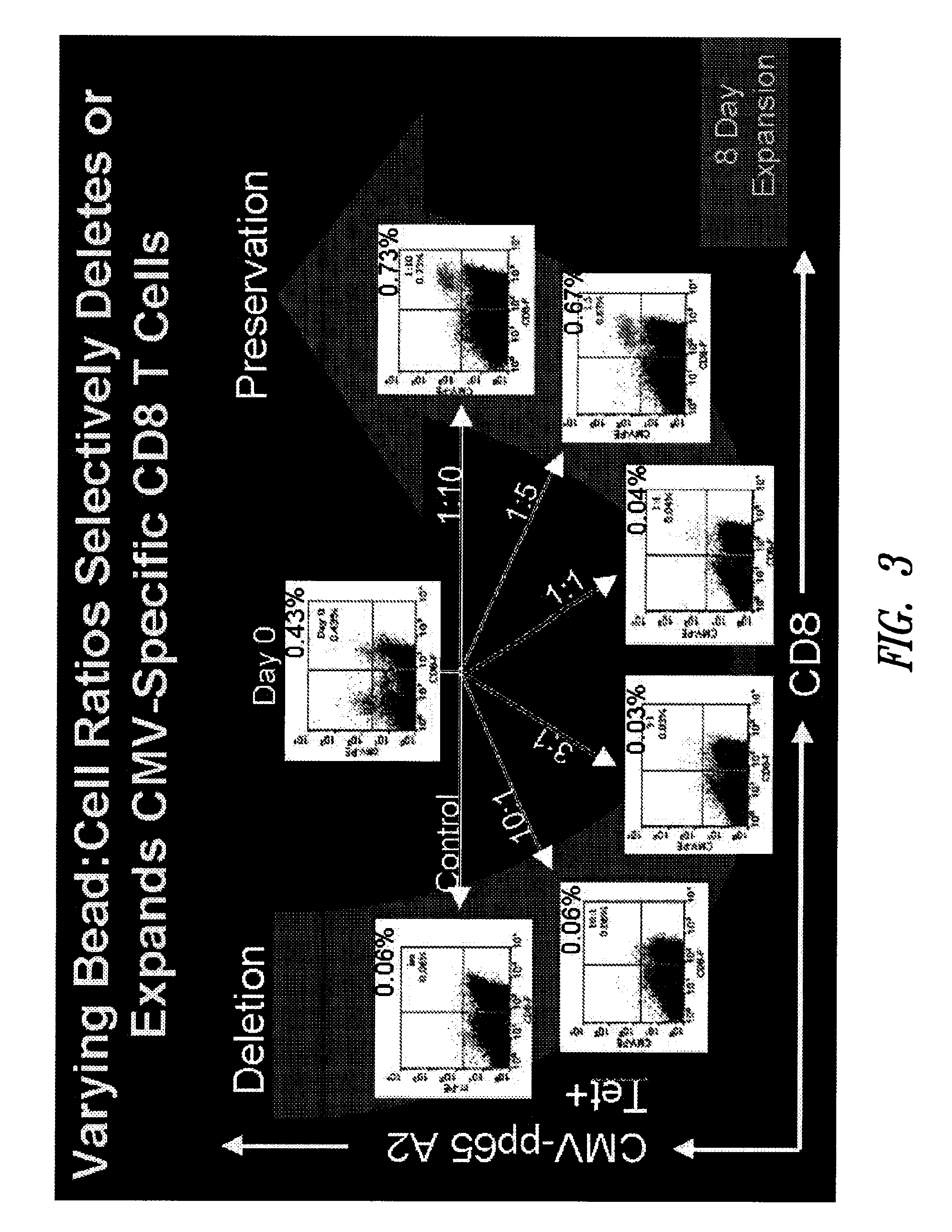
Varying Bead:Cell Ratios can Selectively Expand or Delete Memory CD8 T Cells
[0146]This example shows that the bead:cell ratio can have a profound effect on expansion of different populations of T cells. In particular, a high bead:cell ratio (3:1-10:1, 20:1 and higher) tends to induce death in antigen-specific T cells while a lower bead:cell ratio (1:1-1:10, 1:20, 1:30, 1:40, 1:50 or lower) leads to expansion of antigen-specific T cells. Further, the data described below show that lower bead:cell ratios lead to improved cell expansion in polyclonal cell populations as well. Thus, this example shows that lower bead:cell ratios improve overall cell expansion.
[0147]Cells were prepared and stimulated using the XCELLERATE I™ process essentially as described in U.S. patent application Ser. No. 10 / 187,467 filed Jun. 28, 2002. Briefly, in this process, the XCELLERATED™ T-cells are manufactured from a peripheral blood mononuclear cell (PBMC) apheresis product. After collection from the patien...
PUM
 Abstract
Description
Claims
Application Information
 - R&D
- Intellectual Property
- Life Sciences
- Materials
- Tech Scout
- Unparalleled Data Quality
- Higher Quality Content
- 60% Fewer Hallucinations
Browse by: Latest US Patents, China's latest patents, Technical Efficacy Thesaurus, Application Domain, Technology Topic, Popular Technical Reports.
© 2025 PatSnap. All rights reserved.Legal|Privacy policy|Modern Slavery Act Transparency Statement|Sitemap|About US| Contact US: help@patsnap.com


