
Ophthalmologic apparatus and method for the observation, examination, diagnosis, and/or treatment of an eye

Active Publication Date: 2011-05-19
CARL ZEISS MEDITEC AG
View PDF18 Cites 23 Cited by
  • Summary
  • Abstract
  • Description
  • Claims
  • Application Information

AI Technical Summary

Benefits of technology

[0026]The invention herein is based on the task of developing an ophthalmological device and method for non-contact observation, examination, diagnosis, and / or therapy of an eye. Thereby, independently from diagnosis and / or therapy, observation and / or imaging should be possible with said device which, preferably, is based on the design of a fundus camera and / or an ophthalmoscope. Particularly, the solution developed allows for high reproducibility and automatic documentation from the examination and diagnosis all the way through the therapy process.
[0034]Thereby, said diagnostic and / or therapy option is possible without fixation of the eye. Due to the non-contact approach, the special risk of infections is diminished. For monitoring of the position of the therapy and / or diagnosis spot on the retina, an aiming beam can be used, which indicates said position.
[0035]Moreover, it is important that in the combined application of therapy and diagnostic laser and a fundus camera, all existing tools and adjustment aids, such as positioning aid, focusing aid, and retaining aid of the fundus camera are available for easy and quick operation.
[0038]The invention herein presents a solution in the form of an ophthalmological device and method for non-contact observation, examination, diagnosis, and / or therapy of an eye. Said device, which, preferably, is based in its design on a fundus camera and / or an ophthalmoscope, allows for observation and / or imaging independently from a diagnosis and / or therapy. Therefore, it offers the possibility of executing numerous examinations, diagnoses, and even treatments of an eye with only one device. The high reproducibility and possibility of an automatic documentation from the examination and diagnosis all the way through the therapy process offer an additional essential advantage when compared to separate single devices.

Problems solved by technology

These small, new, anomalous blood vessels grow and proliferate from the choroid layer and can cause acute loss of vision if blood collects in or below the retina.
A more complex diagnosis, which takes place in individual spots on the retina, such as perimetry, optical coherence tomography or a precisely accurate therapy through coagulation, is still not possible with said system.
Thereby, it is disadvantageous that the laser coagulation with a slit lamp requires a contact glass, and its reproducibility is extremely low.
Furthermore, the accuracy of positioning the laser spot on the retina depends greatly on the operator since a precisely accurate therapy or diagnosis is only possible if the scanner is aligned in such a way that the laser spot covers exactly the desired spot on the retina.
In addition, only a local therapy of the retina is possible due to the very small observation area when compared with the fundus camera.
Moreover, it is not possible to perform a fundus image-based positioning for the coagulation laser as well as document the set coagulation points.
Diagnostic procedures, such as perimetry, fluorescence imaging, spectroscopic analyses, or optical coherence tomography are also impossible.

Method used


Image

  • Ophthalmologic apparatus and method for the observation, examination, diagnosis, and/or treatment of an eye
  • Ophthalmologic apparatus and method for the observation, examination, diagnosis, and/or treatment of an eye
  • Ophthalmologic apparatus and method for the observation, examination, diagnosis, and/or treatment of an eye

Examples


Embodiment Construction

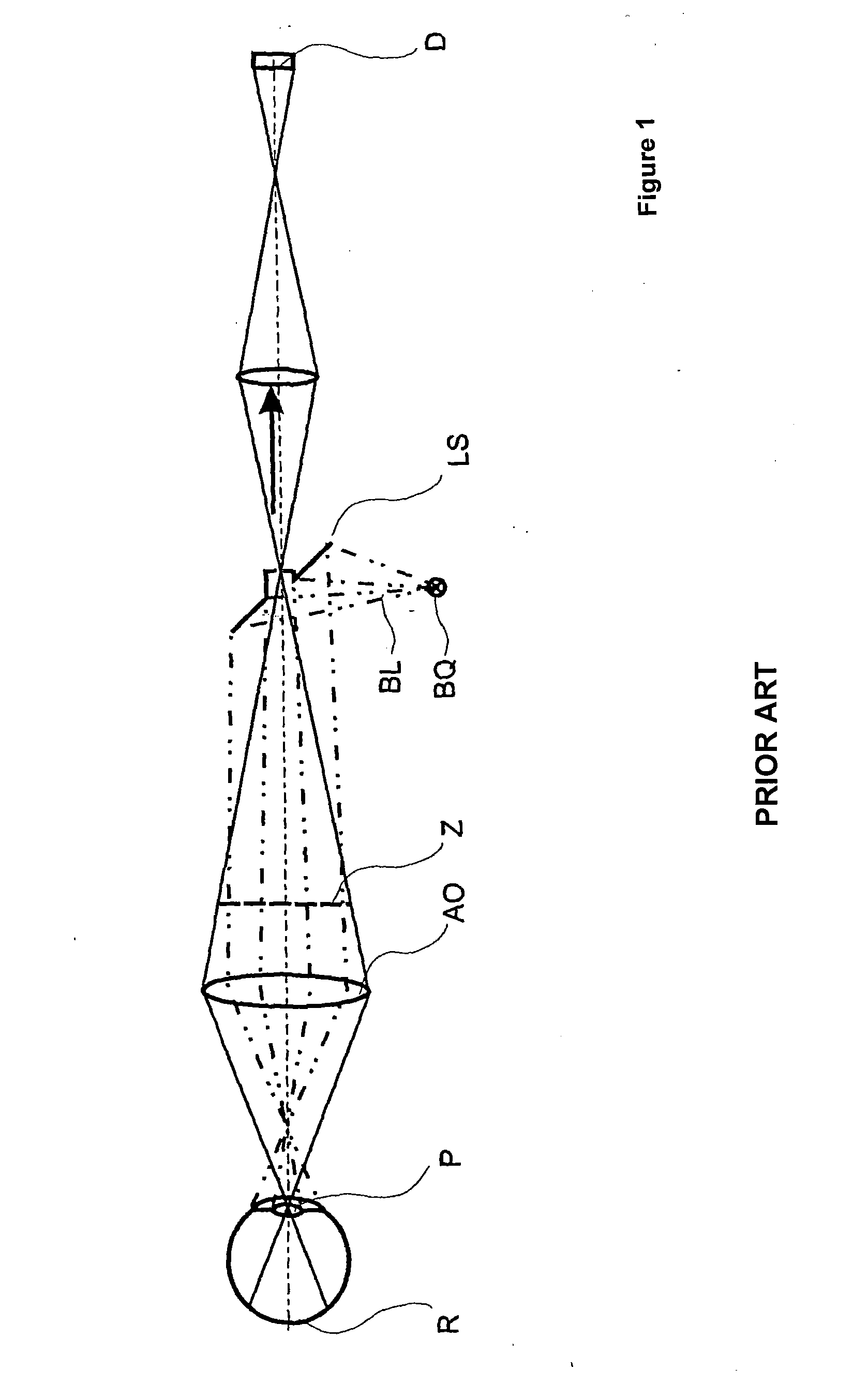
[0044]At first, and in order to better illustrate the suggested technical solution, prior art shall once more be described. Thereto, FIG. 1 shows the schematic design of a known fundus camera.

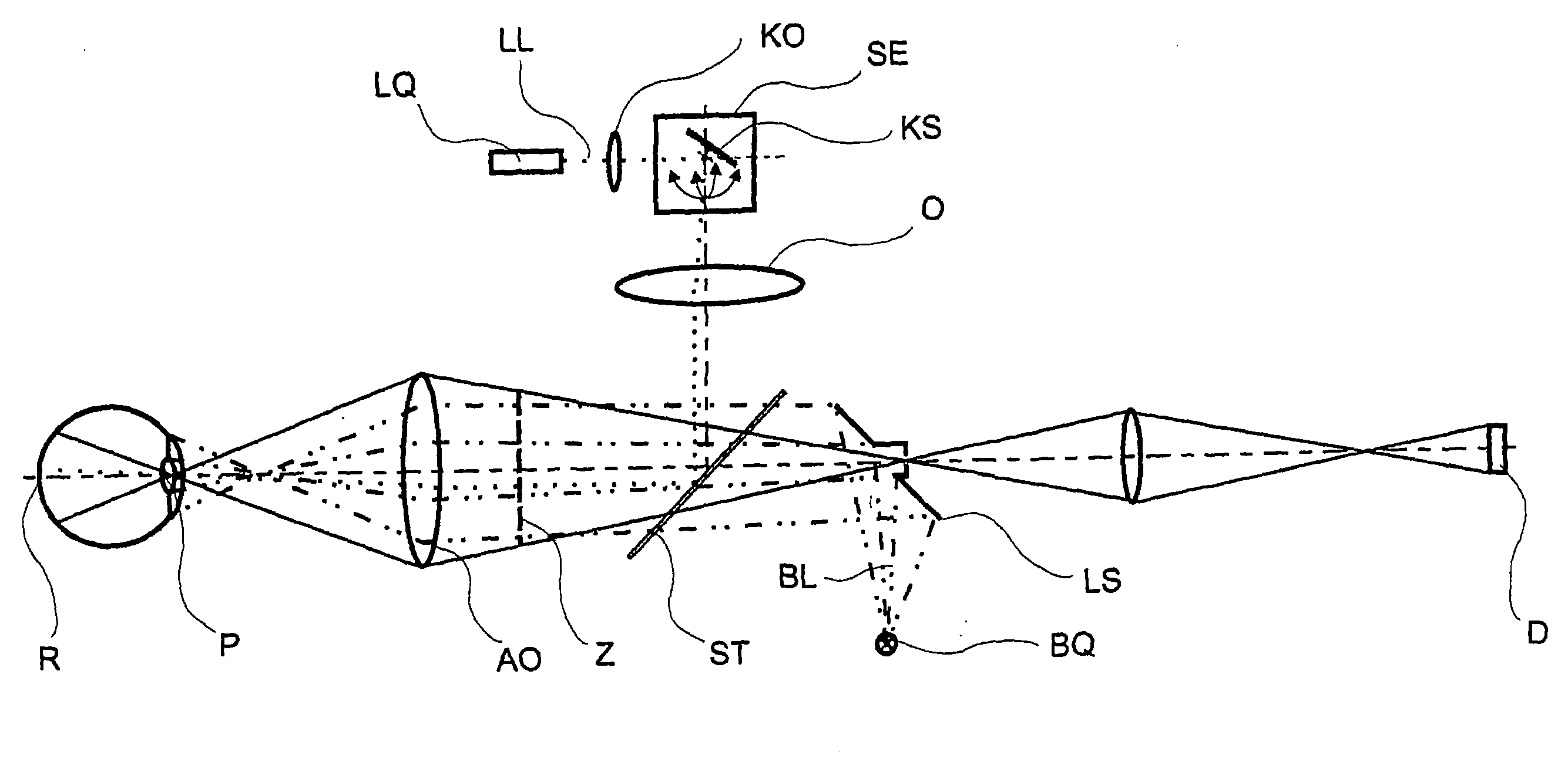
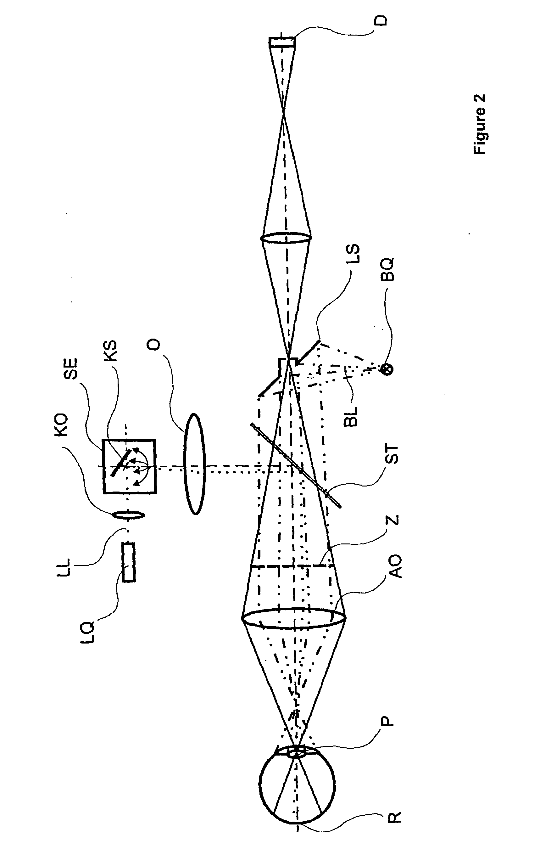
[0045]In a fundus camera, according to prior art, the illumination light BL, originating from an illumination source BQ, is mapped via a perforated mirror LS and imaging optics AO through the pupil P on the retina R of the eye. For the purpose of imaging, the light reflected from the retina R of the eye is mapped on a detector D through the pupil P of the eye via the imaging optics AO and through the perforated mirror LS. Thereby, an intermediate image Z is produced from the image of the retina R between imaging optics AO and perforated mirror LS and mapped through the aperture of the perforated mirror LS on the detector D in the form of a CCD camera.

[0046]Thereby, the illumination source produces white or infrared illumination light BL. The white light can either be emitted as continuous spect...


PUM


Abstract

An ophthalmologic apparatus and a method for the contactless observation, examination, treatment, and / or diagnosis of an eye. The apparatus is structurally based on a fundus camera or an ophthalmoscope. An illumination beam path extends from a first illumination source to the eye and is fitted with a perforated mirror and imaging optics, and an observation beam path extends from the eye to a detector via the imaging optics and through the perforated mirror. The arrangement additionally comprises a beam path for scanning illumination which extends from a second illumination source to the eye and is fitted with a scanning unit, a lens, and a beam splitter in addition to the imaging optics. The scanning unit that is arranged in the beam path for scanning illumination is designed as (an) electrostatically or / and galvanometrically driven bidirectional or unidirectional tilting mirror(s).

Description

PRIORITY CLAIM[0001]The present application is a National Phase entry of PCT Application No. PCT / EP2009 / 001318, filed Feb. 25, 2009, which claims priority from German Application Number 102008011836.2, filed Feb. 28, 2008, the disclosures of which are hereby incorporated by reference herein in their entirety.FIELD OF THE INVENTION[0002]The invention relates to an ophthalmological device and method for non-contact observation, examination, therapy, and / or diagnosis of an eye. In its design, the device is based on a fundus camera and / or an ophthalmoscope.BACKGROUND OF THE INVENTION[0003]Images of the topology of the eyeground (retina or fundus) are important for the diagnosis of many eye diseases. Many of the diseases of the retina can be examined more precisely with the accurate use of diagnostic lasers and treated with the precisely accurate application of therapy lasers.[0004]In the prior art, numerous and diverse solutions are known for the observation, diagnosis, and therapy of t...

Claims


Application Information

Patent Timeline
IPC IPC(8): A61B3/18A61B3/10A61B3/14A61B3/02A61B3/024
CPCA61F9/008A61F9/00821A61F2009/00846G01B9/02091A61F2009/00897A61B3/1025A61F2009/00863
Inventor BIERNAT, DETLEFBUBLITZ, DANIELBRODSCHELM, ANDREASMOHR, THOMASRICHTER, STEFAN
Owner CARL ZEISS MEDITEC AG
Learn More