
Completion of Truncated Attenuation Maps Using MLAA

a technology of attenuation maps and truncated attenuation maps, applied in the field of estimating the attenuation of imaged objects, can solve the problems of increased image noise, image distortion, and loss of counts, and achieve the effects of reducing the number of attenuation maps, increasing image noise, and increasing image nois

Active Publication Date: 2011-05-05
SIEMENS MEDICAL SOLUTIONS USA INC
View PDF9 Cites 33 Cited by
  • Summary
  • Abstract
  • Description
  • Claims
  • Application Information

AI Technical Summary

Benefits of technology

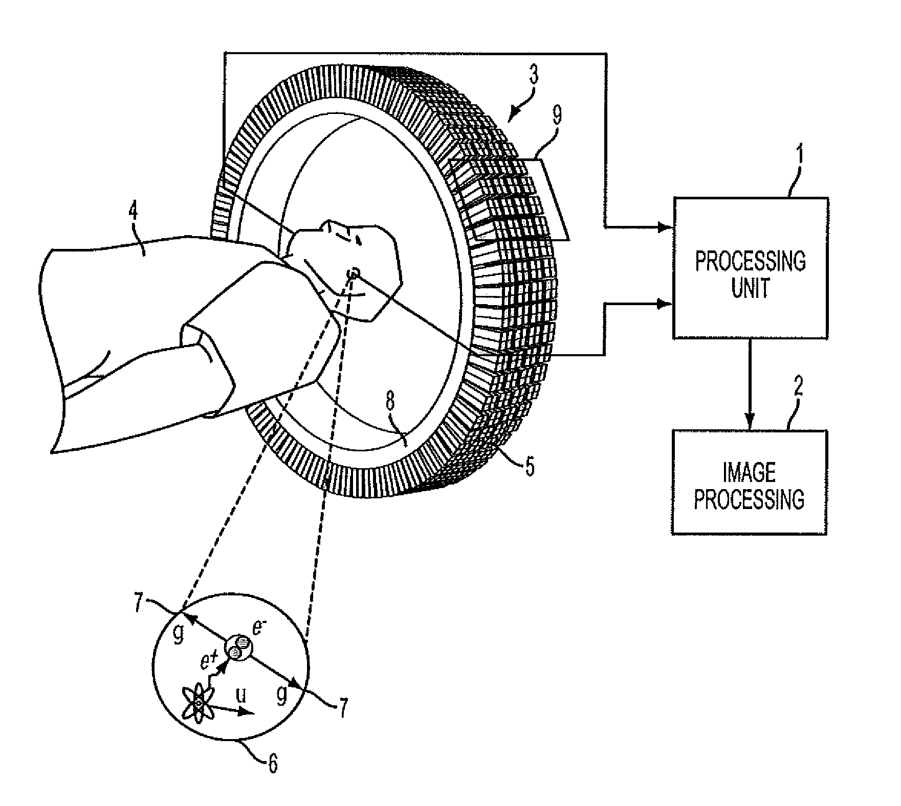
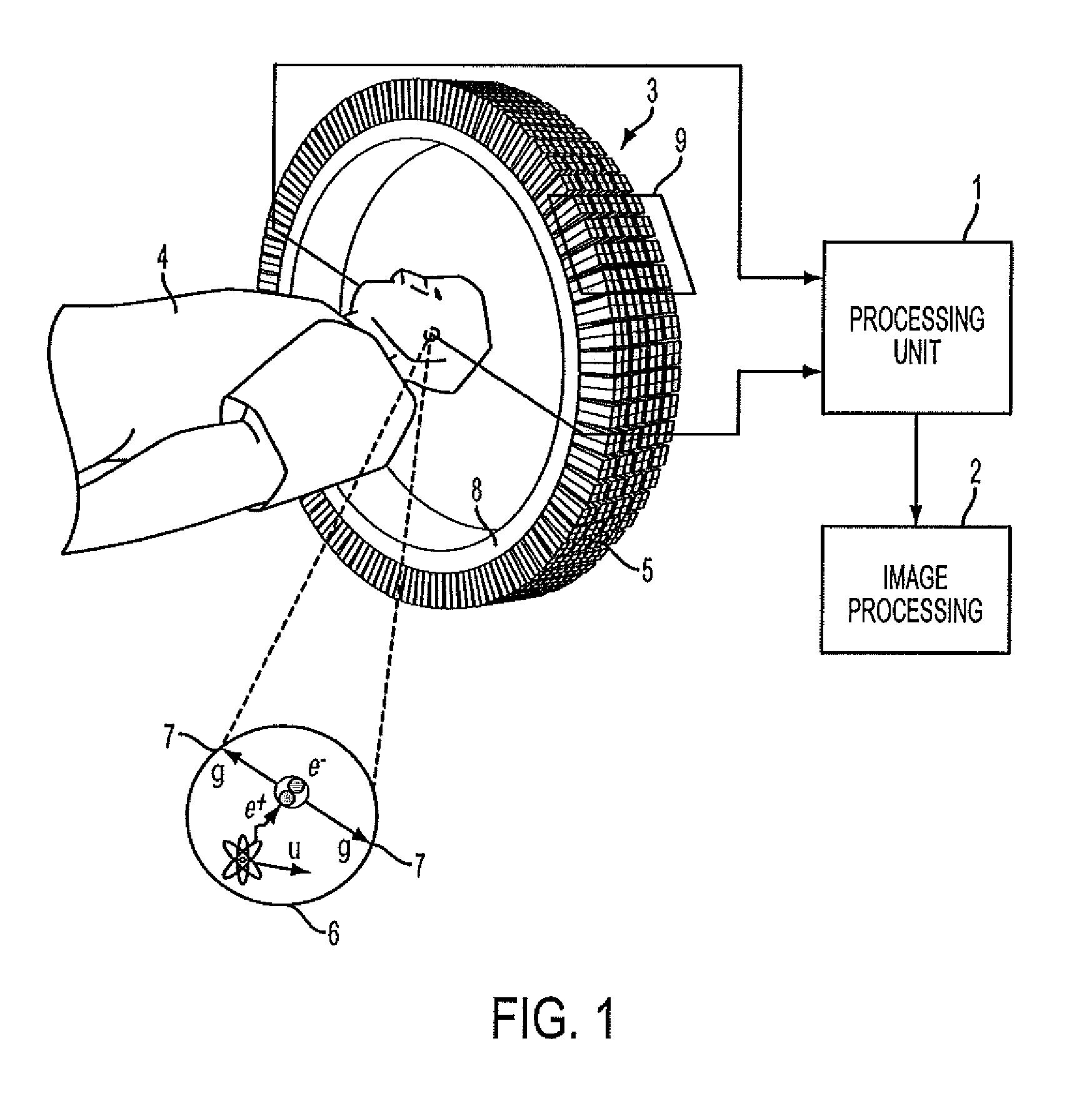
[0019]Consistent with embodiments of the present invention, the present invention comprises a method of generating a best estimate of a complete attenuation map derived from a truncated image and PET emissions data for the object being imaged. The method involves a plurality of steps beginning with the recordation and processing of PET emissions data to determine pixels within a PET imaging system or an integrated hybrid system which includes PET and a morphological imaging modality. Next, the morphological imaging modality as a stand alone system or as part of an integrated hybrid system records image data which is processed to determine an attenuation map. The attenuation maps, are for image modalities such as CT and MR truncated because the field of view of the morphological imaging modality is smaller than the field of view of the PET camera. This results in a truncated image a

Problems solved by technology

Attenuation problems are greater with PET imaging compared to traditional nuclear medicine SPECT imaging.
Loss of counts due to attenuation increases image noise, image artifacts, and image distortion.
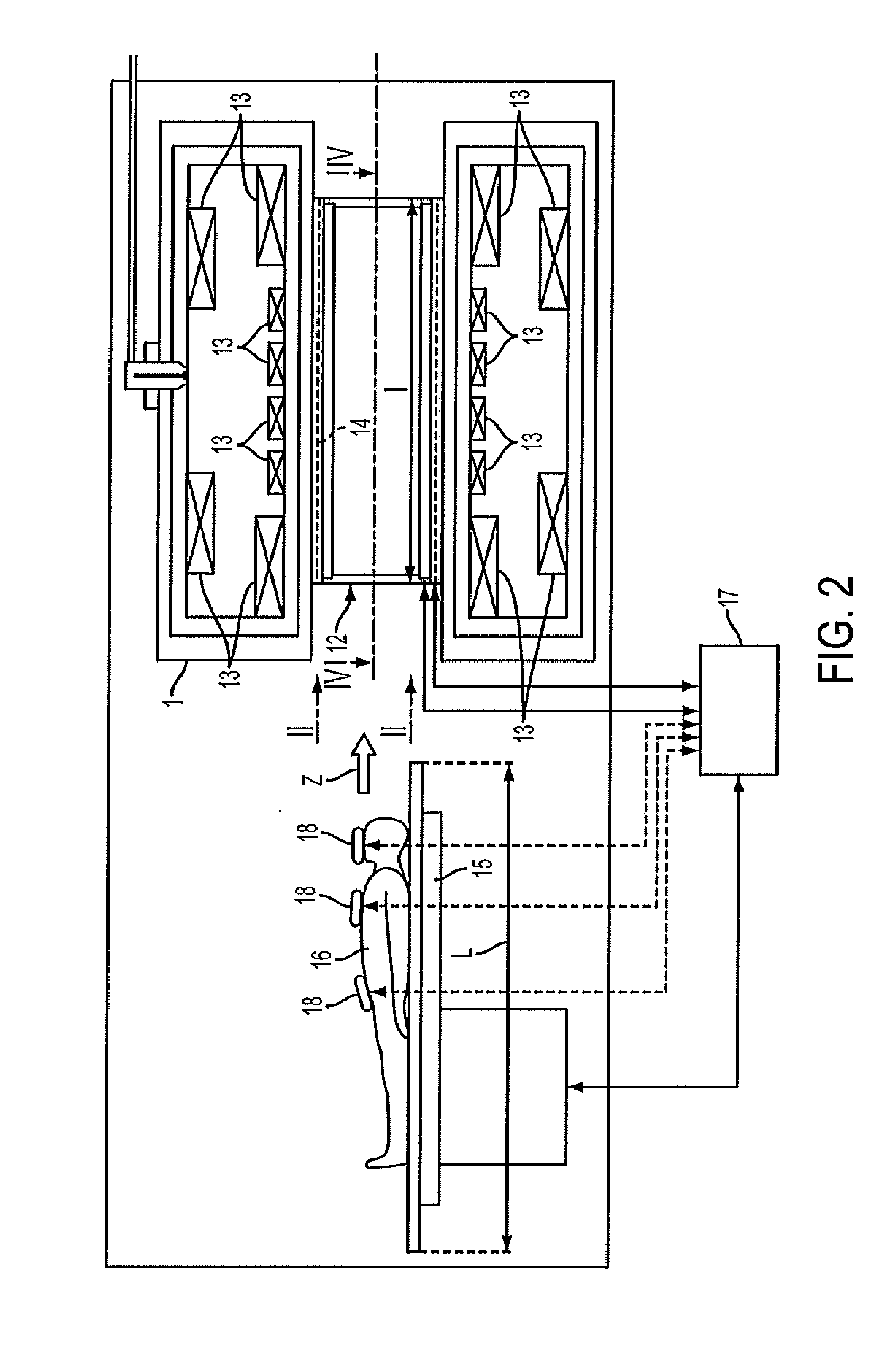
This field of view problem also exists in imaging systems that integrate PET and MR imaging modalities, resulting in an even more truncated MR image than that of a CT image resulting from an integrated PET and CT imaging system.
A patient holding their arms over their head is not a solution for PET-MR systems.
In the MR-PET integrated imaging system the conversion of the MR image values to linear attenuation coefficients at 511 keV adds another layer of complexity, because the MR imaging signal does not correlate with electron density.
The estimation of human attenuation from using MR image data is a difficult task.
However, this approach alone is not sufficient for estimating the attenuation map outside the MR FOV.
The success was limited because there was too much missing information.
Notwithstanding the above, the methodology for deductively generating an attenuation map outside the MR imaging modality field of view using an atlas is not particularly straightforward.

Method used


Image

  • Completion of Truncated Attenuation Maps Using MLAA
  • Completion of Truncated Attenuation Maps Using MLAA
  • Completion of Truncated Attenuation Maps Using MLAA

Examples


Embodiment Construction

[0032]The following detailed description refers to the accompanying drawings. Wherever possible, the same reference numbers are used in the drawings and the following description to refer to the same or similar parts. While several embodiments and features of the invention are described herein, modifications, adaptations and other implementations are possible, without departing from the spirit and scope of the invention. Rather these embodiments are provided so that this disclosure will be complete and will fully convey the invention to those skilled in the art. For example, substitutions, additions or modifications may be made to the components illustrated in the drawings, and the methods described herein may be modified by substituting, reordering or adding steps to the disclosed methods. Accordingly, the following detailed description does not limit the invention. Instead, the proper scope of the invention is defined by the appended claims.

[0033]The present invention solves exist...


PUM


Abstract

The present invention is a method of generating a best estimate of an image attenuation map derived from a truncated image attenuation map and PET emissions data for the object being imaged by a morphological imaging modality. The method involves a plurality of steps beginning with the recordation and processing of PET emissions data. Next, the morphological imaging modality records image data which is processed to determine an attenuation map. The attenuation map, for image modalities such as CT and MR scanning systems integrated with PET, is truncated, resulting in a truncated attenuation map image. Pixels for which attenuation data needs to be determined are identified and attenuation coefficients for these pixels are estimated and combined with the truncated attenuation map to generate a full initial attenuation map for the image, which is iteratively processed together with the PET emission data until the improvement change in the emission image reaches a defined threshold improvement level.

Description

BACKGROUND OF THE INVENTION[0001]I. Field of the Invention[0002]The present invention relates to methods of estimating imaged object attenuation maps and more particularly to the completion of truncated attenuation maps by estimating missing portions using Maximum-Likelihood Expectation Maximization (MLEM) reconstructive techniques.[0003]II. Background Information[0004]Nuclear medicine is a unique medical specialty wherein radiation is used to acquire images, which show the function and anatomy of organs, bones or tissues of the body. Radiopharmaceuticals are introduced into the body, either by injection or ingestion, and are attracted to specific organs, bones or tissues of interest. Such radiopharmaceuticals produce gamma photon emissions, which emanate from the body and are captured by a scintillation crystal, with which the photons interact to produce flashes of light or “events.” Events are detected by an array of photo detectors, such as photomultiplier tubes, and their spatia...

Claims


Application Information

Patent Timeline
IPC IPC(8): G06K9/00
CPCG06T2211/432G06T11/005
Inventor MICHEL, CHRISTIAN J.NUYTS, JOHAN
Owner SIEMENS MEDICAL SOLUTIONS USA INC
Learn More