
Raman-active reagents and the use thereof

a raman-active reagent and raman-active technology, applied in the field of raman-active reagents, can solve the problems of insufficient sensitivity of conventional raman spectroscopy for immunoassay readout, limiting the reliable individual and quantitative detection of multiple analytes in a single sample, and limiting the use of fluorescence spectroscopy. , to achieve the effect of narrowing the spectral bandwidth, maximizing the number of labels

Inactive Publication Date: 2011-03-24
IOWA STATE UNIV RES FOUND
View PDF9 Cites 10 Cited by
  • Summary
  • Abstract
  • Description
  • Claims
  • Application Information

AI Technical Summary

Benefits of technology

The Raman-active reagents provide stable, sensitive, and specific detection of multiple analytes in a single sample, overcoming the limitations of conventional Raman spectroscopy and fluorescence interference, with improved detection limits and reduced labor costs.

Problems solved by technology

Although fluorescence spectroscopy has seen substantial use in scientific research and clinical diagnostics, there are disadvantages in using fluorescence spectroscopy.
Even so, the spectral overlap between the emission of the different fluorescent molecules often limits reliable individual and quantitative detection of multiple analytes in a single sample.
Conventional Raman spectroscopy usually lacks sufficient sensitivity for use as a readout method for immunoassays.
Raman spectroscopy is also unsuccessful for fluorescent materials because the broad fluorescence emission bands tend to swamp the weaker Raman bands.
A major barrier that prohibits using SERS for the direct detection of biological samples is that the surface enhancement effect diminishes rapidly with increasing distance from the metallic surfaces.
In addition, although Raman spectra of biomolecules can be obtained on silver surfaces when coupling SERS and resonance enhanced scattering, the spectra are usually lacking of sufficient chemical content and / or signal amplitude to be used for immunoassay purposes.

Method used


Image

  • Raman-active reagents and the use thereof
  • Raman-active reagents and the use thereof
  • Raman-active reagents and the use thereof

Examples


example 1

Synthesis of Raman-Active Reporter Molecule 4,4′-Dithiobis(Succinimidylbenzoate)

[0078]The Raman-active reporter molecule 4,4′-dithiobis(succinimidylbenzoate) (DSB) was synthesized following a procedure similar to that used for preparing dithiobis(succinimidylundecanoate) as described in Wagner et al., Biophys. J. 1996, 70, 2052, which is incorporated herein by reference. Briefly, 0.50 g of the reporter molecule dithiobisbenzonic acid (DBA) (1.6 mmol) (Toronto Research Chemicals, Inc), 0.67 g of 1,3-dicyclohexylcarbodiimide (DCCD) (3.2 mmol) (obtained from Aldrich), 0.37 g of N-hydroxysuccinimide (NHS) (3.2 mmol) (obtained from Aldrich), and 60 mL of tetrahydrofuran were added to a 100 mL round-bottom flask equipped with a magnetic stir bar and drying tube. The reaction mixture was stirred at room temperature for three days. The solution was then filtered and the solvent was removed under reduced pressure to give an orange residue. The crude product was dissolved in hot acetone and f...

example 2

Preparation of Raman-Active Reagents Using Co-Immobilization

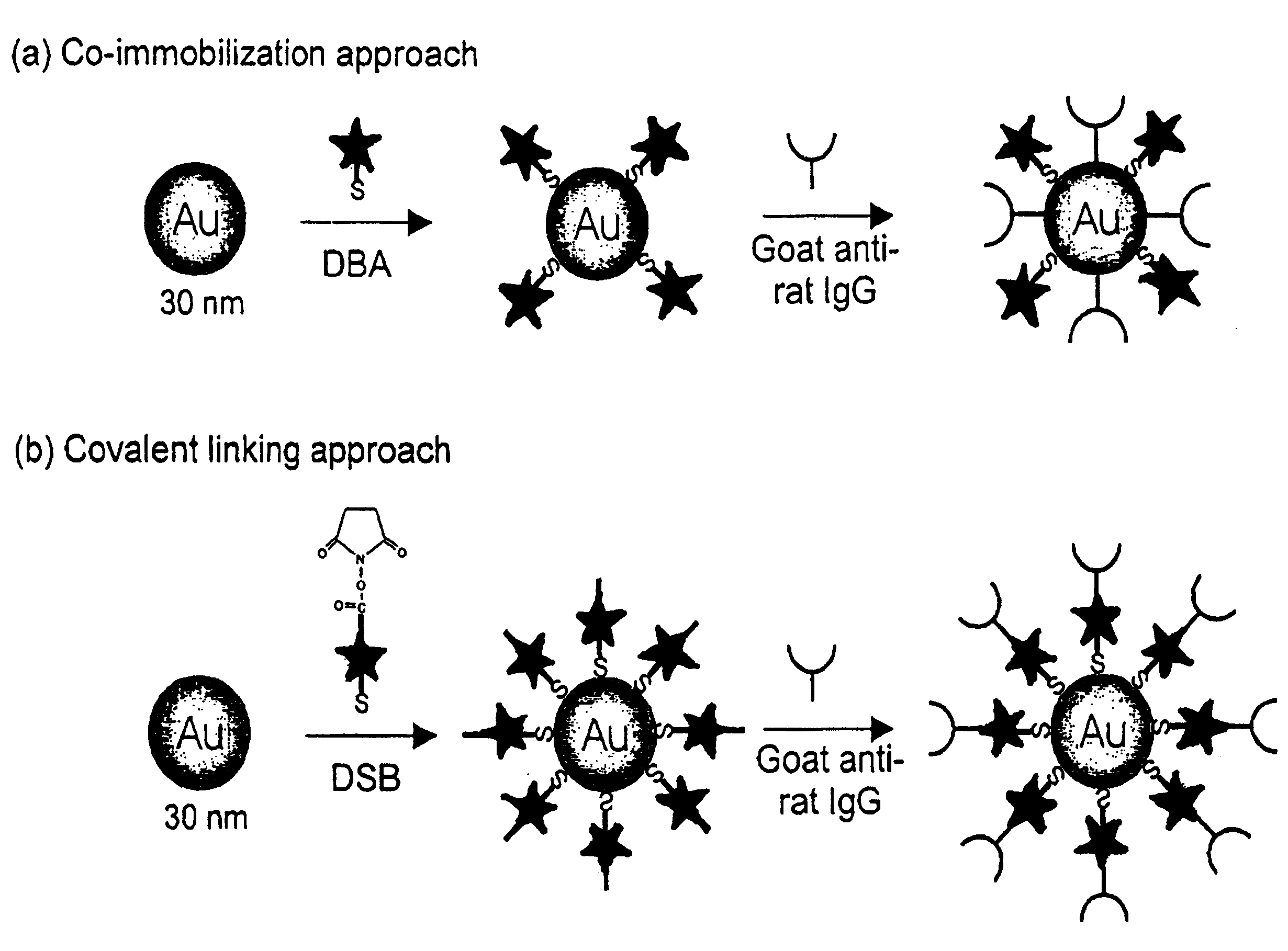
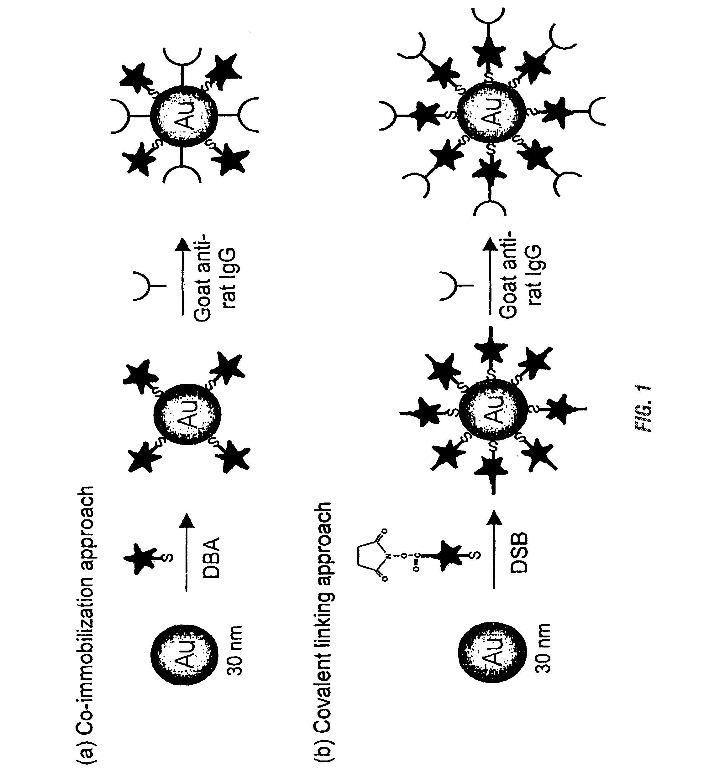
[0079]Raman-active immunogold colloidal reagents were prepared using the coimmobilization approach depicted in FIG. 1(a). First, 25 μL of ethanolic Raman reporter solution (0.5 mM DBA) was added to 10 mL of a suspension of uncoated gold colloids (˜30 nm diameter, 2×10¹¹ particles / mL) (Ted Pella, Inc.). The mixture was allowed to react for 5 hours at room temperature. During this step, the reporter molecules bound via self-assembly onto the colloid surface through the formation of sulfur-gold linkages. We note that this amount of reporter, based on an estimation of the colloidal surface area, will only partially cover the colloid, leaving portions of the uncoated colloidal surface available for protein immobilization. After separating the reporter-labeled colloids from solution by centrifugation at 14,000 g for 4 minutes, the loosely packed, red-colored sediment was resuspended in 10 mL of borate buffer (2 mM, pH 9).

[0080]Th...

example 3

Preparation of Raman-Active Reagents Using a Covalent Linking

[0081]Raman-active immunogold colloidal reagents were prepared using the covalent linking approach depicted in FIG. 1(b). The DSB molecules were used as both Raman reporters and antibody linkers. The succinimide ester group of the DSB molecule can readily react with the primary amine group of an amino acid, such as the lysine, present in antibodies such as IgG to form a covalent bond. As shown in Scheme 2, the preparation of the covalently-linked colloidal reagent follows a process very similar to that used for the co-immobilized reagents. However, with the covalent linking approach, the antibodies indirectly attached to the colloid through the reporter molecules rather than directly adsorbed onto the colloidal surface. Briefly, 25 μL of a reporter-linker solution (5 mM DSB in CHCl3) was added to 10 mL of bare gold suspension (30 nm) under vigorous agitation. The molecules self-assemble onto the colloid surface, with their...


PUM


Abstract

The present invention provides a new class of Raman-active reagents for use in biological and other applications, as well as methods and kits for their use and manufacture. Each reagent includes a Raman-active reporter molecule, a binding molecule, and a surface enhancing particle capable of causing surface enhanced Raman scattering (SERS). The Raman-active reporter molecule and the binding molecule are affixed to the particle to give both a strong SERS signal and to provide biological functionality, i.e. antigen or drug recognition. The Raman-active reagents can function as an alternative to fluorescence-labeled reagents, with advantages in detection including signal stability, sensitivity, and the ability to simultaneously detect several biological materials. The Raman-active reagents also have a wide range of applications, especially in clinical fields (e.g., immunoassays, imaging, and drug screening).

Description

CROSS-REFERENCE TO RELATED APPLICATIONS[0001]This application is a divisional application of U.S. Ser. No. 10 / 931,142, filed Aug. 31, 2004, which is a continuation-in-part of application Ser. No. 09 / 961,628, filed Sep. 24, 2001, which claims the benefit of U.S. Provisional Application 60 / 234,608, filed Sep. 22, 2000. These applications are incorporated herein by reference in their entirety.STATEMENT REGARDING FEDERALLY SPONSORED RESEARCH OR DEVELOPMENT[0002]None.BACKGROUND OF THE INVENTION[0003]Many assays exist for detecting and measuring analytes of small quantity in the presence of a large volume of other substances. Such assays typically make use of the high binding affinity between the analyte (the substance to be detected or measured) and a second molecule having a high degree of specificity for binding to that analyte. These assays are often referred to as ligand-binding assays.[0004]One of the most common ligand-binding assays are immunoassays. Immunoassays typically employ ...

Claims


Application Information

Patent Timeline
Patent Type & Authority Applications(United States)
IPC IPC(8): G01N33/53G01N21/00C07H21/02G01N33/532G01N33/58
CPCC07H21/02G01N21/658G01N33/532G01N33/587G01N33/553G01N33/58G01N33/54373
Inventor PORTER, MARC D.NI, JINGLIPERT, ROBERT J.DAWSON, G. BRENT
Owner IOWA STATE UNIV RES FOUND
Learn More