
Transcranial ultrasound transducer with stereotactic conduit for placement of ventricular catheter

Inactive Publication Date: 2011-01-13
PHILLIPS SCOTT B +1
View PDF27 Cites 11 Cited by
  • Summary
  • Abstract
  • Description
  • Claims
  • Application Information

AI Technical Summary

Benefits of technology

[0011]The present invention is directed to an apparatus, system and method which overcomes the problems in the prior art. In particular, the present invention is an ultrasound probe comprising a handle portion for use in gripping the ultrasound probe and a head portion for directing ultrasound waves emitted from a transducer in a fixed direction into a body of a patient. The head portion is connected to the handle portio

Problems solved by technology

External ventricular drain placement is a highly invasive procedure since the catheter passes through the brain parenchyma before entering the targeted ventricle and can cause iatrogenic hemorrhaging.
In the freehand method, often multiple attempts (or passes) are required before the external ventricular drain enters the targeted ventricle, with each subsequent pass likely increasing the risk of iatrogenic hemorrhaging or other problems.
For example, some external ventricular drains will inevitably fail to drain for various reasons.
This may be remedied by flushing the catheter, but this can increase the risk of infection.
If this and / or other troubleshooting measures fail, the external ventricular drain must be removed and replaced, necessitating an additional invasive procedure.
As a result, greater accuracy during the initial placement of the external ventricular drain can decrease the frequency of obstruction by choroid plexus and parenchymal debris and therefore decrease the overall morbidity of an external ventricular drain.
Currently, there is no method of determining the location of the eyelets during the placement of an EVD.
Although post-procedure head computed tomography (CT) can make such a determination and thus is the only conventional method of determining the viability of tPA therapy via the catheter placed within the ventricle, catheters cannot be repositioned after the procedure is completed and the skin is closed for risk of infection.
However, this approach is still “blind,” and is dependent on an accurately-positioned burr hole, a normocephalic skull shape and a non-distorted intracranial anatomy.
This approach requires a specially-designed and expensive catheter or stylet for performing the ventriculostomy.
One is that the image quality from such a small transducer may be less than optimal.
As a result, the method described in this article requires significant enlargement of the standard-sized burr hole to 15 to 20 mm to properly position both the catheter and probe against the dura in the burr hole opening, requiring either additional steps and the resultant extra time to perform the procedure or additional tools (i.e., additional drilling with multiple drill bits), and is not easily performed bedside.
Another drawback is that although sterile probe covers are available for the Aloka Probe, these covers obscure the groove on the Aloka Probe and make it difficult to insert the catheter.
As a result, it is likely that such covers will not likely be used, necessitating sterilization of the Aloka Probe after every use and a consequent delay in performing a subsequent procedure using the same Aloka Probe of between two to six hours for sterilization.
A still further drawback of the use of the Aloka Probe is the need for the doctor to position the catheter within the shallow groove therein and hold it firmly against the Aloka Probe for the entire procedure.
This additional effort generates fatigue and tremor in the doctor's hand and requires additional time for ensuring that the catheter is being properly inserted while performing the procedure.
Furthermore, the use of a standard probe and standard catheter mated together would likely require a hole in the skull that is larger than that provided by the standard perforator drill bit, thereby either requiring a non-standard drill bit or multiple passes for creating the hole through the skull.
Finally, the use of a separate clip makes it difficult to employ a sterile sheath over the probe.

Method used


Image

  • Transcranial ultrasound transducer with stereotactic conduit for placement of ventricular catheter
  • Transcranial ultrasound transducer with stereotactic conduit for placement of ventricular catheter
  • Transcranial ultrasound transducer with stereotactic conduit for placement of ventricular catheter

Examples


Embodiment Construction

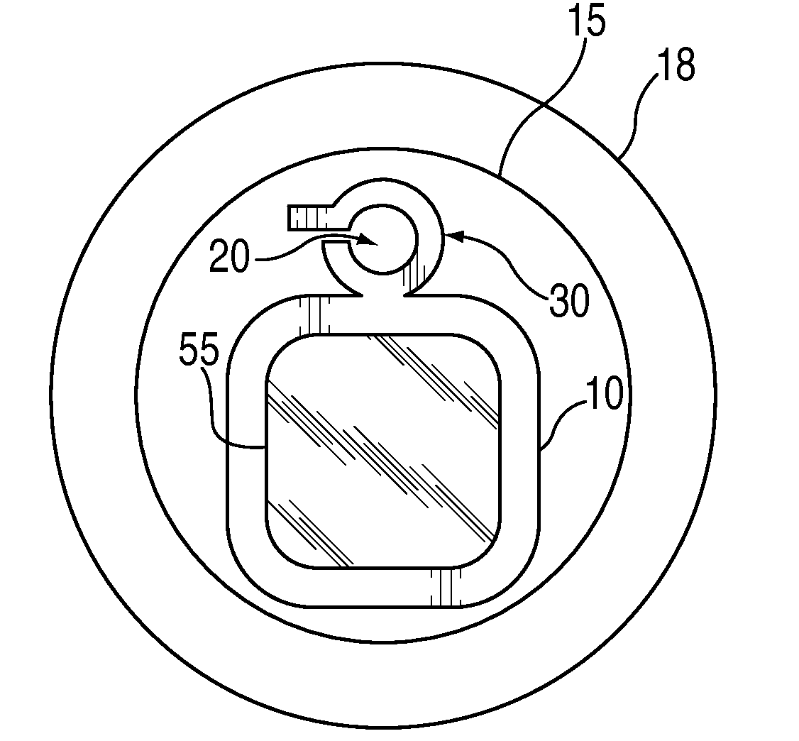
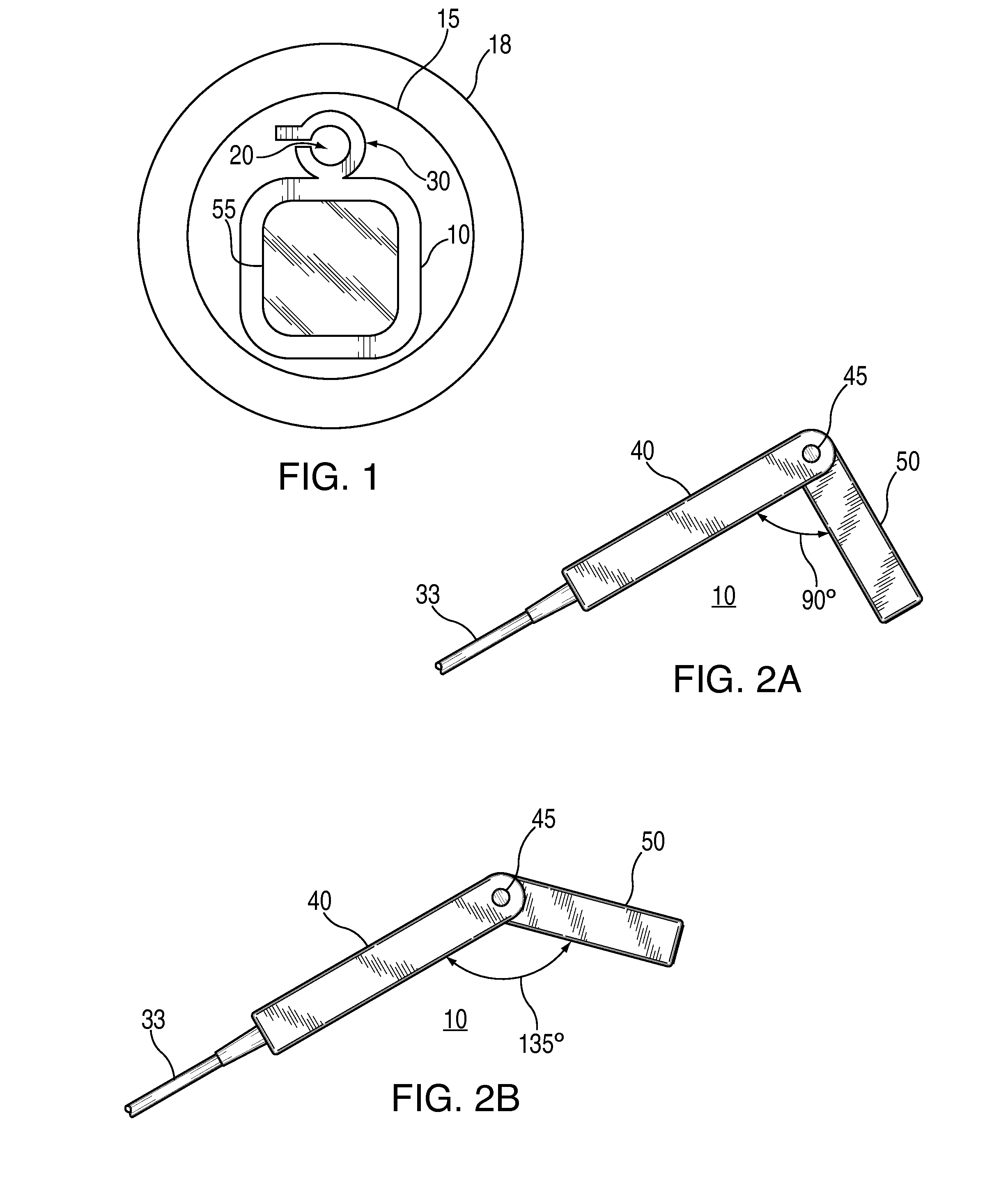
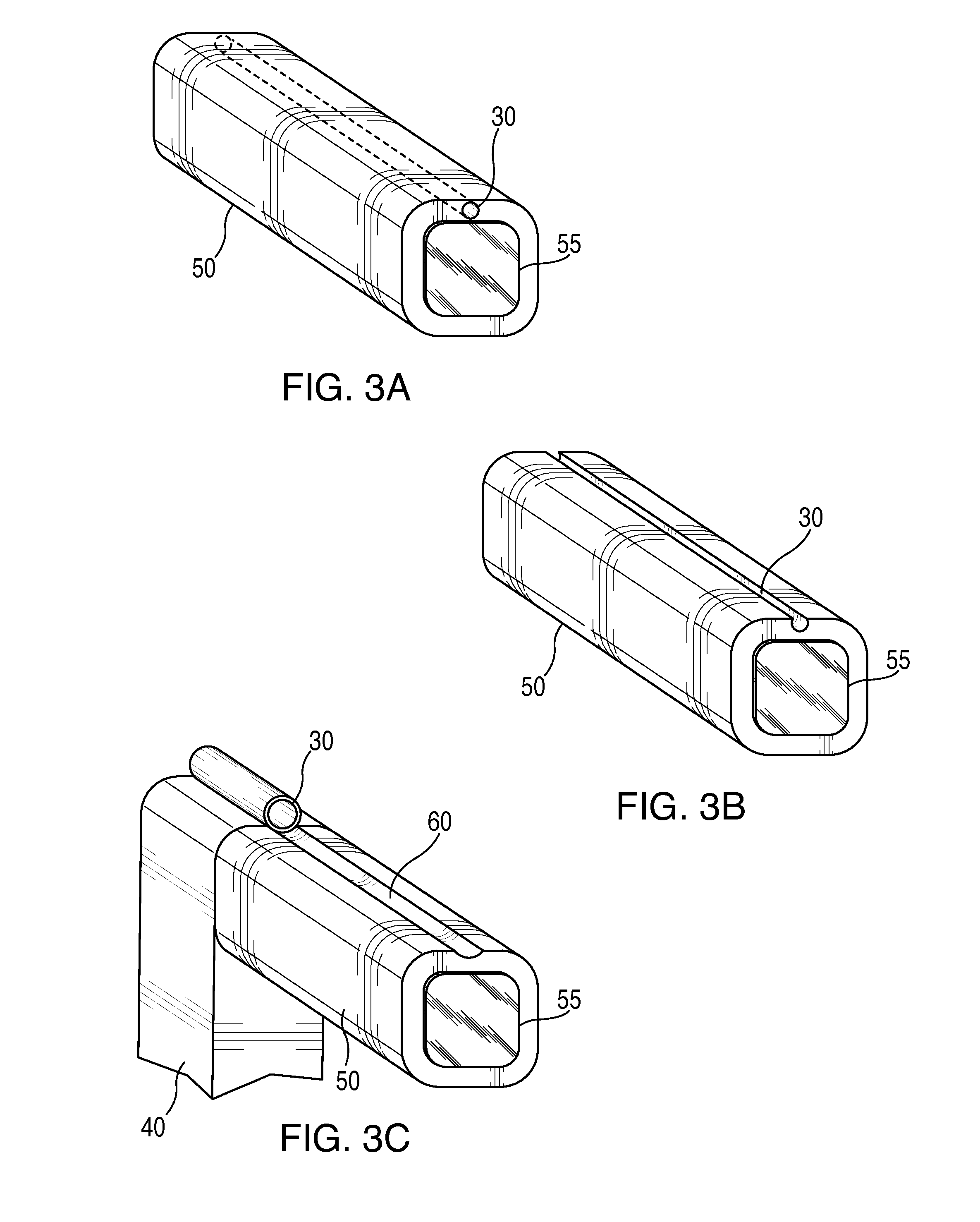
[0028]Referring now to the drawings and in particular FIG. 1, an ultrasound transducer probe 10 for use in guiding the placement of a ventriculostomy catheter via an aperture 20 in a stereotactic conduit 30 mounted on a side of probe 10. Transducer probe 10 is dimensioned to fit within an 11 mm diameter “burr hole” (craniotomy) 15 without requiring an additional step of increasing the diameter of the craniotomy. As discussed above, 11 mm is the current standard size inner cusp diameter craniotomy for ventriculostomies, and 14 mm is the current standard size outer cusp diameter 18. Transducer probe 10 is connected to a conventional portable ultrasound machine (not shown). Conduit 30 serves as a self-contained guide-port for probe 10 for insertion of a standard ventriculostomy catheter (not shown) via a cable 33 (FIG. 2). When the head of probe 10 is placed within the craniotomy and against the exposed surface of the brain (i.e., the dura), the alignment of the phased-array ultrasound...


PUM


Abstract

An apparatus, system and method for performing a ventriculostomy using an ultrasound probe is disclosed. A head portion of the probe is connected to a handle portion at a proximal end of the head portion. A transducer is mounted in a distal end of the head portion to transmit ultrasound waves into the patient. The head portion includes a conduit portion arranged to accept and to accurately direct a catheter into the body of the patient in a direction parallel to the direction of the ultrasound waves. The head portion is sized to fit into a conventionally-sized craniotomy. An adjustment mechanism connects the head portion to the handle portion for selectively adjusting the angle between the handle portion and the head portion. A sterile sheath having an integral conduit assembly for mounting to the head is optionally provided. An illuminator is optionally provided on the probe.

Description

CROSS-REFERENCES TO RELATED APPLICATIONS[0001]The present application claims the benefit of the filing date of U.S. Provisional Patent Application No. 61 / 224,985, filed on Jul. 13, 2009, under 35 U.S.C. §119(e). U.S. Provisional Patent Application No. 61 / 224,985, filed on Jul. 13, 2009, is hereby incorporated by reference.FIELD OF INVENTION[0002]The present invention relates to an apparatus, system and method for accurately positioning a catheter within a body cavity to be treated. More specifically, the disclosed inventive apparatus, system and method provide for the accurate introduction of a catheter into a targeted ventricular cavity of the brain during a ventriculostomy procedure.BACKGROUND OF THE INVENTION[0003]A human brain includes four interconnected cavities, the ventricles, which produce and circulate cerebrospinal fluid. A ventriculostomy procedure involves the placement of a catheter into one of the ventricles. The ventriculostomy may be performed for intracranial press...

Claims


Application Information

Patent Timeline
IPC IPC(8): A61B8/14A61B8/00
CPCA61B8/0833A61B8/0841A61B8/0808A61B8/4455A61B8/4209
Inventor PHILLIPS, SCOTT B.KRISHNAMURTHY, SATISH
Owner PHILLIPS SCOTT B
Learn More