
Medical scanning apparatus and method

a scanning apparatus and ultrasound technology, applied in the field of medical ultrasound scanning devices, can solve the problems of increasing maintenance requirements, reducing reliability, and affecting the operation of motors and associated moving parts, so as to achieve smooth change, reduce cast, and reduce the effect of aging

Inactive Publication Date: 2010-10-14
SIGNOSTICS LTD
View PDF13 Cites 10 Cited by
  • Summary
  • Abstract
  • Description
  • Claims
  • Application Information

AI Technical Summary

Benefits of technology

[0016]Compared to the prior art methods using motor driven transducers or array transducers, a simpler, lower cast way of acquiring a static B scan is to move a single crystal probe through a planar sector by hand. If this is performed in a reciprocating manner, it can produce an image that changes in real time, at a relatively low rate. To provide a smoothly changing image at these low refresh rates, the display is preferably not updated only at the end of each scan. It is advantageous to update the display as soon as each ultrasonic pulse echo returns to the sensor.
[0028]In a further form, the invention may be said to lie in a method of ultrasound scanning to achieve apparent real-time display of a B-mode ultrasound scan. Advantageously, this can be achieved in a low cost manner by employing a single scanline ultrasound scanning apparatus adapted to display a B-mode ultrasound scan image by displaying a series of scanlines, each scanline being a dataset having a series of echo intensity values and a direction.

Problems solved by technology

Since each scan needs to be assembled from its constituent scanlines, processed and displayed, the computer power required for this process is significant.
The motor driven transducer has the disadvantage that the motor and the associated moving parts decrease the reliability and increase maintenance requirements.
The motor also adds cost, and more importantly, weight and bulk to the handpiece.
The motor will also add significantly to the power consumption so of the ultrasound scanning system.
However, the cost of producing transducers with arrays of crystals is high.
There is also a high cost in providing the control and processing circuitry, with a separate channel being required for each crystal.
The power consumption for electronic systems is also high, and is generally proportional to the number of channels to being simultaneously operational.
These problems make the use of such systems in hand held, battery powered applications infeasible.

Method used


Image

  • Medical scanning apparatus and method
  • Medical scanning apparatus and method
  • Medical scanning apparatus and method

Examples


Embodiment Construction

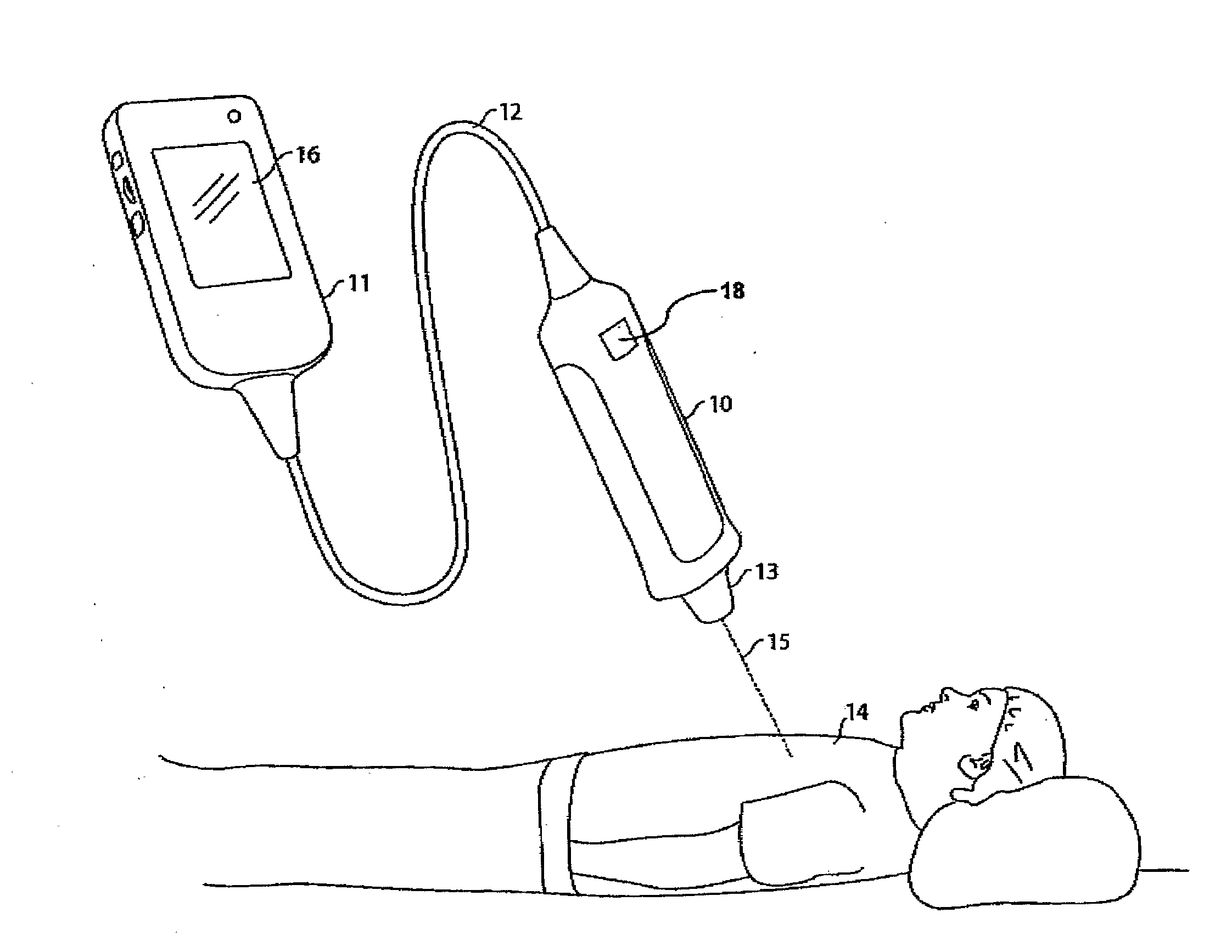
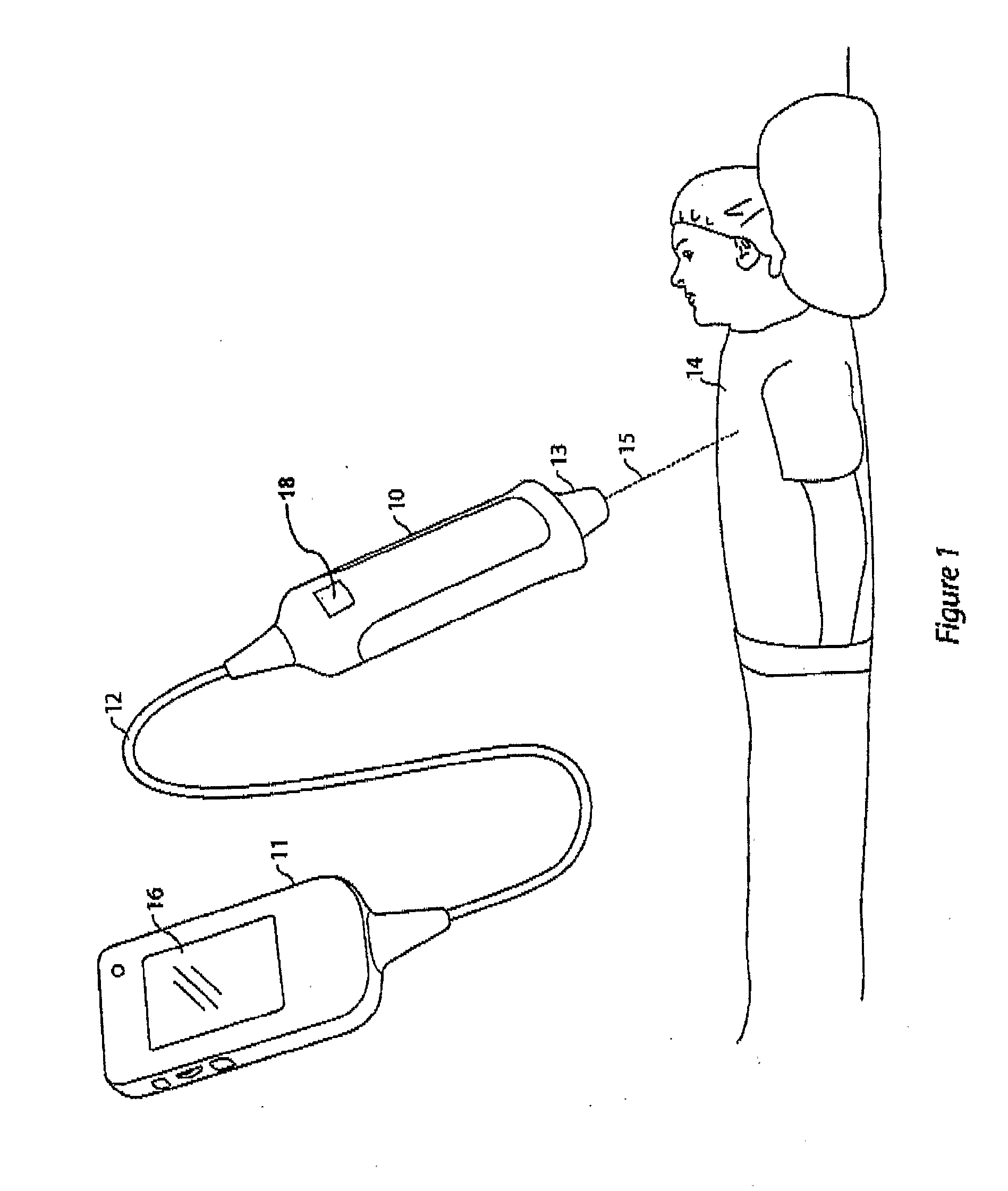
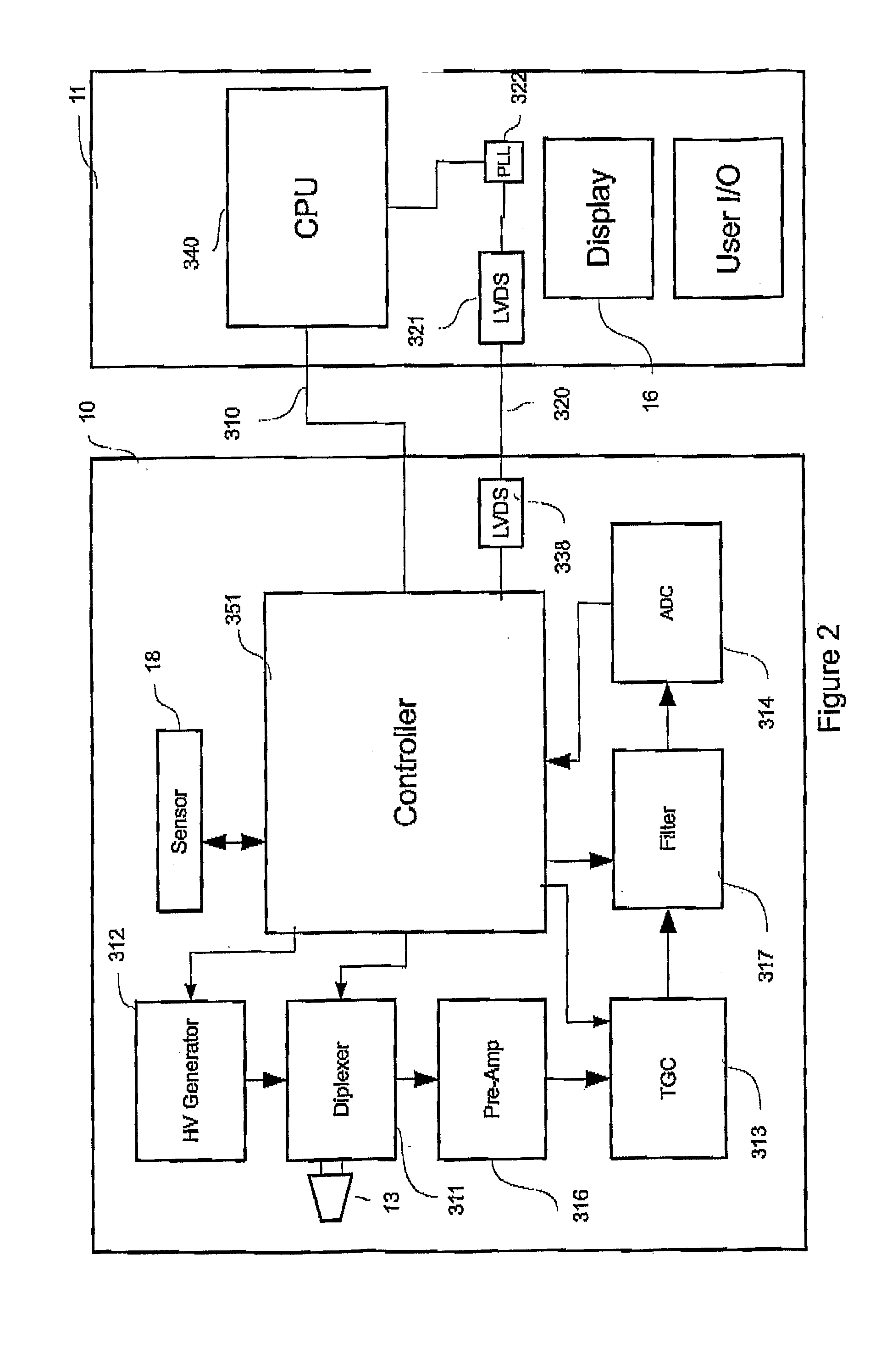
[0036]Referring now to FIG. 1, there is illustrated en ultrasound scanning system incorporating an embodiment of the invention. There is a hand held ultrasonic probe unit 10, a display and processing unit (DPU) 11 with a display screen 16 and a cable 12 connecting the probe unit to the DPU 11.

[0037]The probe unit 10 includes an ultrasonic transducer 13 adapted to transmit pulsed ultrasonic signals into a target body 14 and to receive returned echoes from the target body 14.

[0038]The transducer is adapted to transmit and receive in only a single direction at a fixed orientation to the probe unit, producing data for a single scanline 15. The system is a simple, low cost portable ultrasound scanning system. Additional transducers may be provided, at the expense of increased cost and complexity.

[0039]The probe unit further includes an orientation sensor 18 capable of sensing orientation or relative orientation about one or more axes of the probe unit. Thus, in general, the sensor is abl...


PUM


Abstract

A system and method of ultrasound scanning to achieve apparent real-time display of a sector ultrasound scan Including a single scanline ultrasound scanning apparatus with a sensor adapted to sense the position and / or orientation of the transducer and to output this as data and a screen to display a B-mode ultrasound scan image formed from a series of scanlines, each scanline being a dataset having a series of echo intensity values and a direction captured by reciprocating a transducer probe against a surface of a subject to be scanned such that a planar sector or series of planar sectors of the subject are swept by an ultrasound beam producing a series of scanlines and continuously processing and displaying said scanlines to produce an image that changes in real time In accordance with the features of the subject in the sector being scanned.

Description

TECHNICAL FIELD[0001]This invention relates to the field of medical ultrasound scanning devices, In particular to low cost ultrasound devices which provide moving images of the scan field.BACKGROUND ART[0002]The use of ultrasound scanning of patients for medical diagnostic purposes dates to the mid-20th century. An ultrasound transducer is used to project a beam of ultrasound energy into a patient. The same or another transducer detects the echoes returned from features within the body. These echoes, called a scanline, are then converted to a form suitable for recording or display.[0003]When a series of scanlines, spaced angularly apart, are acquired rapidly and displayed on a display screen, the familiar B-mode sector scan is achieved.[0004]This is a sector of a circle, wherein the brightness of each pixel of the display is proportional to the magnitude of the ultrasound echo received from the corresponding point in the body being imaged.[0005]A method from the prior art for collec...

Claims


Application Information

Patent Timeline
IPC IPC(8): A61B8/14
CPCA61B8/14A61B8/462A61B8/4254
Inventor MEDLIN, ANDREW JOHNBRYDON, JOHNMCCARTHY, MATTHEW
Owner SIGNOSTICS LTD
Learn More