Seprase as a marker for cancer
a technology of seprase and cancer, applied in the field of seprase as a marker for cancer, can solve the problems of cancer risk, occurrence, and/or decrease in the concentration and/or activity of seprase polypeptide and/or fragments in the test sample,
- Summary
- Abstract
- Description
- Claims
- Application Information
AI Technical Summary
Benefits of technology
Problems solved by technology
Method used
Image
Examples
example 1
[0120]As seprase-specific binding molecules rat monoclonal anti-seprase antibodies (clones D8, D28 and D43) were used (Pineiro-Sanchez, M.-L., et al., J. Biol. Chem., 272 (1997) 7595-7601).
[0121]Biotinylation of monoclonal rat IgG
[0122]Monoclonal rat IgG (clones D28 or D43) was brought to 10 mg / ml in 10 mM NaH2PO4 / NaOH, pH 7.5, mM NaCl. Per ml IgG solution 50 μl Biotin —N-hydroxysuccinimide (3.6 mg / ml in DMSO) were added. After 30 min at room temperature, the sample was chromatographed on Superdex 200 (10 mM NaH2PO4 / NaOH, pH 7.5, 30 mM NaCl). The fraction containing biotinylated IgG were collected.
[0123]Digoxygenylation of monoclonal rat IgG
[0124]Monoclonal rat IgG (clones D8 or D43) was brought to 10 mg / ml in 10 mM NaH2PO4 / NaOH, 30 mM NaCl, pH 7.5. Per ml IgG solution 50 μl digoxigenin-3-O-methylcarbonyl-c-aminocaproic acid-N-hydroxysuccinimide ester (Roche Diagnostics, Mannheim, Germany, Cat. No. 1 333 054) (3.8 mg / ml in DMSO) were added. After 30 mM at room temperature, the sampl...
example 2
[0125]ELISA for the measurement of seprase in human serum and plasma samples.
[0126]For detection of seprase in human serum or plasma, a sandwich ELISA was developed. In a first assay format, aliquots of the anti-seprase monoclonal antibodies D28 conjugated with biotin and D8 conjugated with digoxigenin (see Example 1) were used for capture and detection of the antigen (D28×D8 format). In a second assay format, aliquots of the anti-seprase monoclonal antibody D43 (see Example 1) conjugated with biotin and digoxigenin, respectively, were used for capture and detection of the antigen (D43×D43 format).
[0127]Samples (75 μl) were mixed with 150 μl of antibody reagent containing 0.375 μg of each, D28-biotin and D8-digoxigenin antibodies or alternatively D43-biotin and D43-digoxigenin antibodies in incubation buffer (40 mM phosphate, 200 mM sodium tartrate, 10 mM EDTA, 0.05% phenol, 0.1% polyethylene glycol 40000, 0.1% Tween 20, 0.2% BSA, 0.1% bovine IgG, and 0.02% 5-Bromo-5-Nitro-1,3-Dioxa...
example 3
CRC Study Population
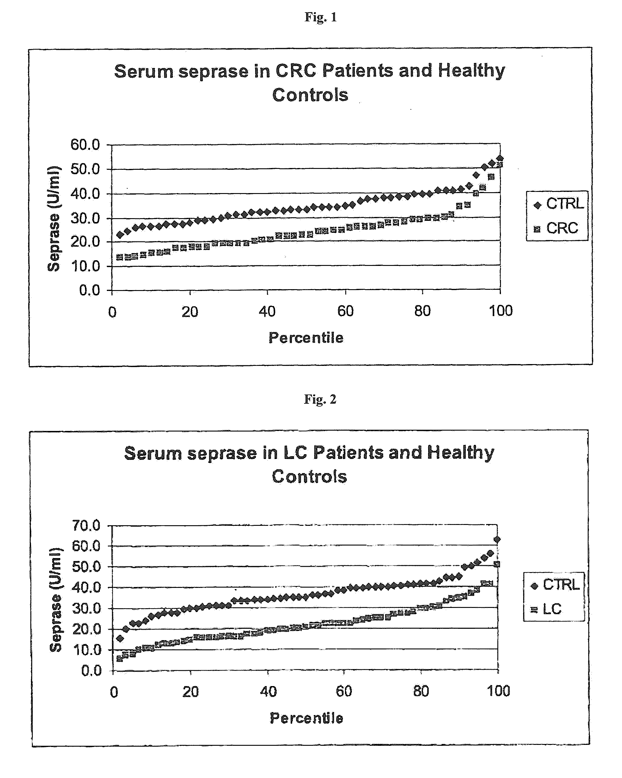
[0134]In a first study, samples derived from 49 well-characterized patients with colorectal cancer (UICC classification given in Table 1) have been used.
TABLE 1CRC study: distribution of samples across UICC stagesStage according to UICCNumber of samplesUICC I8UICC II8UICC III15UICC IV4without staging14total number of CRC samples49
[0135]The samples of Table 1 have been evaluated in comparison with control samples obtained from 50 obviously healthy individuals without any known malignant disease (=control cohort).
PUM
 Abstract
Description
Claims
Application Information
 - R&D
- Intellectual Property
- Life Sciences
- Materials
- Tech Scout
- Unparalleled Data Quality
- Higher Quality Content
- 60% Fewer Hallucinations
Browse by: Latest US Patents, China's latest patents, Technical Efficacy Thesaurus, Application Domain, Technology Topic, Popular Technical Reports.
© 2025 PatSnap. All rights reserved.Legal|Privacy policy|Modern Slavery Act Transparency Statement|Sitemap|About US| Contact US: help@patsnap.com


