
Compositions and methods for detecting oral neoplasm

a technology for oral cancer and compositions, applied in the field of compositions and methods for detecting oral cancer, can solve the problems of difficult to associate the binding of a particular lectin with a specific disease, difficult to understand, and the area of study has received little attention from most cell biologists involved in cancer research, so as to achieve the effect of simplifying and cost-effective oral cancer detection

Inactive Publication Date: 2010-09-16
INTERMED
View PDF7 Cites 8 Cited by
  • Summary
  • Abstract
  • Description
  • Claims
  • Application Information

AI Technical Summary

Benefits of technology

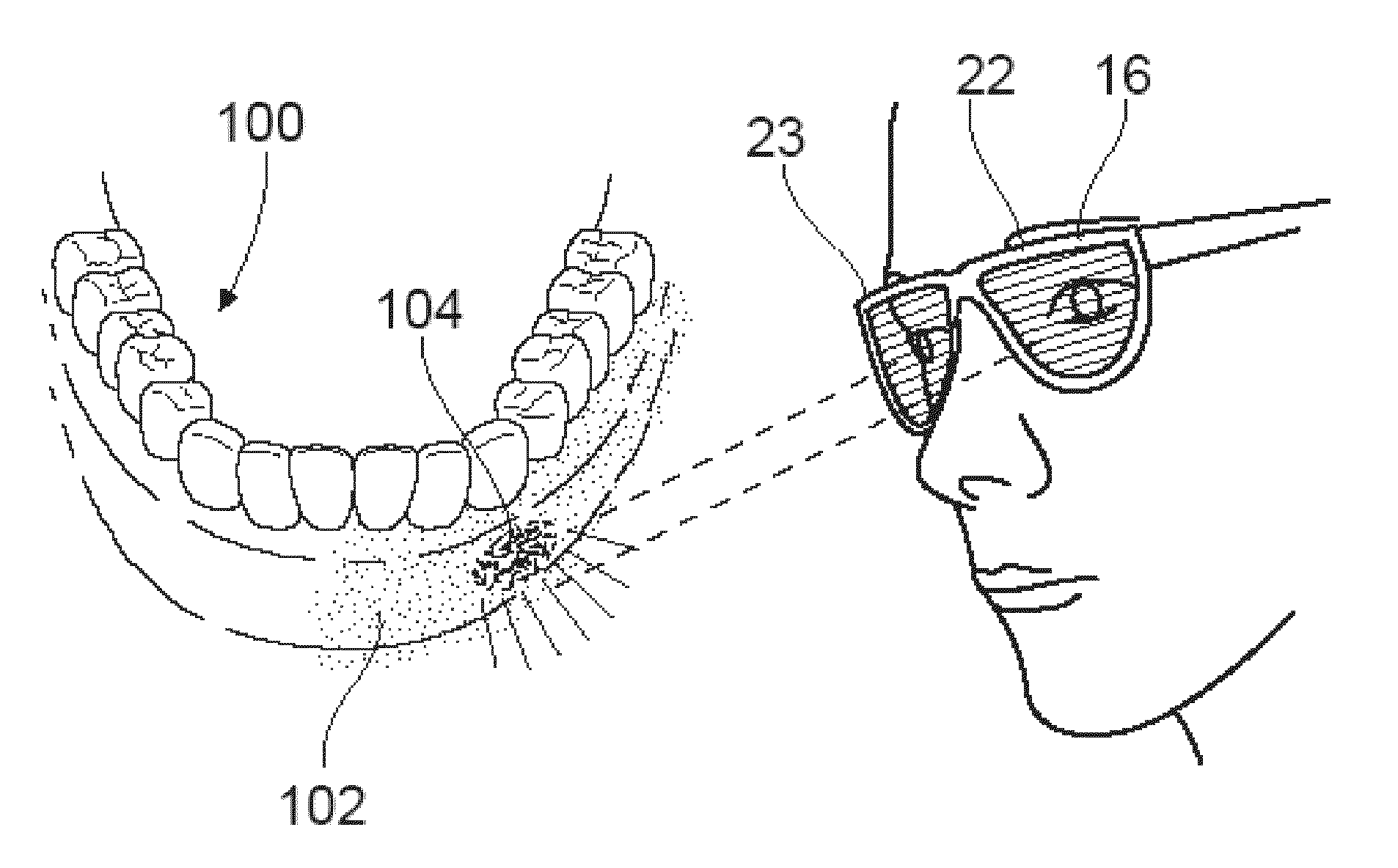
[0035]In a first aspect, the invention encompasses a method for assessing the presence of possible oral cancer in a subject. The method includes the steps of (1) applying a composition comprising one or more lectins operably linked to a detection moiety to the oral mucosa of a subject, wherein said lectins specifically bind to one or more mono and / or oligosaccharides; (2) exposing the oral mucosa of the subject to near infrared light, visible light, or ultraviolet light; and (3) visualizing the oral mucosa to detect possible oral cancer.
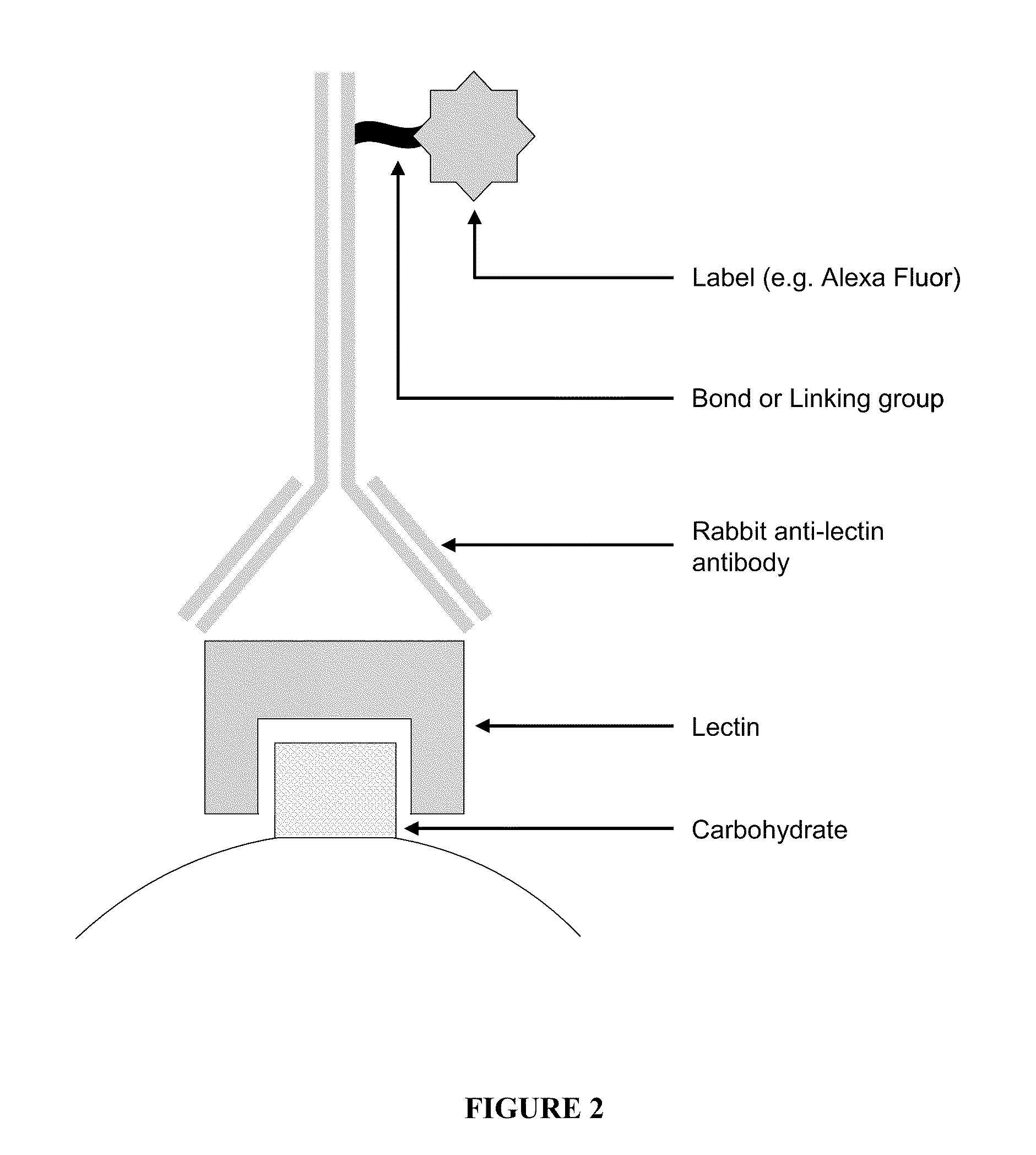
[0036]In a second aspect, the invention encompasses a method for assessing the presence of possible oral cancer in a subject, including the steps of (1) applying one or more lectins to the oral mucosa of a subject, wherein said lectins specifically bind to one or more mono and / or oligosaccharides; (2) applying an antibody or an avidin protein to the oral mucosa of the subject, wherein the antibody or avidin protein is linked to a fluorophore; (3) exposing the oral mucosa of the subject to near infrared light, visible light, or ultraviolet light; and (4) visualizing the oral mucosa to detect possible oral cancer.
[0037]In a third aspect, the invention encompasses a kit for assessing the presence of oral cancer. Such a kit includes (1) one or more lectins operably linked to a fluorophore, wherein said lectins specifically bind to one or more mono and / or oligosaccharides; and (2) a light source emitting light in the ultraviolet, visible, or near infrared spectrum.
[0038]The described embodiments have many advantages, including permitting simplified and more cost-effective detection of oral cancer through routine screening. These and other features, aspects and advantages of the present invention will become better understood from the description that follows. In the description, reference is made to the accompanying drawings, which form a part hereof and in which there is shown by way of illustration, not limitation, embodiments of the invention. The description of preferred embodiments is not intended to limit the invention to cover all modifications, equivalents and alternatives. Reference should therefore be made to the claims recited herein for interpreting the scope of the invention.

Problems solved by technology

Nevertheless, this area of study has received little attention from most cell biologists involved in cancer research, mainly because structural and functional concepts of glycosylation in cancer are more difficult to understand than the functional role of certain proteins and their genes in defining cancer cell phenotypes.
Even so, most lectins interact with more than one group, albeit at a lower association constant, such that it can be difficult to associate binding of a particular lectin with a specific disease.
The presence of fucose linked to C-4 of N-acetylglucosamine on a Type 1 chain results in Le-a activity, but a Type 2 oligosaccharide containing fucose linked to C-3 of N-acetylglucosamine on a Type 2 chain results in very weak Le-a activity.
Tumor cells escape recognition by increasing surface fucosylation levels, which leads to decreased adhesion and uncontrolled tumor growth.
Nevertheless, to the best of our knowledge, EGFR labeling via lectins that specifically target EGFR or VEGFR has not been investigated as a means to detect cancerous tissue in-vitro, ex-vivo, or in-vivo.
Oral cancer, which includes cancers of the lips, tongue, cheeks, floor of the mouth, hard and soft palate, sinuses, and pharynx (throat), is life threatening if not diagnosed and treated early.
This number has not significantly improved over the past few decades, despite continued refinement of surgical techniques and screening technologies.
Because the early signs and symptoms may be misinterpreted by individuals and screening tests tend to be inconsistently employed, oral cancer may not be detected at early stages when it is most treatable.
However, early detection and treatment of cancers can markedly decrease mortality rates and increase survival rates.
However, the effectiveness of these technologies to aid in the detection of oral cancers is inconsistent, and it appears these modalities fail to noticeably improve the detection of oral lesions from routinely preformed standard head and neck exams (Putney JW Jr., Nat Cell Biol 2009; 11: 381-382).
Although such images provide guidance for surgery, these images cannot be generated in real time during surgery to guide the surgeon to the tumor.

Method used


Image

  • Compositions and methods for detecting oral neoplasm
  • Compositions and methods for detecting oral neoplasm
  • Compositions and methods for detecting oral neoplasm

Examples


example

Using Lectin-Fluorophore Probes to Differentiate Normal and Neoplastic Mucosal Tissues

[0137]The inventors here report the results of clinical studies using the methods of the present invention. Specifically, the inventors have collected data from fluorescence imaging of oral mucosa tissue specimens from 11 patients. Fluorescence imaging was performed (1) using conventional tissue autofluorescence, and (2) after applying lectin-fluorophore conjugates to the surface of the tissue. Fluorophore conjugates were used containing the lectins PNA, WGA, and GS-II. Data collected from 9 of the 11 patients demonstrate that aberrant glycosylation changes can be pinpointed via fluorescent lectin conjugates, resulting in high resolution imaging of normal and cancerous tissue in the oral mucosa. Furthermore, these glycosyl changes can differentiate normal, dysplastic, and malignant tissues in the oral cavity with high contrast. This technique holds promise as a non-invasive screening method for pre...


PUM

PropertyMeasurementUnit
wavelengthsaaaaaaaaaa

Abstract

Methods and kits for assessing the presence of, and for detecting cancer, notably oral cancer, are disclosed. Such methods and kits use one or more lectins operably linked to a fluorophore, wherein the lectin binds differentially to cancerous and non-cancerous tissues, and wherein the fluorophore facilitates visualizing the differential binding. The lectins are applied to the oral mucosa of a subject, the fluorophore is exposed to light, and cancerous regions of the mucosa are visualized. In certain embodiments, the lectin specifically binds to one or more of a β-galactoside, an α- or β-N-acetylglucosamine, or a sialic acid moiety.

Description

CROSS-REFERENCE TO RELATED APPLICATIONS[0001]The application claims the benefit of U.S. Provisional Application No. 61 / 208,730 filed on Feb. 27, 2009, which is incorporated herein by reference as if set forth in its entirety.STATEMENT REGARDING FEDERALLY SPONSORED RESEARCH OR DEVELOPMENT[0002]N / ABACKGROUND[0003]The present invention relates to methods, devices and kits for use in screening patients to detect cancers and precancers associated with mucosal tissues, particularly oral cancer, using optical molecular imaging.[0004]Cancer is defined as the uncontrollable growth of cells that invade and cause damage to surrounding tissue. Many properties of mammalian cells are either expressed at or mediated through the cell surface, and cancer is associated with characteristic molecular changes on the cellular surface.[0005]Specifically, membrane proteins and lipids having attached carbohydrate side chains project from the external surface of a cell and form the cell glycocalyx. The glyco...

Claims


Application Information

Patent Timeline
Patent Type & Authority Applications(United States)
IPC IPC(8): A61K49/00A61B1/24A61B10/02
CPCG01N2333/42G01N33/57407A61B5/0071A61B5/0088A61B50/33A61B2562/0233A61K49/0056
Inventor POND, GARYBAETEN, JOHN
Owner INTERMED
Learn More