
Apparatus and method to determine functional lung characteristics

Inactive Publication Date: 2010-09-09
DRAGERWERK AG
View PDF20 Cites 69 Cited by
  • Summary
  • Abstract
  • Description
  • Claims
  • Application Information

AI Technical Summary

Benefits of technology

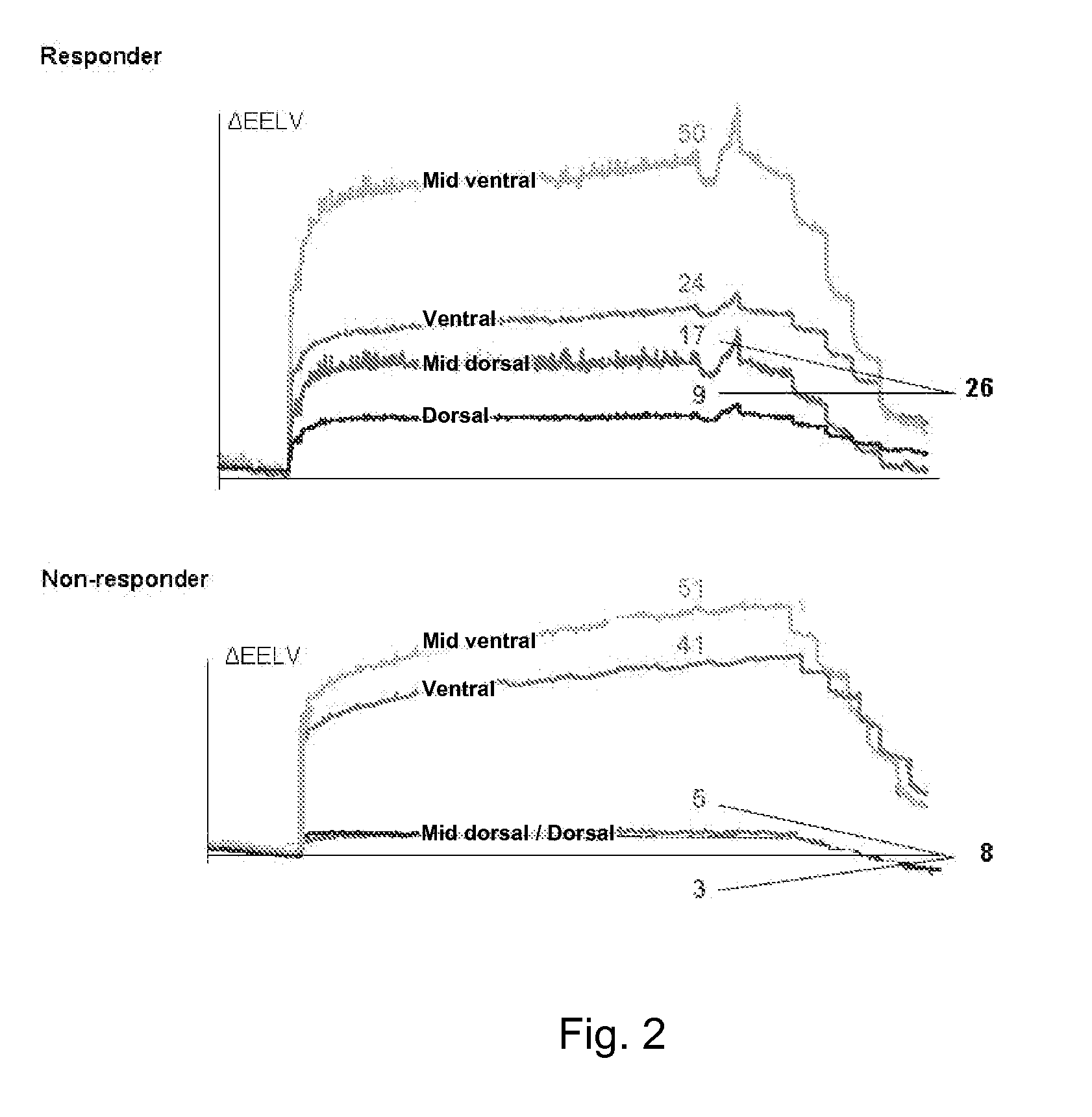
[0014]It is the object of this invention to provide an apparatus and a method to determine functional lung characteristics, which are indicative of the breathing activity of lung areas, which may be used to define such things as PEEP and tidal volume as well as appropriateness and susceptibility to recruitment maneuvers (RM). In particular, it would be preferred if the apparatus and the method would be applicable in a non-invasive manner and would be usable continuously at the bedside. In particular, the apparatus and the method should allow determining the PEEP level and tidal volume that is required to open up collapsed lung regions and to keep the balance between the goals to avoid atelectotrauma and overdistension. Furthermore, it would be preferred if the apparatus and the method are able to provide an indication in which patients a recruitment maneuver (RM) will be beneficial to differentiate responders from nonresponders to RM. Finally, the apparatus and the methods should allow displaying functional lung parameters in value which is easily perceived and interpreted.

Problems solved by technology

While the phase lag mode analyzes the delay of the regional beginning of inspiration in a complete breath it is not sensitive to changes occurring within the inspiratory and expiratory phase, therefore does not allow a detailed analysis of functional lung characteristics.
Selecting optimal PEEP and appropriate tidal volume is a difficult task.
Selection of optimal PEEP is a real challenge with no satisfying practical answer today.
Therefore, with the use of these settings, there is no guarantee that cyclic opening and closing are not accompanied by overstretching as well.
A complicated way to set PEEP has been suggested by using CT images, but obviously this is a very “unclinical” method that cannot be used routinely.
Crit Care Med 2007; 35:214), but this has so far only been done in a surfactant deficient pig model, and furthermore dynamic compliance is a fairly insensitive indicator to properly set PEEP in ALI / ARDS patients.
The peak pressure (60 cmH2O) applied to the lung is quite high and could be dangerous as it involves the risk of developing pneumothorax in patients.
However, as recruitment and overstretching may occur simultaneously, these conditions might remain undetected due to the fact that these concurrent effects are masked in the global pressure curve, which only reflects the overall behavior and resulting effects of the inhomogeneous lung as a whole.

Method used


Image

  • Apparatus and method to determine functional lung characteristics
  • Apparatus and method to determine functional lung characteristics
  • Apparatus and method to determine functional lung characteristics

Examples


Embodiment Construction

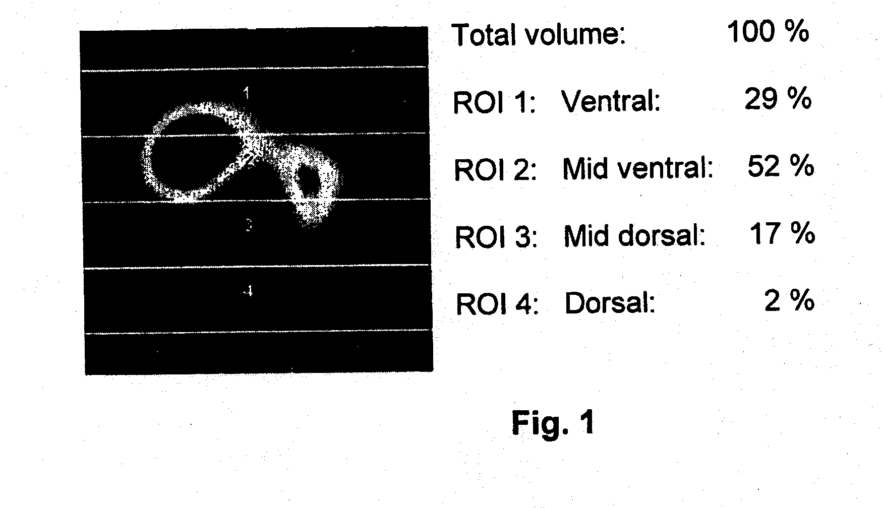
[0034]Referring to the drawings in particular, in FIG. 1 an EIT image of a cross-section of a lung is shown wherein the reconstructed impedance levels are represented by different grey levels. Such EIT image of the cross-section of the lung which is typically taken within the juxtadiaphragmatic plane corresponding to the fifth intercostal space is subdivided into regions of interest (ROI). In FIG. 1 the cross-sectional EIT image of the lung is subdivided into four segments (ROI 1-ROI 4) corresponding to the ventral, midventral, mid-dorsal and dorsal portions of the lung.

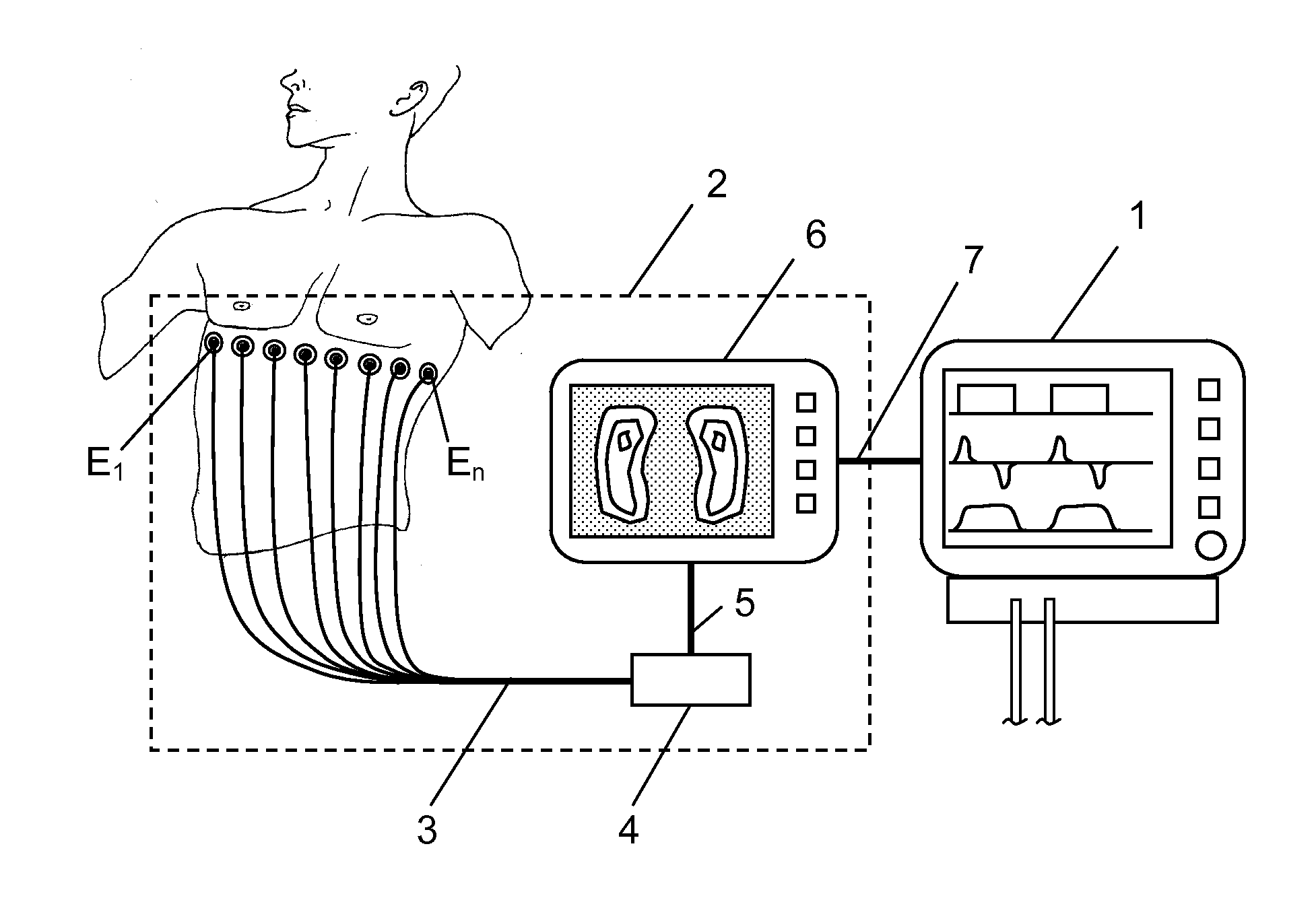
[0035]FIG. 9 schematically shows an apparatus according to the present invention, optionally connected via data-interface 7 to a ventilator / expirator 1. The EIT imaging device 2 includes a display unit 6 and a control and analysis unit 4 which is connected to the display unit 6 on the one hand and to a plurality of electrodes E1, . . . , En which are to be placed around the thorax of a patient. The control and analys...


PUM


Abstract

An apparatus for determining functional lung characteristics of a patient includes an electrical impedance tomography (EIT) imaging device adapted to record the impedance distribution within a plane of the thorax of the patient. The EIT imaging device includes a control and analysis unit for performing the impedance measurement and deriving the impedance distribution within the plane of the thorax. The control and analysis unit automatically performs steps including determining a global impedance change, defined as the impedance change with respect to an earlier measured reference impedance distribution integrated over the electrode plane, and recording the global impedance change curve as a function of time, performing breath detection in order to identify a breathing cycle, subdividing each breathing cycle to define a plurality of intratidal intervals, subdividing an EIT image from each interval into a plurality of regions of interest and calculating for each region of interest the ratio of the integrated impedance change within this region of interest to the global impedance change of this EIT image, for each intratidal interval presenting indications of the ratios determined for the regions of interest to provide an intratidal gas distribution representation for each interval.

Description

CROSS REFERENCE TO RELATED APPLICATIONS[0001]This application claims the benefit of priority under 35 U.S.C. §119 of European Patent Application EP 09 003 376.2 filed Mar. 9, 2009, the entire contents of which are incorporated herein by reference.FIELD OF THE INVENTION[0002]The present invention relates to an apparatus and a method for determining functional lung characteristics of a patient utilizing an electrical impedance tomography (EIT) imaging device adapted to record the impedance distribution within a plane of the thorax of the patient, the EIT imaging device including a control and analysis unit for performing the impedance measurement and deriving the impedance distribution within the plane of the thorax (EIT image).BACKGROUND OF THE INVENTION[0003]Electrical impedance tomography (EIT) is an imaging technique in which an image of the impedance distribution of a part of the body is inferred from surface electrical measurements. EIT is a non-invasive method in which an alter...

Claims


Application Information

Patent Timeline
IPC IPC(8): A61B5/053
CPCA61M16/0051A61B5/0536A61M16/021
Inventor TESCHNER, ECKHARDSTENQVIST, OLA
Owner DRAGERWERK AG
Learn More