
Angiogenesis Mechanism and Method, and Implantable Device

a technology of angiogenesis and angiogenesis, which is applied in the direction of prosthesis, peptide/protein ingredients, and metabolism disorders, etc., can solve the problems of pancreas transplantation, absolute deficiency of insulin, and limited success, so as to facilitate vessel formation and facilitate vessel formation

Inactive Publication Date: 2010-08-05
MEDTRONIC INC
View PDF42 Cites 81 Cited by
  • Summary
  • Abstract
  • Description
  • Claims
  • Application Information

AI Technical Summary

Benefits of technology

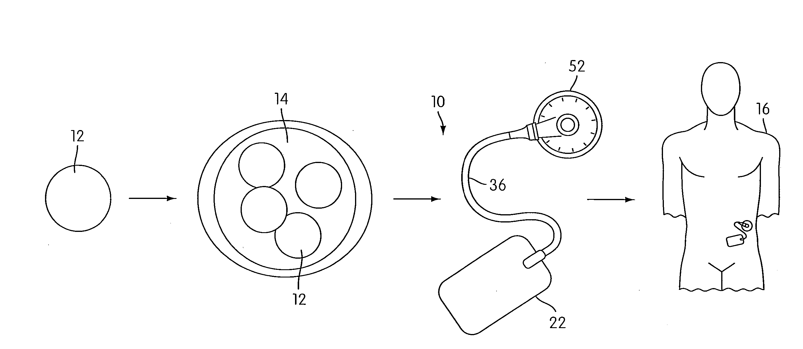
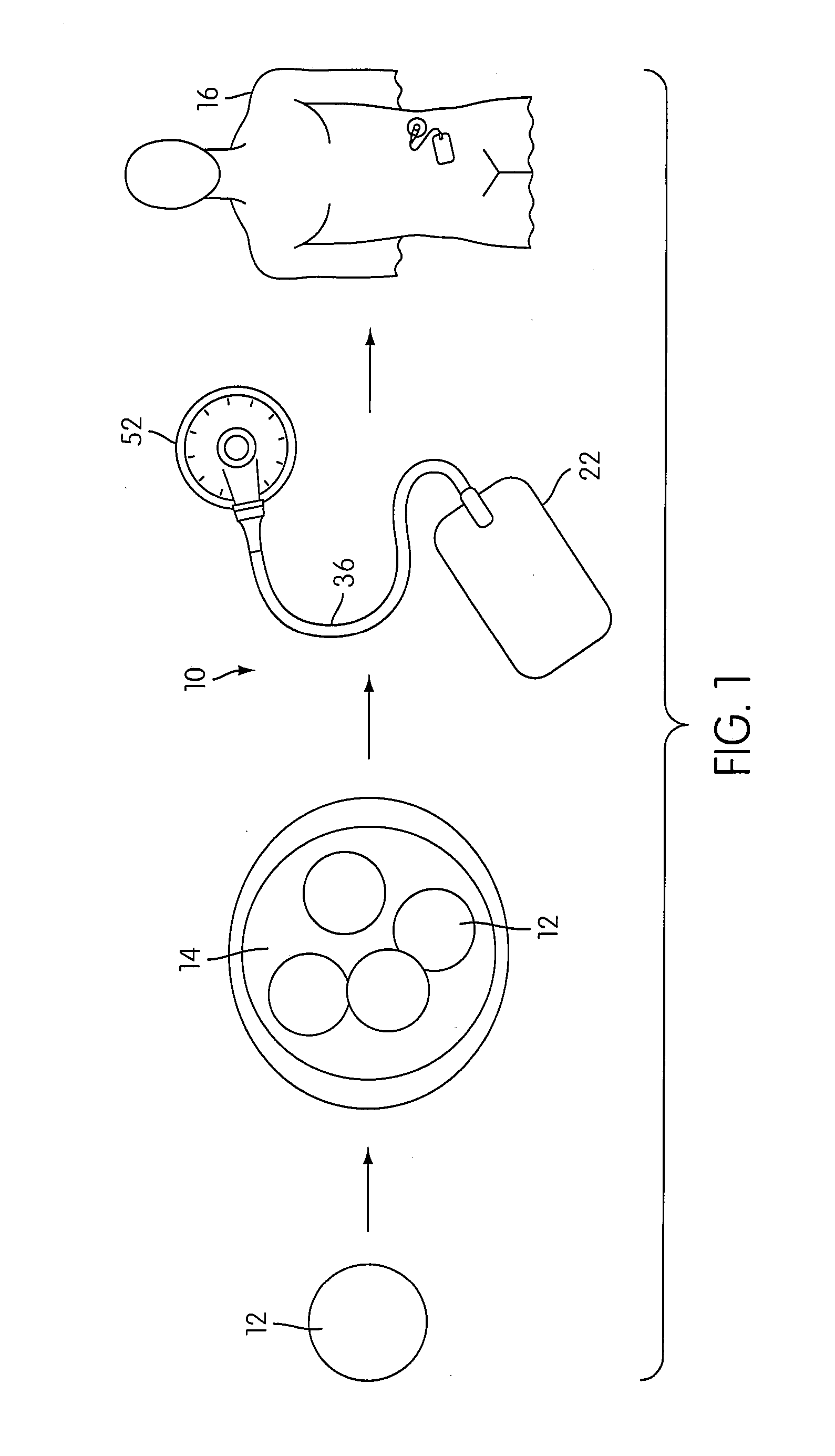
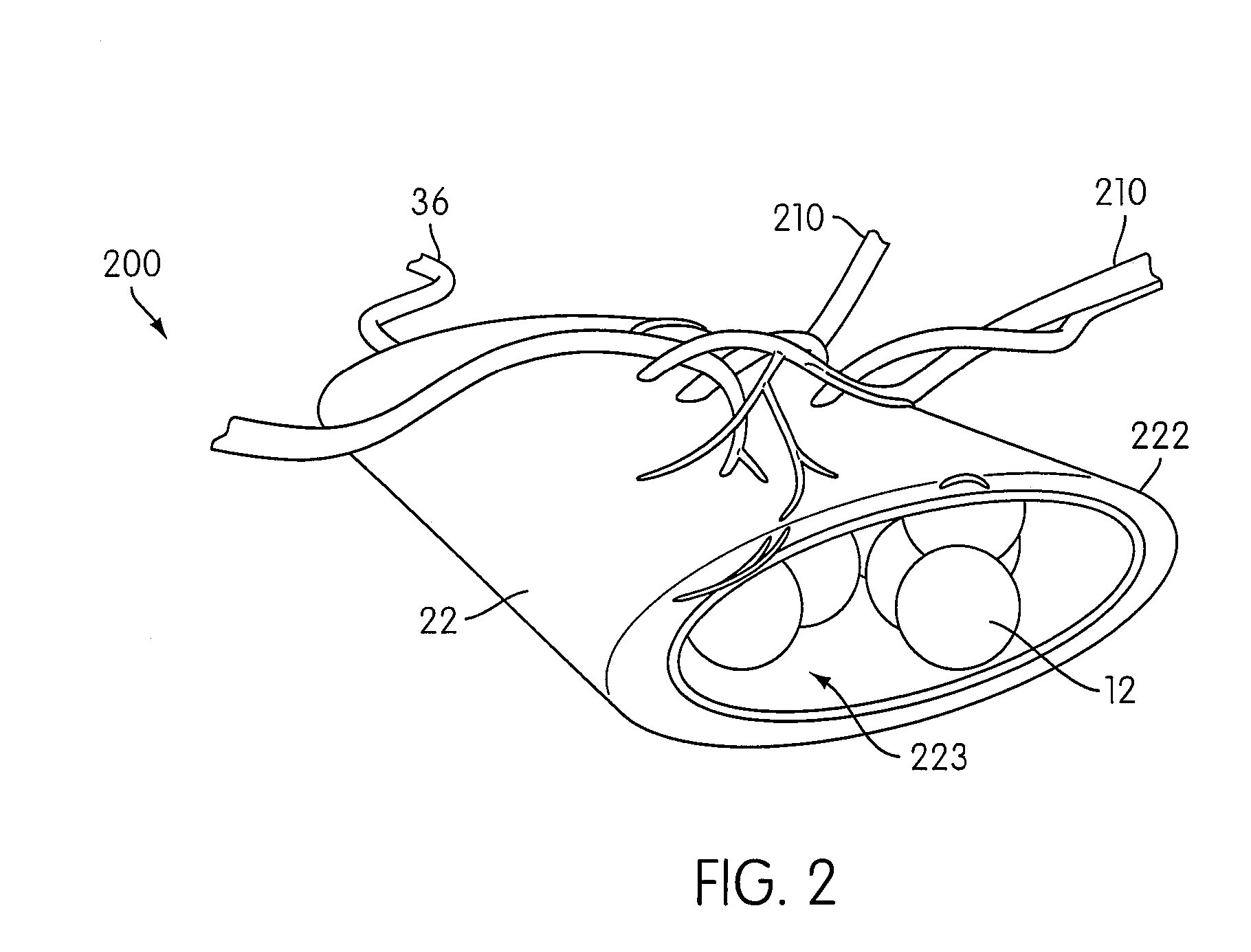
[0011]In accordance with another embodiment of the present invention, an implantable device for providing therapy to a living body is provided that includes a permeable container and therapeutic living cells carried by the container. The living cells are capable of generating cell products that are emitted from the permeable container to the living body. A void space within the container receives agents that enhance living conditions for the living cells.
[0029]In accordance with another embodiment of the present invention, an assembly for providing a vasularized, implanted device is provided, comprising an implantable permeable container, a removable barrier disposed within the container that is permeable to growth factors and impermeable to vessels, and an agent source in communication with the permeable container and configured to provide growth factors into the container, the growth factors permeating through the barrier and to the permeable container to facilitate vessel formation on the permeable container.

Problems solved by technology

It is characterized by β-cell destruction, which usually leads to an absolute deficiency of insulin.
Pancreas transplants, however, have been met with limited success because introducing a new, functioning pancreas to a patient with diabetes can have negative effects on the patient's normally functioning kidney.
Additionally, the availability of pancreases is limited because an organ donor is necessary.
For example, cells within a living cell containing device may not be provided with a sufficient blood supply from the recipient, such that the device may perform poorly.
In addition, the device may also perform poorly due to a lack of nutrients or oxygen provided to the cells.
Alternately, the device may perform poorly due to an accumulation of waste products in or around the device.

Method used


Image

  • Angiogenesis Mechanism and Method, and Implantable Device
  • Angiogenesis Mechanism and Method, and Implantable Device
  • Angiogenesis Mechanism and Method, and Implantable Device

Examples


example 1

Preparation and Evaluation of APGS

[0288]A. Preparation of APGS

[0289]Whole blood was drawn from 4 human donors (2 male and 2 female) to give a starting sample of sixty (60) ml of whole blood for each donor, which was then used to obtain generate preparations of normal serum and APGS. The blood was mixed with sufficient sodium citrate to act as an anticoagulant. Sixty (60 ml) of blood from each donor was processed using a Medtronic Magellan™ Autologous Platelet Separator (Medtronic Inc., Minneapolis Minn.) to obtain 6 ml of platelet rich plasma (PRP) from each donor. Approximately 6000 units of thrombin (at 1000 U / ml) was added to the PRP and mixed by repeated pipetting. The thrombin / PRP mixture was allowed to clot for about 15 minutes at room temperature, to generate autologous platelet gel (APG, clotted PRP). The APG was then centrifuged at 13,000 rpm for 5 minutes. The supernatant was removed and saved as autologous platelet gel serum (APGS). A total volume of approximately 6 ml of...

example 2

Silicone Tubing O2 Permeability Experiments

[0319]The capacity for silicone tubing to supply oxygen (O2) was measured by determining the O2 permeability of silicone tubing. Calculated (theoretical) and experimental (actual) flux values were determined, and these values were evaluated in terms of the predicted O2 requirements of islet cells.

[0320]A round-bottom flask was filled to the level of the bottom of the stopper with water (235 ml, i.e., no headspace) and equipped for magnetic stirring. An oxygen sensor was threaded through the stopper and immersed in the water. A sealed silicone catheter (tube) was also threaded into the flask and connected to an external O2 supply. The silicone cathether had a total length of 10 cm in the flask, a wall thickness of 292 microns, and an internal diameter (ID) of 1.204 mm. The flask was then immersed in a water bath at 37° C.

[0321]The water was deoxygenated by purging with nitrogen (N2) until the sensor reading was zero for oxygen, after which t...

example 3

Formation of a PEG-Based Hydrogel and Encapsulation of Cells

[0334]Cells were encapsulated in a PEG-based hydrogel as described below. PEG having an average of 8 arms, at approximately 20,000 g / mol was functionalized with vinyl sulfone (VS) groups to form PEGVS solution, and further modified using a solution of RGD peptide, and crosslinked with dithiothreitol (DTT), to form a hydrogel having a nominal PEG concentration of approximately 10% (m / v), where the ratio of RGD peptide to VS was 50:1, and the ratio of thiol groups (SH) to VS groups was 1:1, in a total gel volume of 50 μl.

[0335]In a first step, pendant groups were added to vinyl-sulfone-functionalized PEG (PEG-VS). Twenty-five (25)μl of PEG-VS was centrifuged at 13000 rpm for 30 seconds, 6.15 μl RGD solution was added to PEGVS, and the mixture was rotary mixed (vortexed) for 3 seconds and incubated for 30 minutes at 37° C.

[0336]In a second step, cells were prepared for encapsulation. The required number of cells (suspending in...


PUM

PropertyMeasurementUnit
Volumeaaaaaaaaaa
Volumeaaaaaaaaaa
Volumeaaaaaaaaaa

Abstract

Various methods and devices are provided for providing therapy to a living body. In one embodiment, an implanted permeable container has living cells provided therein, and various nutrients and / or agents are provided into the container to promote cell life.

Description

CROSS-REFERENCE TO RELATED APPLICATIONS[0001]The present application claims the benefit of priority from PCT Application Serial No. PCT / US07 / 088,407, published as WO 2008 / 079997, filed Dec. 20, 2007, which claims priority to U.S. Provisional Application Ser. No. 60 / 876,532, filed Dec. 22, 2006, U.S. Provisional Application Ser. No. 60 / 890,041, filed Feb. 15, 2007 and U.S. Provisional Application Ser. No. 60 / 890,326, filed Feb. 16, 2007, the entire contents of which are all incorporated herein by reference.FIELD OF THE INVENTION[0002]The present invention relates to therapeutic implantable medical devices and, more particularly, to implantable cell therapy devices that facilitate improved cell viability.BACKGROUND OF THE INVENTION[0003]Type I diabetes (formerly known as insulin-dependent diabetes, childhood diabetes, or juvenile-onset diabetes) is most commonly diagnosed in children and adolescents, but can occur in adults, as well. It is characterized by β-cell destruction, which us...

Claims


Application Information

Patent Timeline
IPC IPC(8): A61K9/00A61K38/18A61K35/12A61P3/10
CPCA61F2/022A61L27/3616A61L27/38A61L2300/434A61L2300/414A61L2300/416A61L2300/426A61L27/54A61P3/10
Inventor BECK, MICHAEL THEODOREBEZUIDENHOUT, DEONBONNEMA, KELVINCHAFFIN, KIMBERLY A.DAVIES, NEILFERNANDES, BRIAN C.A.FOGT, ERIC J.GROVENDER, ERIC A.HAMLEN, ROBERT CUSHINGJOLLY, MATTHEWMCANDREW, EAMONN J.PAKALA, SYAMASUNDAR V.VANANTWERP, WILLIAM P.ZILLA, PETER P.KOULLICK, EDOUARD A.
Owner MEDTRONIC INC
Learn More