Electrical methods for detection and characterization of abnormal tissue and cells
a technology of abnormal tissue and cell, applied in the direction of diagnostic recording/measuring, instruments, applications, etc., can solve the problems of inability to use x-ray machines inside the body, inability to detect cellular abnormalities, and further limit the use of ultrasound
- Summary
- Abstract
- Description
- Claims
- Application Information
AI Technical Summary
Problems solved by technology
Method used
Image
Examples
example 1
Intra-Operative Assessment in Breast Conservation Surgery
[0047]Experiments were conducted using three prophylactic mastectomy fixated tissue samples. The samples were pre-diagnosed with either invasive carcinoma, ductal carcinoma in situ (DCIS), or benign tissue. Measurements were obtained using the electrical device described herein. Measurements were made at several locations including transition areas between malignant and benign tissue to assess sensitivity and resolution of margins detection. In the tissue sample from one patient, measurements indicated that the entire sample was a benign tissue sample. The sample showed a large frequency spread in frequency response typical to benign tissue. The electrical device was able to differentiate the benign tissue and margins from cancerous and fibrous tissue is a sample from a second patient. The tissue showed electrical responses about 100 times lower than responses from benign tissue.
example 2
Detection of Breast Cancer In Vitro
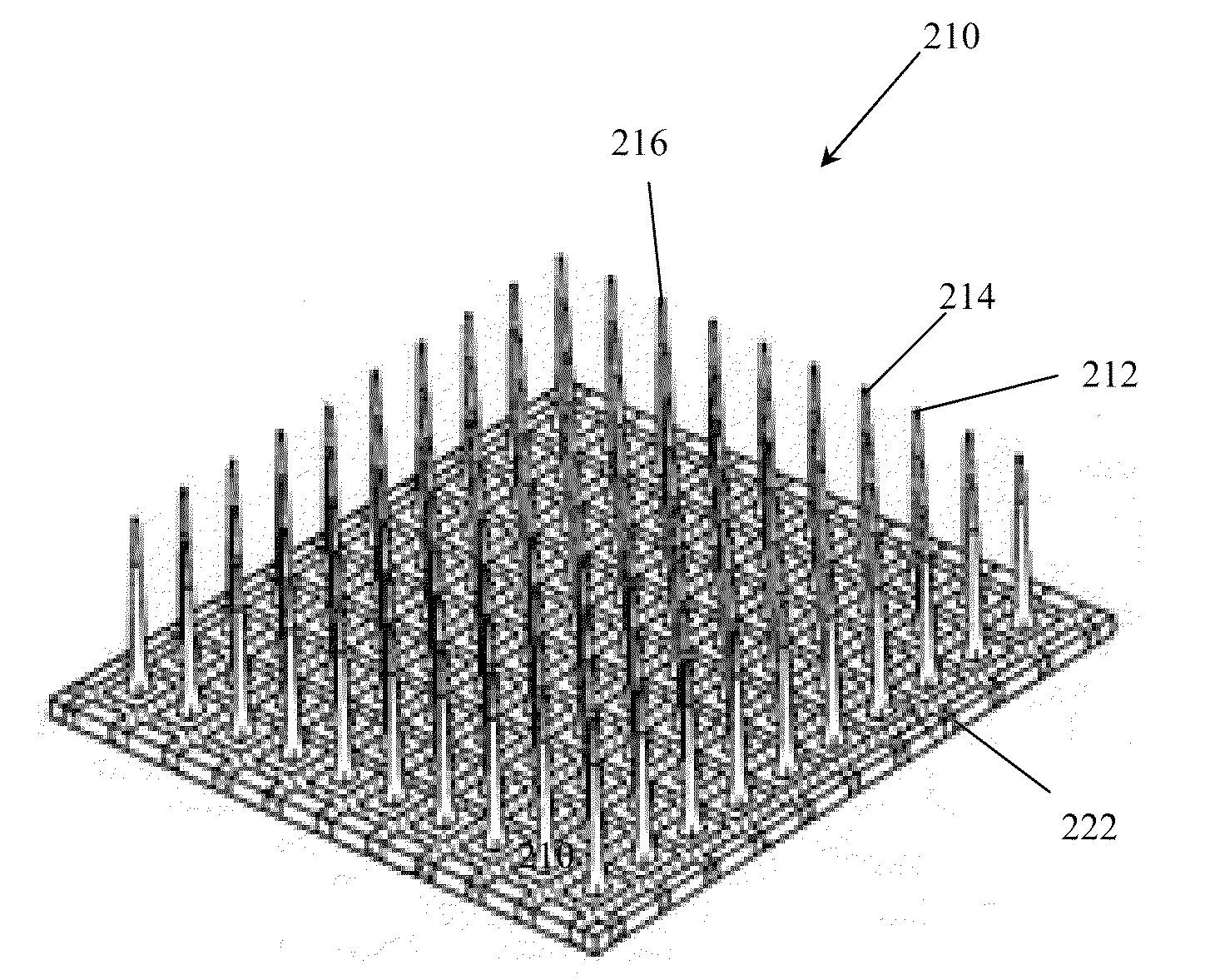
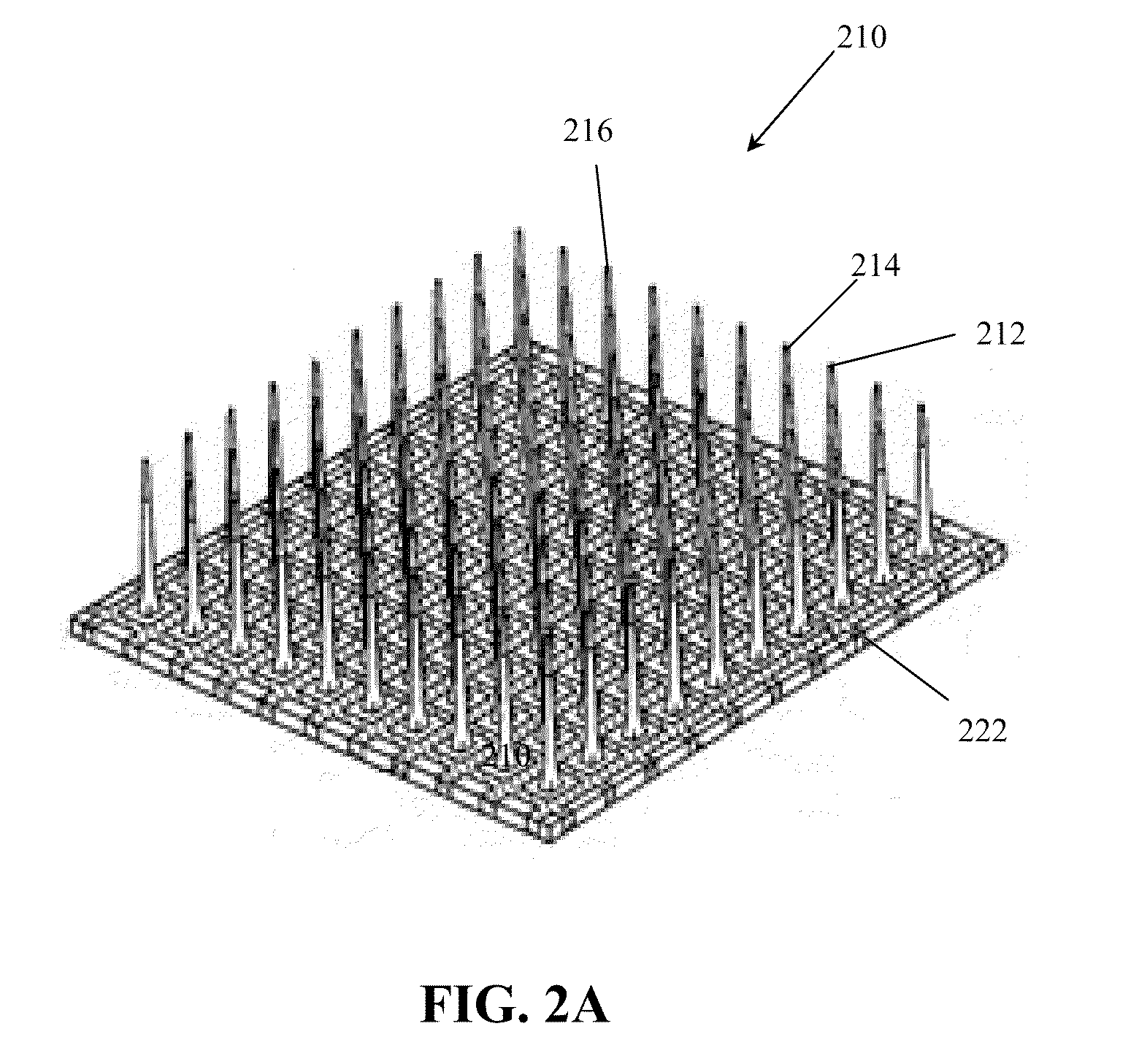
[0048]The electrical device can be used to detect suspect areas of tissue during a lumpectomy. Once the surgeon excises the suspect breast tissue, either the surgeon, a pathologist, or lab technician in the operating room can use the handheld device as shown in FIG. 3 to investigate if the margins of the tissue are free of malignant tissue, the steps of which are shown in the FIG. 7. The user approaches the tissue with the handheld device and inserts the electrode array into the tissue at the desired location. The user than uses the device to detect and measure the electrical response from the suspected areas of tissue. A reference signal specific to the patient is collected from a clear benign portion of the tissue and the results of the benign tissue response are compared to the measurements obtained from the suspect portions of the sample. Cancerous tissue has been shown to exhibit lower resistance and higher capacitance as compared to benign ti...
example 3
Detection of Breast Cancer In Vivo
[0049]The electrical device can also be used to detect cancerous tissue in vivo during a lumpectomy. During the lumpectomy, the surgeon will use the probe in the body cavity of the patient to assess if, once the suspect tissue has been excised, the remaining tissue in the breast is free of malignancy. Once the excision procedure is complete, the surgeon will use the device inside the body to scan the surface of the cavity. The depth of the electrode will be adjusted based on the surgeon and hospital convention and the assigned depth will be measured using the probe. The results obtained from a benign tissue sample will be registered to form a baseline for the patient tissue response and the results from the suspect areas will be compared with the benign values. The console will display the variations in the tissue response. If a larger deviation is observed in the impedance vector, or resistance, and / or capacitance compared to the benign signals, th...
PUM
| Property | Measurement | Unit |
|---|---|---|
| size | aaaaa | aaaaa |
| area | aaaaa | aaaaa |
| area | aaaaa | aaaaa |
Abstract
Description
Claims
Application Information
 - R&D
- Intellectual Property
- Life Sciences
- Materials
- Tech Scout
- Unparalleled Data Quality
- Higher Quality Content
- 60% Fewer Hallucinations
Browse by: Latest US Patents, China's latest patents, Technical Efficacy Thesaurus, Application Domain, Technology Topic, Popular Technical Reports.
© 2025 PatSnap. All rights reserved.Legal|Privacy policy|Modern Slavery Act Transparency Statement|Sitemap|About US| Contact US: help@patsnap.com


