
Method and system for detection and/or characterization of a biological particle in a sample

a biological particle and sample technology, applied in the field of methods and systems for monitoring, detecting and/or characterizing samples, can solve the problems of limited sensitivity of this approach, standard automated blood culture instruments, and lack of information about the characterization or identity of microorganisms, so as to improve efficiency and safety

Active Publication Date: 2010-03-18
BIOMERIEUX INC
View PDF27 Cites 88 Cited by
  • Summary
  • Abstract
  • Description
  • Claims
  • Application Information

AI Technical Summary

Benefits of technology

[0007]In another embodiment, the invention provides an automated system for detecting and / or characterizing a biological particle that may be present in a sample, said system comprising: (1) a growth chamber comprising a sample and a growth composition; (2) a measurement device comprising a reflectance and / or fluorescence spectrometer to enable a measurement of reflectance and / or fluorescence from said growth chamber taken at two or more time points; and (3) a means for relating said measurement to detecting and / or characterizing said biological particle if present in said sample, wherein said growth chamber is located in the same system as the measurement device and the measurement is non-invasive to the growth chamber. In this system, no manual sampling of the composition comprising a sample is required to provide early detection and / or characterization, thus providing improved efficiency and safety that may be particularly useful in both clinical and non-clinical applications.

Problems solved by technology

Standard automated blood culture instruments are restricted to a detection system that gives either a positive or negative result but does not provide any information about the characterization or identity of the microorganism.
Aside from testing with molecular based assays, which are expensive and generally limited to specific microorganisms, the identity of the microorganism is typically established by taking a sample from a positive blood culture bottle and conducting a separate identification test, usually after an overnight subculture step to prepare the organisms for identification testing.
The sensitivity of this approach is limited because the quenching, fluorescence, and reflectance properties of the surrounding medium within which the biological sample of interest is contained can interfere with the measurement.
However, for turbid, optically dense, or other complex samples, such as blood, the system is inefficient because the variability of the complex background complicates the microorganism spectrum when measured at only a single time point.
Thus, sensitive detection and early characterization or identification of the biological entity in a complex sample is generally not practical with a single measurement approach as taught by this reference.

Method used


Image

  • Method and system for detection and/or characterization of a biological particle in a sample
  • Method and system for detection and/or characterization of a biological particle in a sample
  • Method and system for detection and/or characterization of a biological particle in a sample

Examples


example 1

Blood Culture (Culture Medium and Blood Sample) in Quartz Cuvette in Waterbath Adapter

[0066]Seeded blood cultures were set up in autoclaved 1.0 cm screw-capped quartz cuvettes (Starna, Inc.) containing a stir bar for agitation. To the cuvette was added 2.4 mL of standard blood culture medium, 0.6 mL of fresh normal human blood and 0.05 mL of a 103 / mL suspension of test microorganism (approx. 10 CFU / cuvette). A sterile, septum screw cap was placed on the cuvette, and it was inserted into the front-face adapter previously described. The culture was maintained at approximately 36° C. by connecting the adapter to a recirculating water bath heated to 36° C. The cuvette was read every 45 minutes by the Fluorolog 3 fluorescence spectrophotometer that was software controlled. A full EEM spectra was collected at each time point with an Excitation wavelength range of 260-580 nm (every 5 nm) and an Emission wavelength range of 260-680 nm (every 5 nm) for a total of 3,139 data-points per scan. ...

example 2

Blood Culture in Acrylic Cuvette in Waterbath Adapter

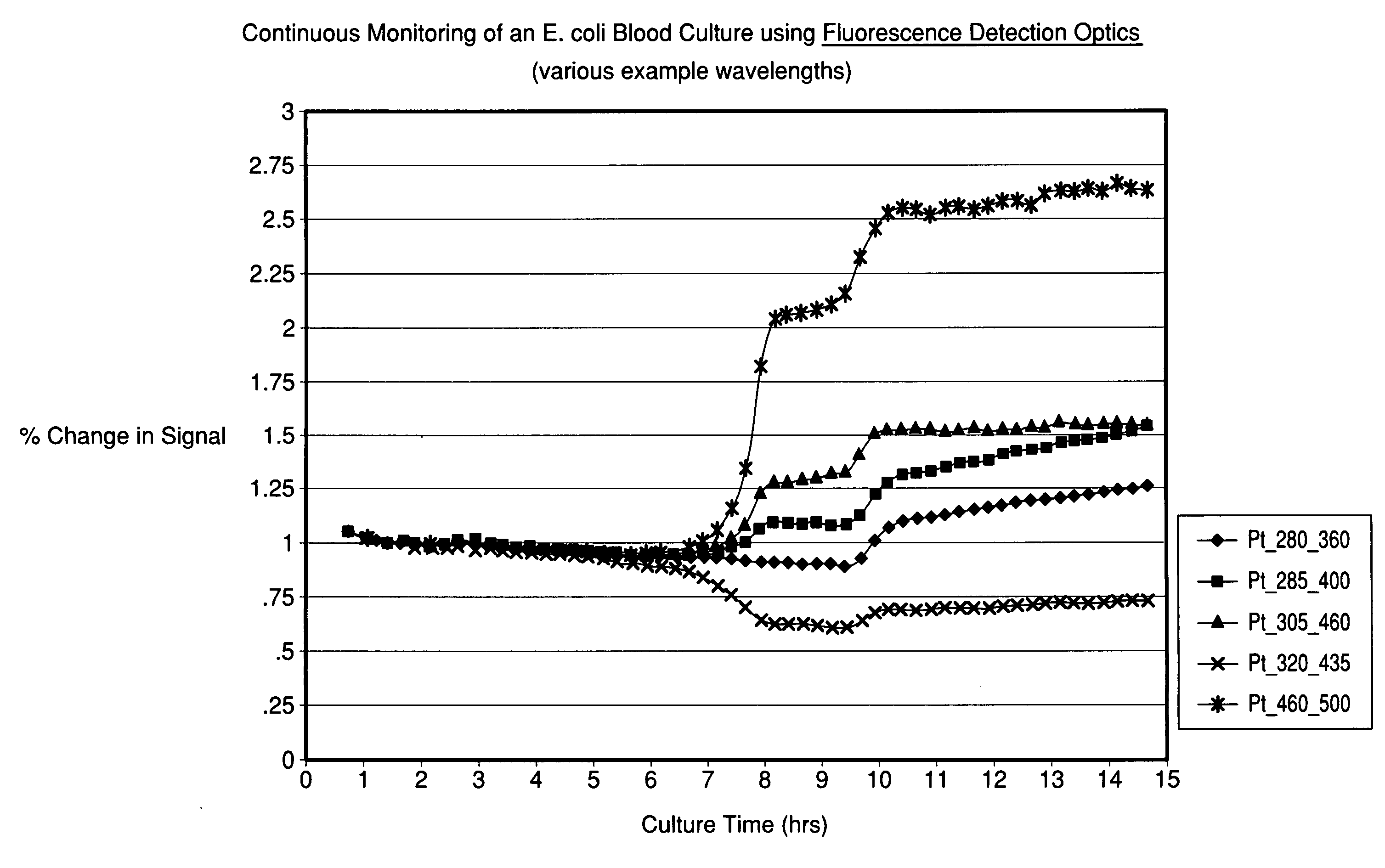
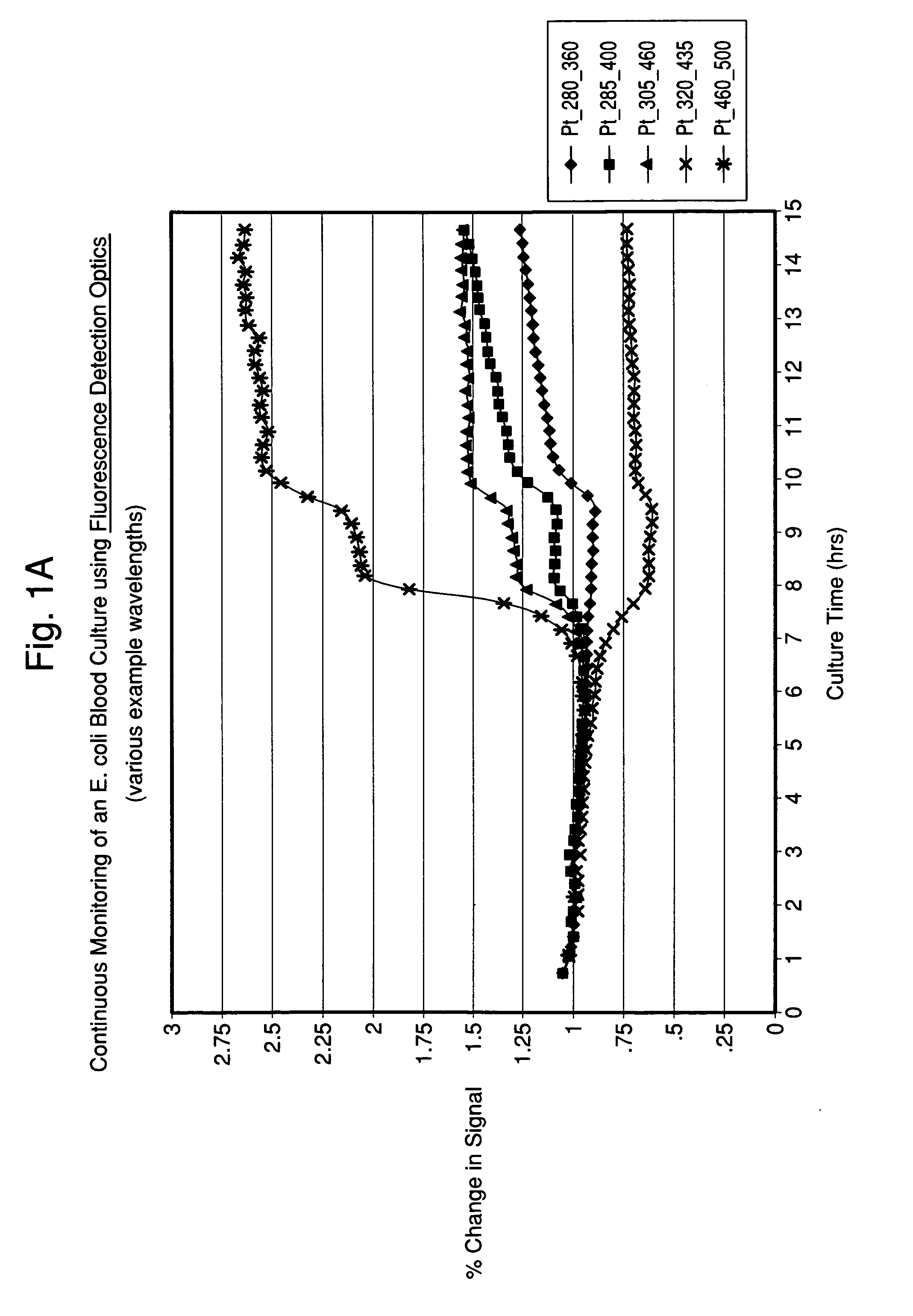
[0069]An E. coli blood culture was set up as described in Example 1 with the exception that the cuvette was constructed of a UV-transparent acrylic (Sarstedt 67.755), and sterilized by ethylene oxide treatment. The change in the autofluorescence or intrinsic fluorescence of the blood-media mixture over time with multiple wavelength pairs is shown in FIG. 5.

example 3

Blood Culture in Acrylic Cuvette in Multi-Station Carousel Adapter

[0070]An E. coli blood culture was set up as described in Example 2 with the exception that the cuvette was loaded into a custom-built, temperature-controlled carousel adapter for the Fluorolog 3 system. The change in the autofluorescence or intrinsic fluorescence of the blood-media mixture over time with multiple wavelength pairs is shown in FIG. 6.

[0071]The experiments described in Examples 2 and 3 demonstrate that time-dependent changes in fluorescence were measured in readily available plastic containers and in a manner amenable to larger scale automation, respectively. The same is true for the diffuse reflectance measurements collected in these experiments (data not shown).


PUM


Abstract

The present invention provides a method and system for monitoring, detecting, and / or characterizing a biological particle that may be present in a sample whereby the method may be accomplished non-invasively by utilizing a time-dependent spectroscopic technique to obtain at least two measurements of a growth composition comprising a sample and correlating said measurements for the detection and / or characterization of a biological particle, that may be present in the sample.

Description

FIELD OF THE INVENTION[0001]The present invention relates to a method and system for monitoring, detecting and / or characterizing a sample for biological particle growth.BACKGROUND OF THE INVENTION[0002]Methods to detect and identify microorganisms are typically conducted on separate automated systems in clinical laboratory and industrial settings. Standard automated blood culture instruments are restricted to a detection system that gives either a positive or negative result but does not provide any information about the characterization or identity of the microorganism. Aside from testing with molecular based assays, which are expensive and generally limited to specific microorganisms, the identity of the microorganism is typically established by taking a sample from a positive blood culture bottle and conducting a separate identification test, usually after an overnight subculture step to prepare the organisms for identification testing.[0003]WO2007 / 019462 generally relates to a m...

Claims


Application Information

Patent Timeline
IPC IPC(8): C12Q1/04C12M1/34
CPCC12Q1/04G01N21/31G01N21/47C12Q1/10G01N21/6486G01N2021/6419G01N2021/6421G01N21/64
Inventor WALSH, JOHN D.HYMAN, JONES M.CLAY, BRADFORD G.THORPE, THURMAN C.
Owner BIOMERIEUX INC
Learn More