
Method for inducing differentiation of pluripotent stem cells into cardiomyocytes

a technology cardiomyocytes, which is applied in the field of inducing differentiation of pluripotent stem cells into cardiomyocytes, can solve the problems of cardiac failure, cardiac failure, and further exhaustion and death of cardiomyocytes, and achieve the effect of efficient and selective production

Inactive Publication Date: 2009-12-31
DAIICHI SANKYO CO LTD
View PDF5 Cites 10 Cited by
  • Summary
  • Abstract
  • Description
  • Claims
  • Application Information

AI Technical Summary

Benefits of technology

[0080]Myocardial precursor cells and cardiomyocytes can be very efficiently and selectively produced from ES cells and other pluripotent stem cells using the method of the present invention. Cardiomyocyte (precursor) cells prepared by the method of the present invention can be used to search for and develop effective drugs for treating heart disease, and could potentially be applied to myocardial transplantation therapy for severe heart disease.

Problems solved by technology

In general, cardiomyocytes undergo active cell division with beating autonomously before birth, but immediately after birth they lose the ability to divide, and since they have very few undifferentiated stem cells and precursor cells whose growth and differentiation abilities are extremely low, when cardiomyocytes die due to exposure to various forms of stress including myocardial infarction, myocarditis and the like, the lost cardiomyocytes cannot be regenerated.
As a result, the surviving cardiomyocytes try to maintain myocardial function through compensatory hypertrophy and the like, but if the stress continues and exceeds an allowable threshold, it leads to further exhaustion and death of cardiomyocytes and a consequent lowering of myocardial function (that is, heart failure).
Heart failure and other types of heart disease are the second leading cause of death in Japan, and prognoses are very poor, with a 5-year survival rate of only about 50% for patients with heart diseases.
Conventional therapeutic drugs for heart failure include digitalis preparations that increase the contractive force of the myocardium and xanthine preparations and other heart stimulants, but long-term administration of these drugs is known to make the condition worse because there is too much expenditure of myocardial energy.
More recently, mainstream therapy has shifted to β-blockers and ACE inhibitors, which reduce the excess burden on the heart due to stimulation of the sympathetic nervous system and renin-angiotensin system, but these methods only deal with the immediate symptoms and cannot restore damaged cardiac tissue.
By contrast, heart transplantation is a fundamental treatment for severe heart failure, but it is one that is difficult to apply commonly because of such problems as the shortage of heart donors, ethical concerns, the physical and financial burden on patients and the like.
However, it is difficult to obtain sufficient cardiomyocytes for this method, and application to clinical medicine is also difficult from an ethical standpoint.
However, one serious problem remains to be elucidated to use pluripotent stem cell-derived cardiomyocytes for cell transplantation therapy and other purposes.
However, although the purity of the cardiomyocytes is extremely high in this method, the final number of cardiomyocytes obtained is only a few percent of the total cell count, making it difficult to obtain enough amounts of cardiomyocytes for transplantation.
Moreover, 5-azacytidine is a demethylation agent that alters the expression of genes by removing methyl groups bound to DNA, and because it acts directly on the chromosomes, it is not a suitable drug for preparing cells for cell transplantation.
However, cardiomyocyte-specific or selective differentiation has not been achieved with any of these methods.
However, P19 cell lines do not have the ability to differentiate into a variety of cells or the ability to form chimeras, unlike ES cells.
Thus, it was not possible to predict, based on scientific grounds, whether the findings obtained in this experimental system could be directly extrapolated to myocardial differentiation-inducing systems for ES cells and other pluripotent stem cells.
Under these circumstances, no optimum culture method has been established for inducing myocardial differentiation.

Method used


Image

  • Method for inducing differentiation of pluripotent stem cells into cardiomyocytes
  • Method for inducing differentiation of pluripotent stem cells into cardiomyocytes
  • Method for inducing differentiation of pluripotent stem cells into cardiomyocytes

Examples


example 1

Study on Expression Patterns of Various

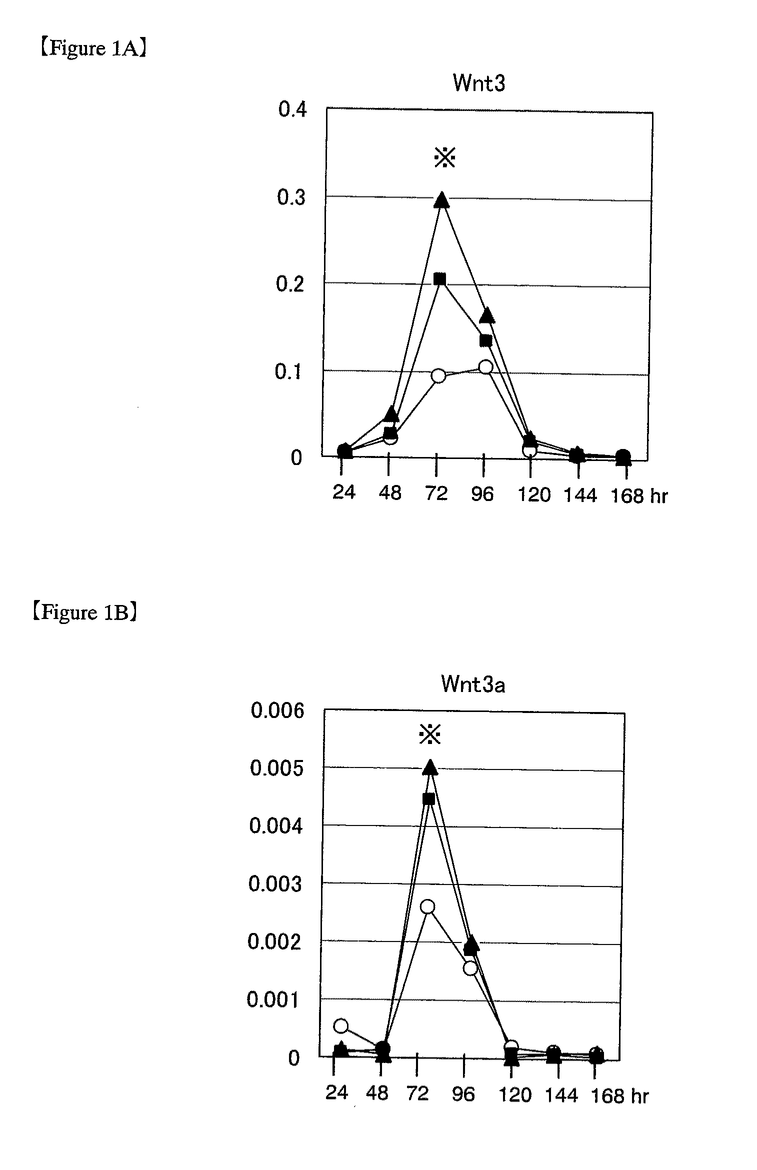
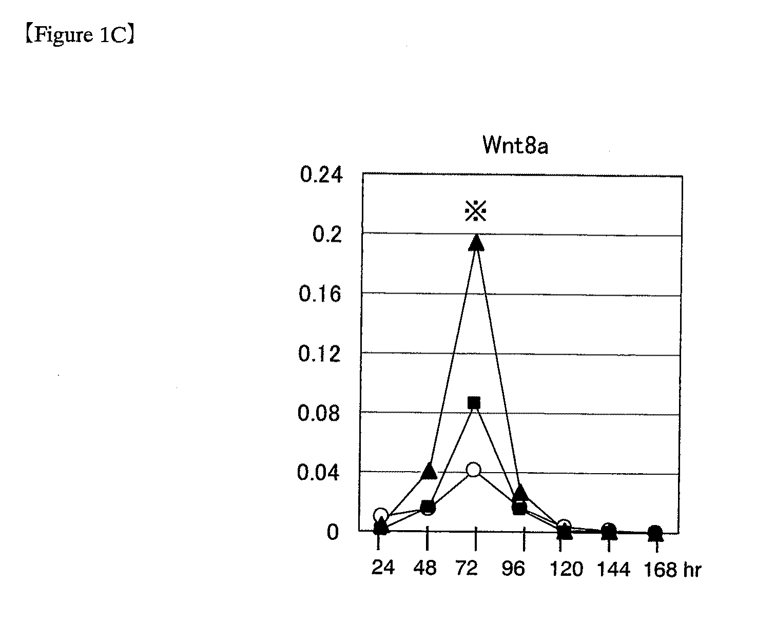
[0141]Wnt genes during induction of differentiation in ES cells (1) Various Wnt genes were studied for their expression during differentiation in mouse ES cells. For use in experiments, mouse ES cells were passaged and maintained in an undifferentiated state according to the methods as described in “Manipulating the Mouse Embryo: A Laboratory Manual” (Hogan et al., eds., Cold Spring Harbor Laboratory Press, 1994) and “Embryonic Stem Cells: Methods and Protocols” (Turksen ed., Humana Press, 2002) by using Knockout-DMEM (Invitrogen) medium containing 20% fetal bovine serum, 2 mmol / L L-glutamine and 0.1 mmol / L 2-mercaptoethanol (hereinafter referred to as ESM), supplemented with 1000 U / mL LIF (ESGRO; Chemicon). ES cells passaged under these conditions are hereinafter referred to as “ES cells passaged under ordinary culture conditions.” The mouse ES cells used in the following experiments were D3 cells, R1 cells and 129SV cells (purchased from Dain...

example 2

Enhancing Effect of Recombinant Wnt Protein Treatment on the Appearance of Cardiomyocytes Derived from ES Cells (1)

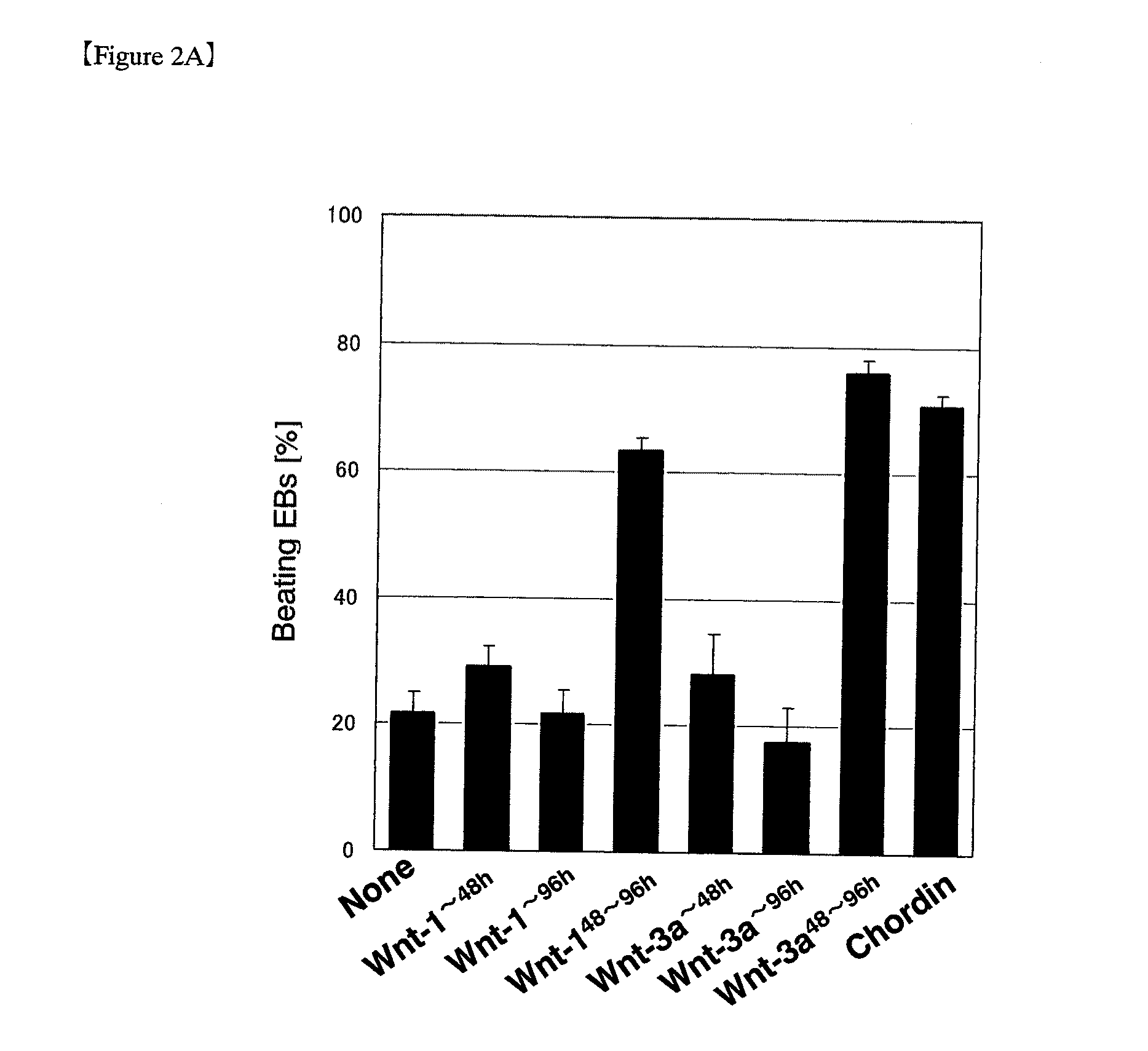
[0149]In the early stage of differentiation in ES cells, transient elevations in expression of various Wnt genes were observed prior to the appearance of cardiomyocytes. Then, ES cells at this stage were treated with recombinant Wnt proteins to study the myocardial differentiation-inducing effect of the proteins. Induction of ES cell differentiation was accomplished in the same manner as used in Example 1, except that some of the experimental groups were cultured in medium containing a commercially available recombinant WNT-1 (Peprotech), Wnt-3a (R&D systems) or Wnt-5a (R&D systems) protein. Treatment of ES cells in medium supplemented with a recombinant protein of canonical Wnt such as WNT-1 is hereinafter referred to as “Wnt treatment.”

[0150]The appearance rate of EBs exhibiting spontaneous beating was investigated periodically as one of a useful index of the differen...

example 3

Properties of Cardiomyocytes Derived from Wnt-Treated ES Cells

[0155]As shown in Example 2, beating of EBs prepared from ES cells was increased significantly by Wnt treatment, and to confirm that the beating cells in these EBs were cardiomyocytes, further studies were performed to investigate gene expression and protein production of various myocardial-specific marker molecules. In the same manner as used in Example 2, ES cells were induced to differentiate and the resulting EBs were collected at 10 days after induction of differentiation to prepare cDNA. Real-time PCR quantification was performed by the TaqMan probe method, i.e., by using the above cDNA (1 μL) as a template and using a TaqMan Universal PCR Master Mix (PE Applied Biosystems) according to the instructions attached thereto. TaqMan probes for detection of various genes were designed using primer design software (ABI PRISM Primer Express) on the basis of the nucleotide sequence information of the genes. The nucleotide se...


PUM


Abstract

The present invention provides a method for inducing differentiation of cardiomyocytes efficiently and selectively from stem cells.A method for inducing differentiation of cardiomyocytes from pluripotent stem cells, which comprises: (i) culturing the pluripotent stem cells in a culture medium containing no substance that promotes activation of the canonical Wnt signaling pathway during the time period between initiation of differentiation induction and 24 hours before the period of elevated canonical Wnt gene expression; and then (ii) culturing the pluripotent stem cells in a culture medium containing a substance that promotes activation of the canonical Wnt signaling pathway during a time period of 24 to 96 hours, starting from 24 to 0 hours before the period of elevated canonical Wnt gene expression.

Description

TECHNICAL FIELD[0001]The present invention relates to a method for preparing cardiomyocytes selectively and efficiently from ES cells and other pluripotent stem cells.BACKGROUND ART(1) Preparation of Cardiomyocytes Using Pluripotent Stem Cells[0002]In general, cardiomyocytes undergo active cell division with beating autonomously before birth, but immediately after birth they lose the ability to divide, and since they have very few undifferentiated stem cells and precursor cells whose growth and differentiation abilities are extremely low, when cardiomyocytes die due to exposure to various forms of stress including myocardial infarction, myocarditis and the like, the lost cardiomyocytes cannot be regenerated. As a result, the surviving cardiomyocytes try to maintain myocardial function through compensatory hypertrophy and the like, but if the stress continues and exceeds an allowable threshold, it leads to further exhaustion and death of cardiomyocytes and a consequent lowering of my...

Claims


Application Information

Patent Timeline
IPC IPC(8): C12N5/08C12N5/02C12N5/077
CPCC12N5/0657C12N2506/02C12N2501/415C12N5/0602C12N5/00C12N5/0606
Inventor KOSHIMIZU, UICHITANAKA, TOMOFUMIKAWASHIMA, KAYOKOKADOKURA, MICHINORI
Owner DAIICHI SANKYO CO LTD
Learn More