
Methods and Apparatus for Locating the Fossa Ovalis and Performing Transseptal Puncture

a technology of fossa ovalis and transseptal puncture, which is applied in the field of methods and apparatus for and methods and apparatus for performing transseptal punctures. it can solve the problems of insufficient and additional tools, needle and catheter puncture, etc., and achieve the effect of locating the fossa ovalis using these techniques

Inactive Publication Date: 2009-12-03
ST JUDE MEDICAL ATRIAL FIBRILLATION DIV
View PDF5 Cites 1 Cited by
  • Summary
  • Abstract
  • Description
  • Claims
  • Application Information

AI Technical Summary

Benefits of technology

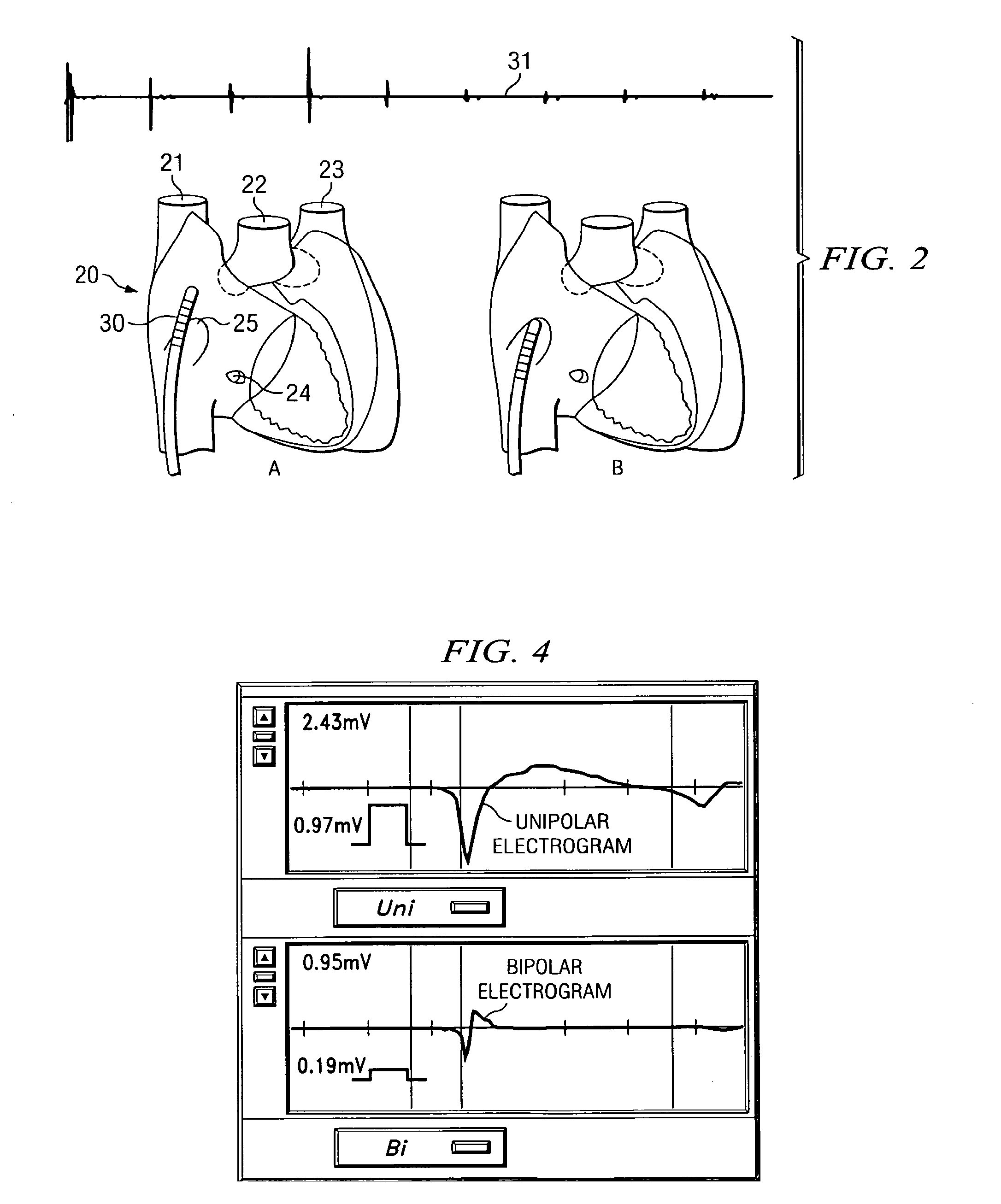
The present invention provides a method for identifying the fossa ovalis in a patient's heart and a catheter for use in transseptal punctures. The method involves positioning electrodes against the tissue of the interatrial septum and acquiring electrograms while moving the electrodes to multiple positions. The fossa ovalis can be identified based on parameters such as unipolar voltage reduction, signal fractionation, broadened signal, reduced signal slew rate, reduced local myocardial impedance, increased phase angle, and increased pacing threshold. The catheter comprises a hollow lumen with a distal end and two electrodes positioned on the catheter.

Problems solved by technology

The danger of the transseptal approach lies in the possibility that the needle and catheter will puncture an adjacent structure such as the aorta, the coronary sinus or the free wall of the atrium.
Due to the potentially life threatening complications of the procedure, many operators feel that fluoroscopy, which at best represents an epicardial shadow of the heart, is not enough and additional tools are needed.
While echocardiography can be useful, there are potential problems in using these techniques to locate the fossa ovalis.
If transesophageal echocardiography (TEE) is used to guide the puncture, a different operator has to operate the TEE system and therefore errors can occur especially in the interpretation of the data.
Cardiac tamponade and other complications are known to have occurred during transseptal punctures performed in electrophysiology laboratories despite the routine use of ultrasound guidance.
The additional time and expense that the use of ultrasound catheters and sheaths incurs is not inconsiderable and this can make it impractical to use them routinely.

Method used


Image

  • Methods and Apparatus for Locating the Fossa Ovalis and Performing Transseptal Puncture
  • Methods and Apparatus for Locating the Fossa Ovalis and Performing Transseptal Puncture
  • Methods and Apparatus for Locating the Fossa Ovalis and Performing Transseptal Puncture

Examples


Embodiment Construction

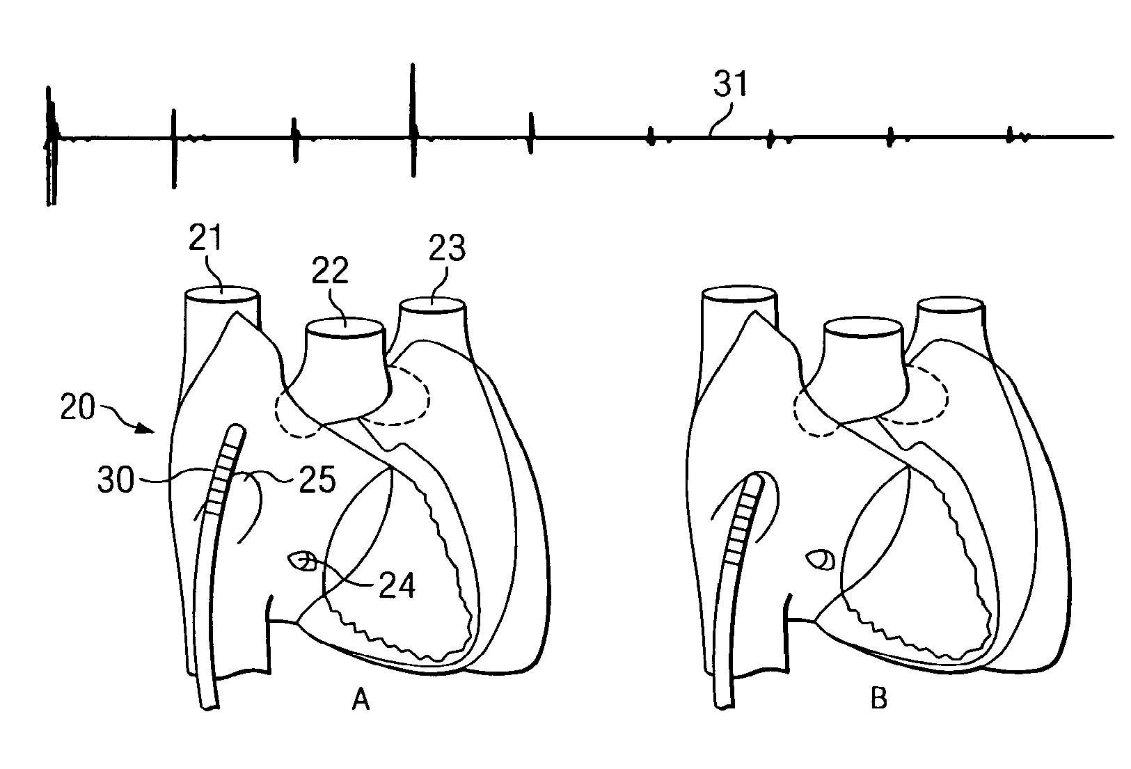
[0049]Applicant has discovered that the fossa ovalis can be located by measuring electrophysiological activity of the tissue of the interatrial septum. In addition, Applicant has also developed an apparatus which may be used not only for locating the fossa ovalis, but also for performing transseptal punctures through the fossa ovalis.

[0050]The fossa ovalis is the depression at the site of the fetal interatrial communication termed the foramen ovale. In fetal life, this communication allows richly oxygenated IVC blood coming from the placenta to reach the left atrium and has a well-marked rim or limbus superiorly. The floor of the fossa ovalis is a thin, fibromuscular partition. In fact, because the fossa ovalis is thinner than the rest of the interatrial septum, light may even be used to selectively transilluminate the fossa ovalis.

[0051]However, in addition to being thinner than the surrounding tissue of the interatrial septum, Applicant has discovered that the fossa ovalis has con...


PUM


Abstract

A method of identifying the fossa ovalis in a patient by positioning one or more electrodes against the tissue of the interatrial septum of the patient and acquiring unipolar and / or bipolar electrograms of the tissue of the interatrial septum while moving the electrodes to a plurality of positions against the tissue of the interatrial septum. The fossa ovalis is identified on the basis of unipolar voltage reduction, signal fractionation, broadened signal, reduced signal slew rate, reduced local myocardial impedance, increased phase angle and / or increased pacing threshold. An apparatus for identifying the fossa ovalis is also provided.

Description

CROSS-REFERENCE TO RELATED APPLICATIONS[0001]This application is a continuation of U.S. application Ser. No. 10 / 648,844, filed 25 Aug. 2003, now pending, which claims benefit of U.S. provisional application No. 60 / 405,849, filed 24 Aug. 2002. The foregoing applications are hereby incorporated by reference as though fully set forth herein.BACKGROUND OF THE INVENTION[0002]a. Field of the Invention[0003]The present invention relates to methods and apparatus for locating the fossa ovalis, as well as methods and apparatus for performing transseptal punctures.[0004]b. Description of Related Art[0005]The transseptal puncture for left atrial and left ventricular catheterization was initially described simultaneously by Ross and Cope in 1959. In the 1960s, Brockenbrough and colleagues modified the design of the transseptal needle and the guiding catheter. In the late 1960s and 70s, its use declined because of the occurrence of complications and because of the development of selective coronar...

Claims


Application Information

Patent Timeline
Patent Type & Authority Applications(United States)
IPC IPC(8): A61B5/04A61M29/00A61B17/34A61B5/107A61B5/296A61B17/00A61M25/00A61M25/08
CPCA61B5/042A61B5/0422A61B2017/00243A61B5/1076A61B5/05A61B5/4836A61B5/0036A61B5/283A61B5/287A61B5/068A61B17/3478A61B2017/00044A61B2017/00247A61M25/0662A61M2025/0166
Inventor KRISHNAN, SUBRAMANIAM C.
Owner ST JUDE MEDICAL ATRIAL FIBRILLATION DIV
Learn More