
Modulation of bio-electrical rhythms via a novel engineering approach

Active Publication Date: 2009-11-26
THE JOHN HOPKINS UNIV SCHOOL OF MEDICINE
View PDF0 Cites 11 Cited by
  • Summary
  • Abstract
  • Description
  • Claims
  • Application Information

AI Technical Summary

Benefits of technology

[0011]We now provide gene transfer and cell administration methods that can create a “bio-battery” to exert pacemaking function, and / or to modulate the activity of an endogenous or induced cardiac pacemaker function using genetically modified HCN constructs.
[0014]In a further aspect, a composition is administered to a subject to alter the frequency of (i.e. to tune) an existing endogenous or induced cardiac pacemaker function. Polynucleotides of or modified cells containing a genetically modified HCN construct that displays a particular phenotype are preferred agents for administration. The method can be applied to virtually any cell types that exhibit bioelectrical rhythms (e.g. certain neuronal and pancreatic cells) other than cardiac cells so as to subsequently modify / improve their physiological functions. For instance, the activity of electrically active neuronal cells in the case of neuropathic pain can be decreased using a different HCN construct (e.g. one that is less ready for opening or even a dominant-negative construct) to reduce or eliminate the sensation of pain. (i.e. to correct a particular defect due to diseases or traumas).

Problems solved by technology

For instance, spontaneous neuronal electrical discharges in damaged dorsal root ganglions underlie neuropathic pain but limited useful therapy is available.
In the heart, abnormal pacing leads to various forms of arrhythmias and electrical disorders that necessitate traditional pharmacologic interventions and implantation of costly electronic devices that are associated with various side effects, inherent risks, and expenses.
Thus, native If is difficult to reproduce by simple expression of a single HCN isoform.
Indeed, previous attempts to overexpress wild-type HCN2 in adult ventricular cardiomyocytes failed to induce automaticity19, presumably due to its negative activation relative to the voltage range of cardiac pacing.

Method used


Image

  • Modulation of bio-electrical rhythms via a novel engineering approach
  • Modulation of bio-electrical rhythms via a novel engineering approach
  • Modulation of bio-electrical rhythms via a novel engineering approach

Examples


example 1

Expression of an Engineered If in Ventricular Cardiomyocytes

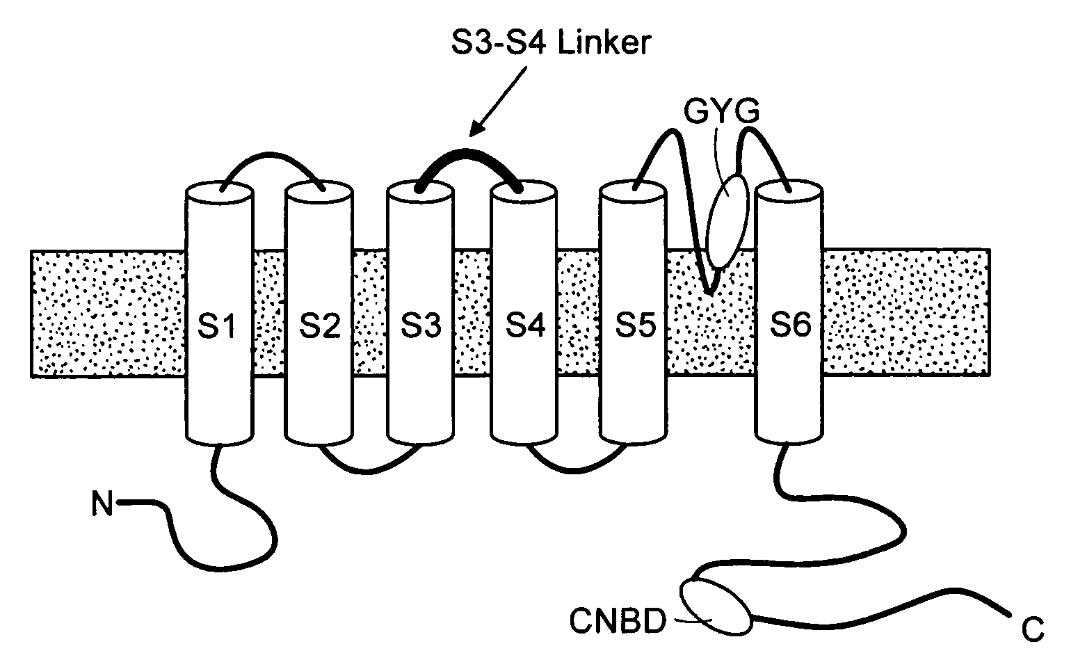
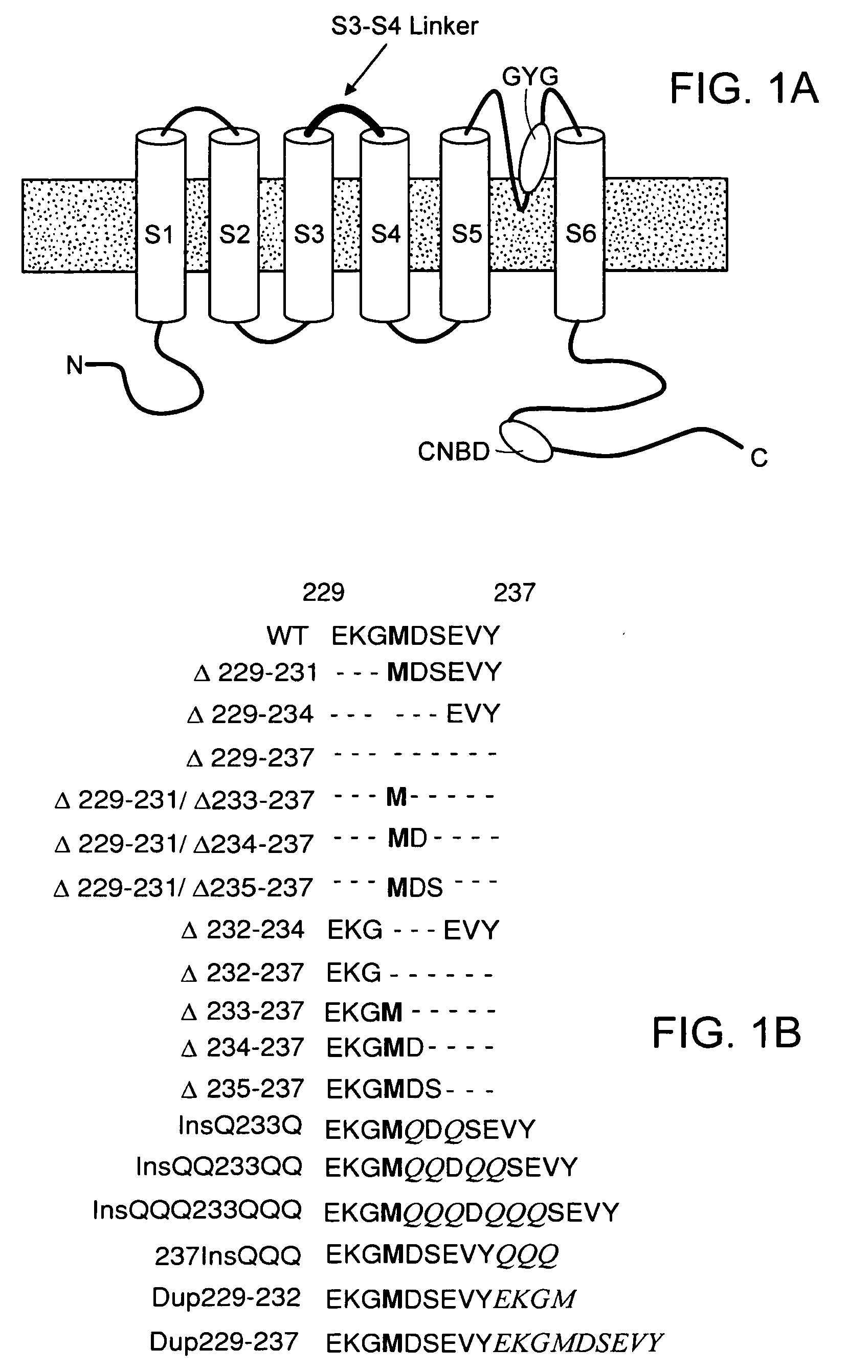
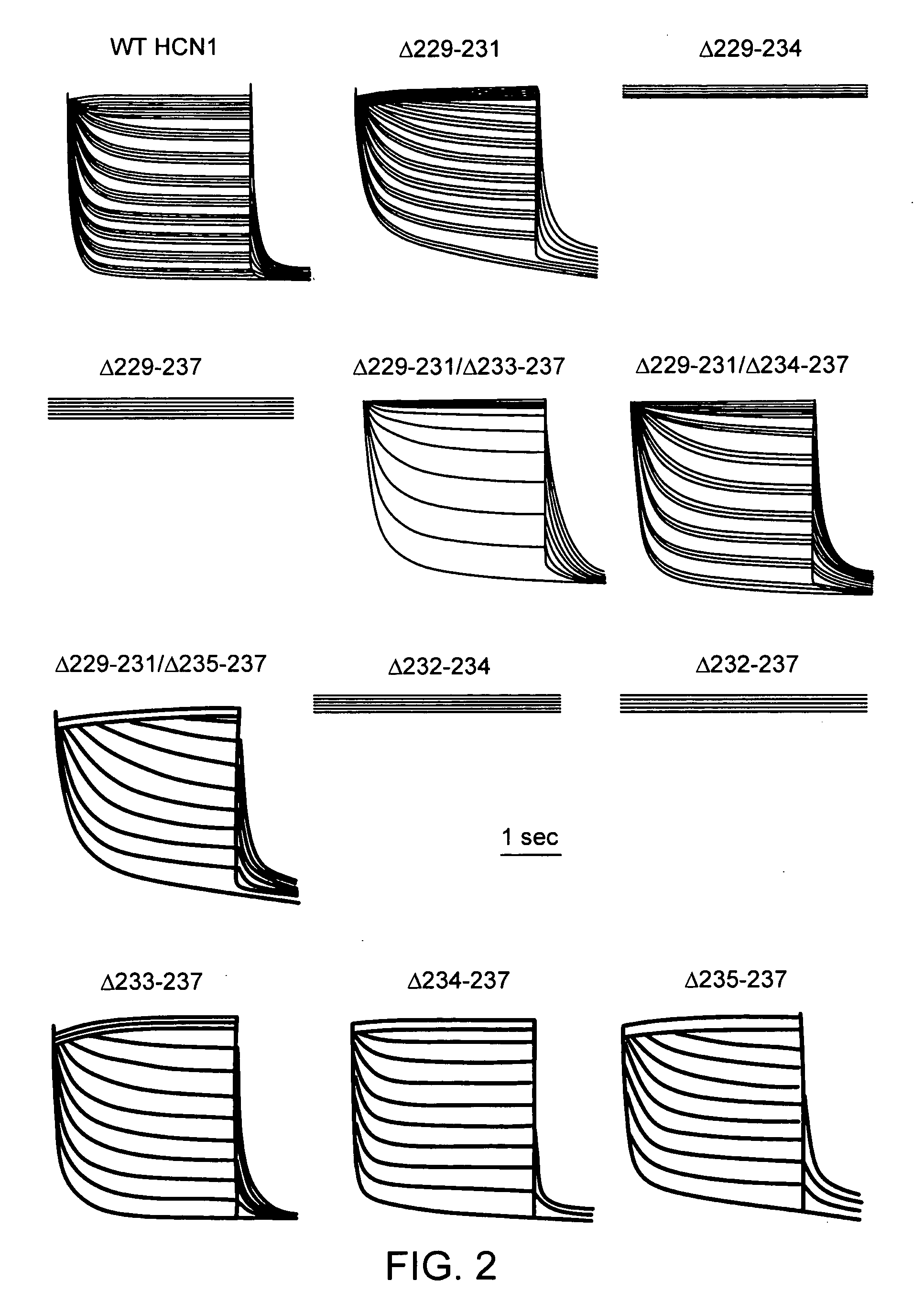
[0060]We first created the recombinant adenoviruses Ad-CGI and Ad-CGI-HCN1-ΔΔΔ to overexpress the green fluorescent protein (GFP) and the engineered HCN1 construct EVY235-7ΔΔΔ, respectively. HCN1-EVY235-7ΔΔΔ channels, whose S3-S4 linker has been shortened by deleting residues 235-237, was chosen because they open more readily (with a ˜20 mV depolarizing shift of activation V1 / 2) than any of the wild-type HCN1-4 isoforms when expressed in heterologous expression systems23,24. Therefore, we conjectured that overexpressing EVY235-7ΔΔΔ channels alone in cardiomyocytes would sufficiently mimic the heteromultimeric native nodal If without having to simultaneously express or manipulate multiple HCN isoforms.

[0061]FIG. 9 shows that IK1(A and E), which could be completely blocked by 1 mM Ba2+ (B and F) was robustly expressed in control (non- or Ad-CGI-transduced) adult guinea pig left ventricular (LV) cardiomyocytes (n=36). For LV c...

example 2

Induced Automaticity by HCN-Encoded If Overexpression

[0062]Shown in FIG. 10A (left) is a typical control ventricular cell (same from FIGS. 9A-B) that was normally electrically-quiescent with no spontaneous activity. The resting membrane potential, RMP, was −76±5 mV (n=7). Upon injection of a stimulating current (˜0.8 nA for 5 ms), the same cell generated a single action potential (AP), indicating normal excitability. Addition of 1 mM Ba2+ to block IK1 destabilized the normal RMP and subsequently resulted in spontaneous firing with an average cycle length of 689±132ms that was ˜3-fold slower than that of guinea pig nodal cells (FIG. 10A, right) similar to that induced by IK1 genetic suppression (personal communication with Dr. H B Nuss). Collectively, these observations indicate that although IK1 suppression unleashes latent pacemaker activity of ventricular cardiomyocytes, it is insufficient to reproduce the normal frequency of endogenous nodal pacing.

[0063]We next investigated the ...

example 3

The HCN Approach Functionally Substitutes an Electronic Device In Vivo

[0064]To better test the functional efficacy and therapeutic potential of our gene-based approach in vivo, we next switched to a large animal model. Swines were chosen because they have anatomy and baseline sinus rate (78±14 bpm) similar to those of humans. We first developed a swine model of the SSS by ablating the SA node to cause sinus dysfunction (FIG. 11A-B). To prevent asystole or bradycardias after ablation, we implanted a dual-chamber electronic pacemaker with one electrode positioned at the high anterolateral wall of the RA and another at the right ventricular apex. Electrocardiogram (ECG) recordings after ablation demonstrated the presence of junctional escape rhythm (FIG. 11C) or ventricular pacing rhythm (FIG. 11D) as a result of the implanted device's programmed electrical stimulation when the spontaneous HR fell below 30 bpm.

[0065]To further validate our swine SSS model, we closely monitored the hea...


PUM

PropertyMeasurementUnit
Fractionaaaaaaaaaa
Frequencyaaaaaaaaaa

Abstract

The present invention relates to novel compositions and methods to induce, and / or modulate bio-electrical rhythms (e.g. in cardiac, neuronal and pancreatic cells) by fine-tuning the activity of HCN-encoded pacemaker channels via a novel protein- and genetic-engineering approach to augment or attenuate the associated physiological responses (e.g. heart beat, neuronal firing, insulin secretion, etc) for achieving various therapeutic purposes (e.g. sick sinus syndrome, epilepsy, neuropathic pain, diabetes, etc).

Description

CROSS REFERENCE TO RELATED APPLICATIONS[0001]This application claims priority to U.S. Provisional Patent Applications Ser. No. 60 / 598,170 filed Aug. 2, 2004, the entire disclosure of which are incorporated herein by reference in its entirety.STATEMENT OF FEDERALLY SPONSORED RESEARCH[0002]Funding for the present invention was provided in part by the Government of the United States by virtue of Grant No. HL72857 by the National Institutes of Health. Thus, the Government of the United States has certain rights in and to the invention claimed herein.FIELD OF THE INVENTION[0003]The present invention relates to novel approaches for engineering the generation of bioelectrical impulses (such as those from the cardiac, neuronal and pancreatic cells, etc) using genetically-modified membrane channels.BACKGROUND OF THE INVENTION[0004]Spontaneous cellular electrical rhythms (or pacing) govern numerous biological processes from the autonomous beating of the heart, pain transmission, to respirator...

Claims


Application Information

Patent Timeline
IPC IPC(8): A61K35/12C12N5/06A61K48/00
CPCA61K48/005A61K35/34A61K35/30A61K35/12A61P25/00A61P25/04A61P3/04A61P9/06A61P3/10
Inventor MARBAN, EDUARDOLI, RONALD A.TSANG, SUK-YINGCHO, HEE CHEOLXUE, TIAN
Owner THE JOHN HOPKINS UNIV SCHOOL OF MEDICINE
Learn More