
Method of deriving a quantitative measure of the instability of calcific deposits of a blood vessel

a blood vessel and calcific deposit technology, applied in image enhancement, instruments, image data processing, etc., can solve the problems of high risk of cardiovascular disease for patients, high risk of cardiovascular disease, and relatively unstable calcific deposits

Inactive Publication Date: 2009-08-13
BIOCLINICA
View PDF42 Cites 4 Cited by
  • Summary
  • Abstract
  • Description
  • Claims
  • Application Information

AI Technical Summary

Benefits of technology

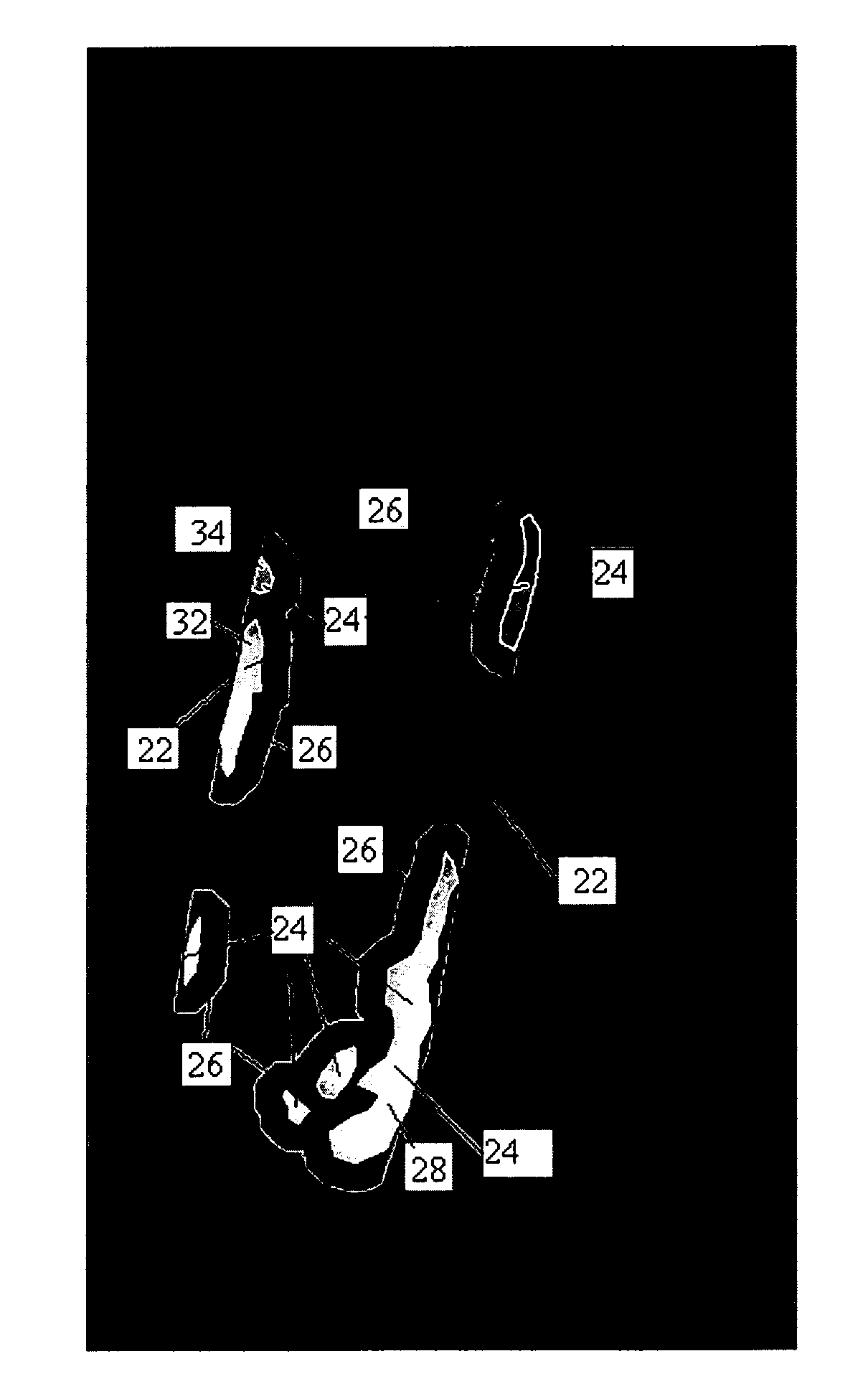
[0009]The present inventors have found that, in biological terms, a greater number of small calcific deposits distributed over a large portion of a blood vessel indicate a greater risk of developing cardiovascular disease than fewer larger deposits over the same area. The inventors have also found that the risk that a patient may suffer an episode of cardiovascular disease is high while calcific deposits are growing as during growth, the calcific deposits are relatively unstable. Because of its size relative to the size of a blood vessel a big, dense calcific deposit might be thought to be of grave concern, but could be quite stable and safe in terms of resulting in an episode of cardiovascular disease. By contrast, several small calcific deposits might seem not to be severe because of their size, but may carry a greater risk of resulting in an episode of cardiovascular disease as they would be associated with a greater risk of growth. The scope for growth of individual calcifications also increases as the periphery of a calcification becomes more irregular and deviates from being round.
[0014]Preferably, the method further comprises counting the number of calcific deposits and weighting said comparative index by said number. Weighting the number of calcifications with the comparison between the area of expanded calcific deposits and the area of unexpanded calcific deposits provides an enhanced measure of the degree of calcification in a blood vessel and the associated risk of developing cardiovascular disease.
[0020]In an embodiment, points along the boundary of each respective calcific deposit are moved outwards by the fixed distance x or, if closer, up to an aortic wall or unexpanded boundary of an adjacent calcific deposit. Preventing expansion of the boundaries of respective areas of calcification beyond either the arterial walls or adjacent calcific deposits gives a realistic prediction of the likely growth of the calcific deposits.

Problems solved by technology

The inventors have also found that the risk that a patient may suffer an episode of cardiovascular disease is high while calcific deposits are growing as during growth, the calcific deposits are relatively unstable.
By contrast, several small calcific deposits might seem not to be severe because of their size, but may carry a greater risk of resulting in an episode of cardiovascular disease as they would be associated with a greater risk of growth.

Method used


Image

  • Method of deriving a quantitative measure of the instability of calcific deposits of a blood vessel
  • Method of deriving a quantitative measure of the instability of calcific deposits of a blood vessel
  • Method of deriving a quantitative measure of the instability of calcific deposits of a blood vessel

Examples


Embodiment Construction

[0046]The present invention will hereinafter be described with particular reference to the analysis of x-ray images of an aorta. It will, however, be appreciated that the described method could be applied to other medical images of an aorta for example, DXA, Computer Tomography (CT) or Magnetic Resonance. Furthermore, the invention is not limited to analysis of images of an aorta and may also be applied to other blood vessels.

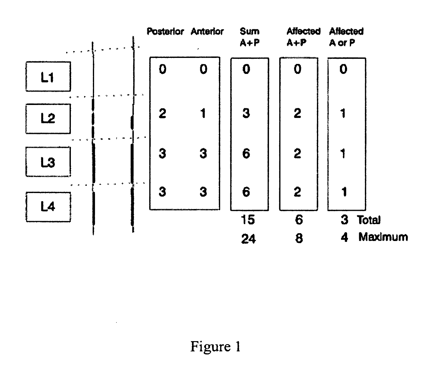
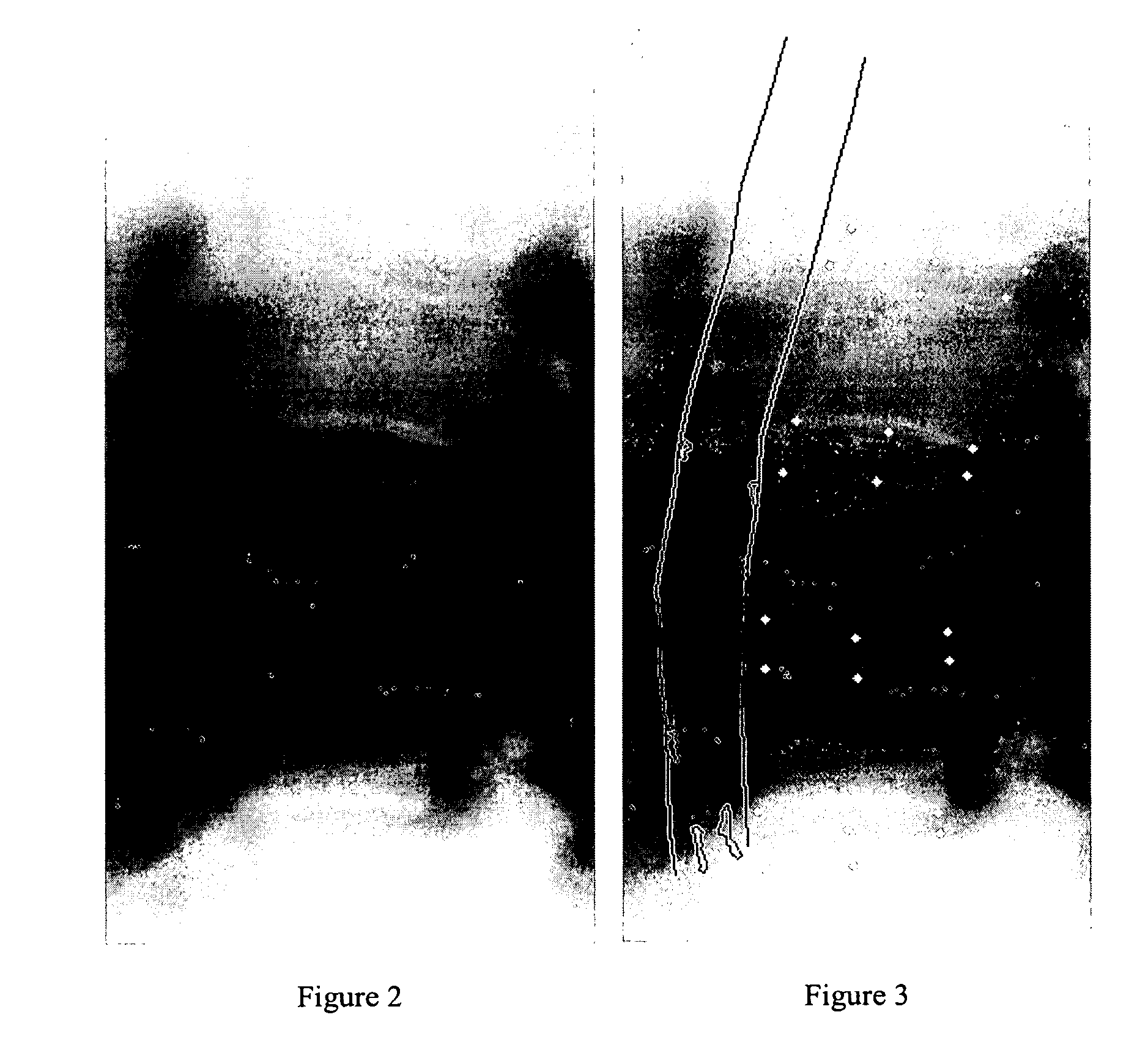
[0047]The first step in preparing the image for analysis is to outline the walls of the lumbar aorta in an image. FIG. 2 shows an image of part of a lumbar spine and lumbar aorta where there are calcific deposits 4 in the lumbar aorta. The six points for vertebral height measurements are annotated on L1 to L4 of the lumbar vertebrae as shown in FIG. 3 and from this the lumbar aorta can be identified and annotated. Further information about how the outline of the aorta is found is given by Lauze F et al. in (“Towards automated detection and segmentation of aorti...


PUM


Abstract

A computer-implemented method of processing an image of at least part of a blood vessel to derive a measure indicative of the instability of calcific deposits in the blood vessel, said blood vessel containing at least one calcific deposit, comprises locating and annotating one or more calcific deposits. Using information derived from the annotation of said calcific deposits, the method further comprises calculating a measure reflecting either one or both of a) the aggregate of the deviations from roundness of individual calcific deposits, and b) up to at least a threshold value, the extent to which the separate calcific deposits are spaced from one another.

Description

FIELD OF INVENTION[0001]The present invention relates to a method of deriving a quantitative measure of the instability of calcific deposits of a blood vessel.BACKGROUND OF THE INVENTION[0002]Cardiovascular disease (CVD) is at present the most common cause of death in the developed world and almost one million deaths are caused by CVD annually. Despite vast epidemiologic and interventional studies that demonstrate significant declines in CVD incidence and prevalence with adherence to a healthy lifestyle and identification and treatment of risk factors, CVD mortality remains significant. Two thirds of women who die suddenly of CVD have no previously recognised symptoms.[0003]An overwhelming range of potential risk factors for assessing CVD risk have already been identified. It is therefore unlikely that identification of additional independent risk factors will adequately identify patients at risk, because the more dominant factors are likely to have already been identified. For this...

Claims


Application Information

Patent Timeline
IPC IPC(8): G01N33/48
CPCG06T7/0012G06T2207/30101G06T2207/10116G06T7/602G06T7/62
Inventor NIELSEN, MADSLAUZE, FRANCOIS B.DE BRUIJNE, MARLEENDAM, ERIK B.KARSDAL, MORTEN A.CHRISTIANSEN, CLAUS
Owner BIOCLINICA
Learn More