
Flexible endoscope for endo luminal access radio frequency tumor ablation

a flexible, radio frequency tumor technology, applied in the field of endoscopic surgery, can solve the problems of limited surface treatment, inadequacies for effective tumor resection and destruction, and inability to penetrate deeply in the tissu

Active Publication Date: 2009-06-25
TROD MEDICAL NV
View PDF8 Cites 11 Cited by
  • Summary
  • Abstract
  • Description
  • Claims
  • Application Information

AI Technical Summary

Problems solved by technology

More recently, some new endoscopic devices introduced some new therapeutic approaches, but still inadequate for an effective tumor resection and destruction.
For example, the U.S. Pat. No. 4,920,978 and No. 6,238,392 as well as patent applications n 20050131402, n 20050107829 and n 20040230190 describe flexible endoscopes using radio frequency for endo luminal treatment, but are unable to penetrate deeply in the tissue and are limited to surface treatment.
This problem is particularly limiting in the lungs, where a tumor can obstruct an airway and, at the same time, spread deeply in the pulmonary parenchyma.
In addition, these devices' electrodes are unable to adapt to a tumor shape and, consequently, cannot guarantee the destruction of deeply spread tumors.
However, it does not allow a flexible endo luminal access, obliging the operator to penetrate through the patient's skin to reach the tumor, causing important bleeding and surrounding tissue damage.
Moreover, the electrodes described are placed at the tip a handle, not allowing it to access tumors laterally, from a non tumor obstructed lumen.

Method used


Image

  • Flexible endoscope for endo luminal access radio frequency tumor ablation
  • Flexible endoscope for endo luminal access radio frequency tumor ablation
  • Flexible endoscope for endo luminal access radio frequency tumor ablation

Examples


Embodiment Construction

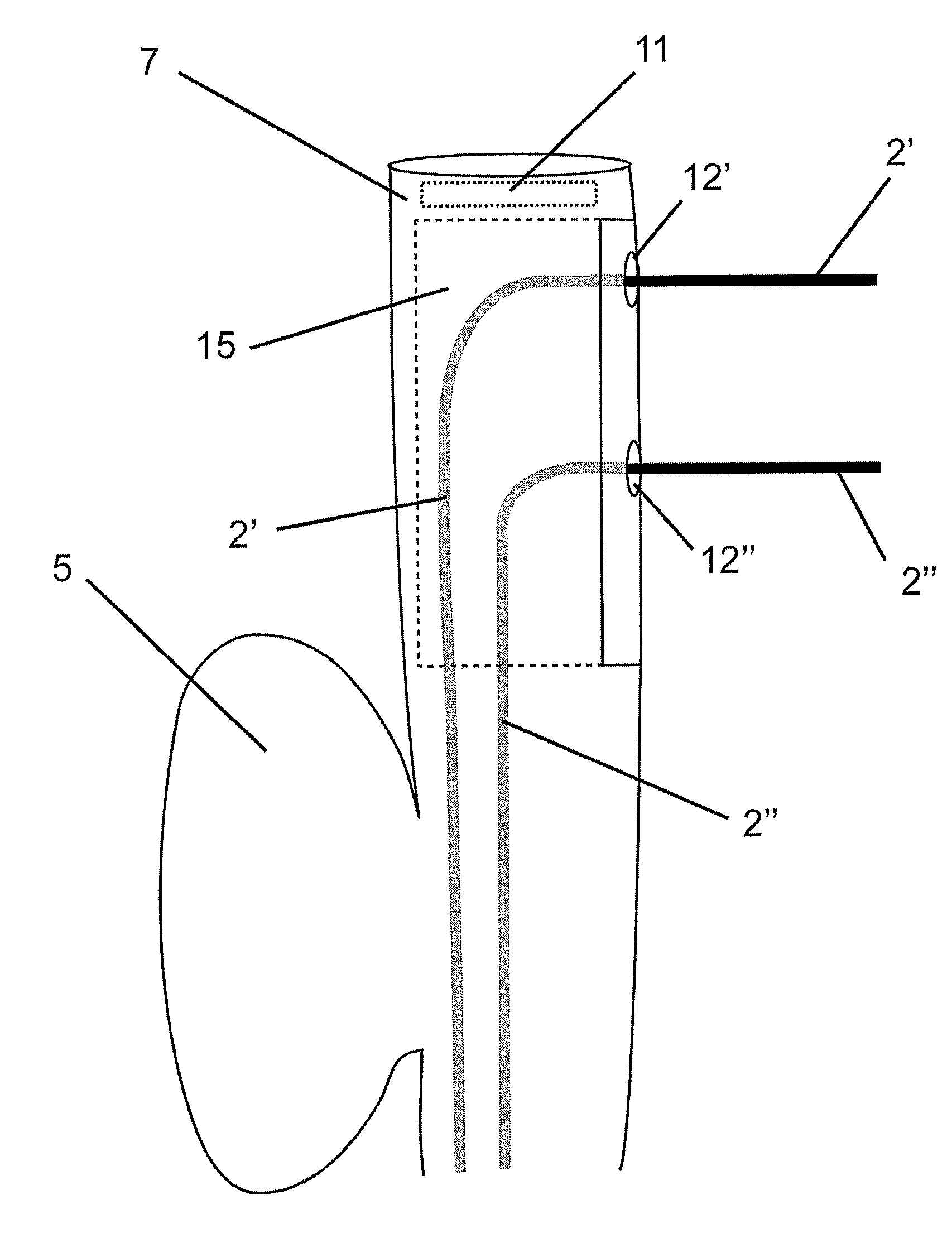
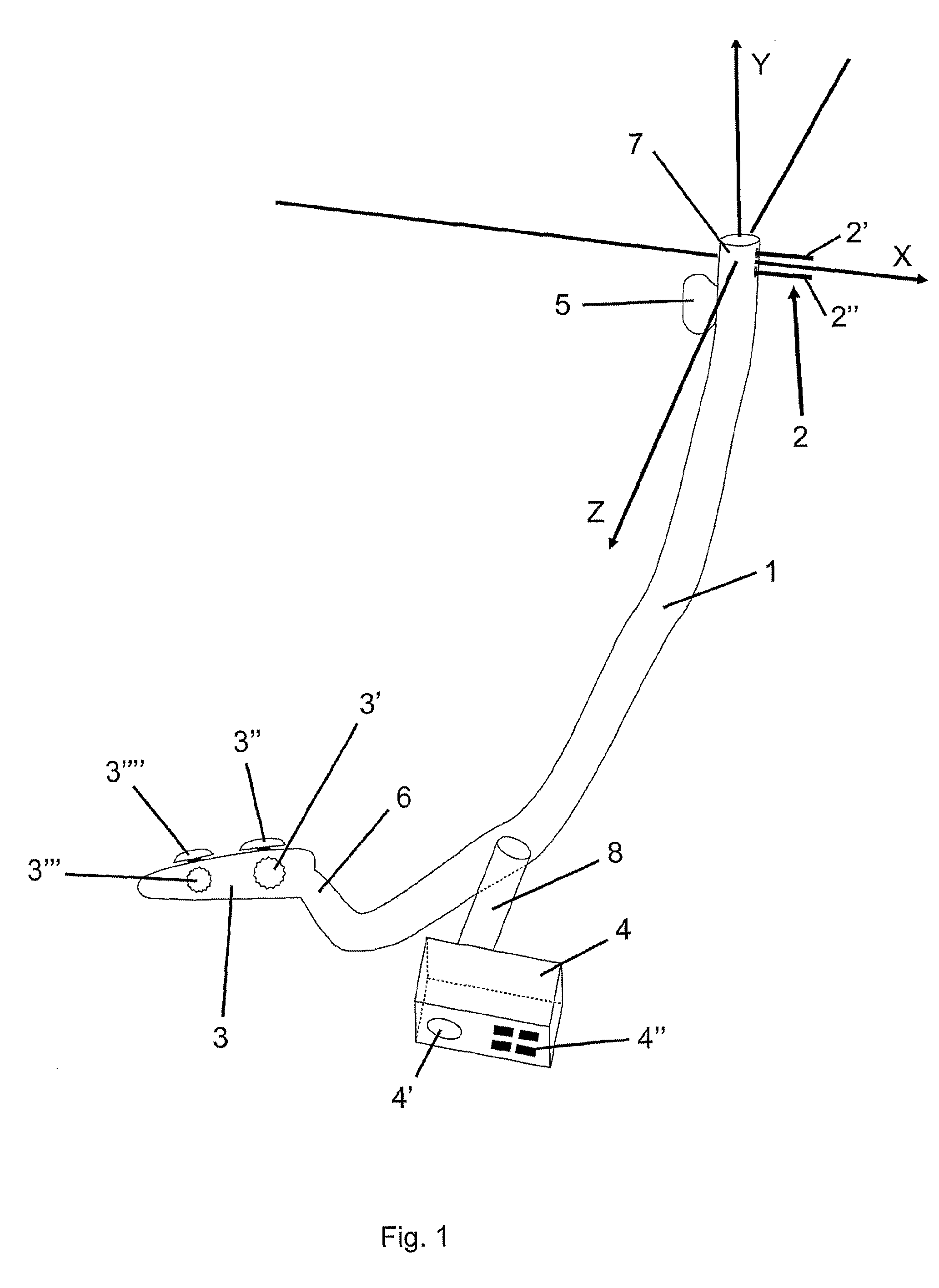
[0040]As illustrated on FIG. 1, the medical device according to the invention comprises as elements a main body 1, a handle 3, a control box 4 and a bipolar electrode 2.

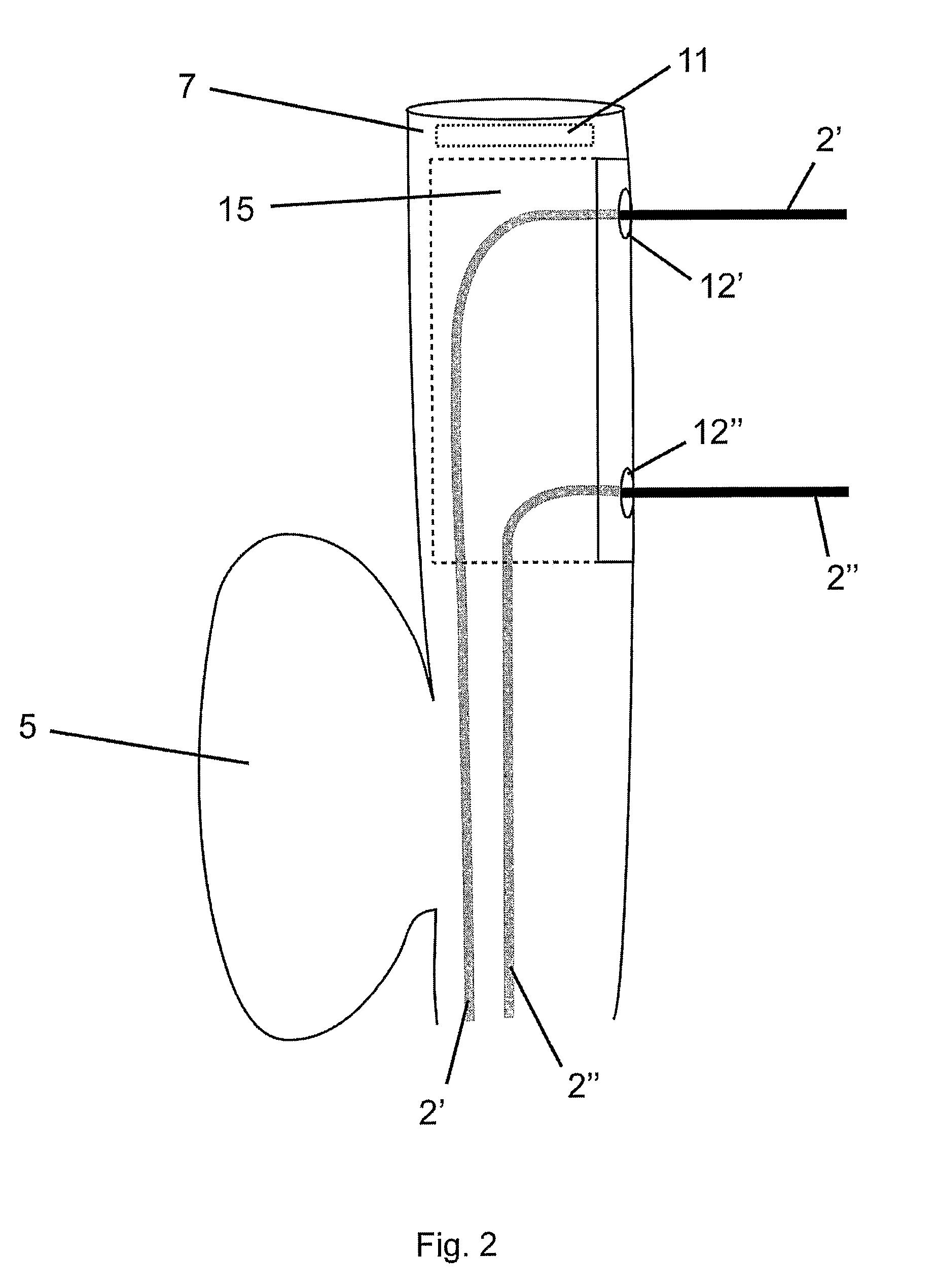
[0041]The bipolar electrode 2 has typically two flexible wires 2′ and 2″, but can have a different number of wires.

[0042]The main body 1 has a proximal end 6 and a distal end 7.

[0043]The main body 1 is an elongated and cylinder shaped flexible tube, of a total length between 30 cm and 80 cm, roughly.

[0044]The distal end 7 of the main body 1 has, roughly, a diameter between 4 and 15 mm.

[0045]The motor box 4 is connected to the main body 1 via the motor box tube 8. The motor box 4 can be disconnected from the motor box tube 8, allowing the main body 1, as well as the motor box tube 8, to be cleaned and sterilized before each procedure.

[0046]FIG. 13 shows a cross section of the main body 1 where we can see the following components crossing the main tube from the proximal end 3 to the distal end 7: more internally, two c...


PUM


Abstract

A radio frequency (RF) treatment flexible endoscope, for the in depth destruction of tumors by endo luminal access, like in a lung, esophagus, colon, rectum, etc . . . The electrodes (2′,2″) working in bipolar mode, deploy laterally and in parallel from the endoscope main body (1), reaching a tumor deeply located in the organ parenchyma. The use of different disposable distal end portions (15) of the endoscope allows the deployment of the electrodes (2′,2″) at different distances one from each other, in order to fit the destruction are to the tumor's size and shape. The deployment of the parallel electrodes (2′,2″) from the endoscope's main body (1) is controlled by motor drives (4) or a mechanical handled system (3), in order to reach the tumor with near a millimeter precision. In order to fix the endoscope's distal end to the tissue, so to allow a perfect parallel deployment of the electrodes, an inflating cuff (5) is placed at the opposite side of the distal end, as well as a suction system, with its holes (21) placed near the electrodes' deployment location. An external RF generator and a imaging system are associated to the use of the device.

Description

CONTINUING APPLICATION DATA[0001]This application claims benefit of pending prior U.S. Provisional Patent Application Ser. No. 60 / 582,449 filed Jun. 23, 2004 and entitled “Endocavitary access flexible and adjustable endoscope for tumor ablation using radio frequency”, by Faure A. which is incorporated herein by reference.BACKGROUND OF THE INVENTION[0002]1. Field of Invention[0003]The present invention relates to endoscopic surgery, more specifically, to an endoscopic flexible device, able to ablate (destruct) a tumor, using radio frequency, by an endo luminal access.[0004]2. Description of the Related Art[0005]Since the beginning of endo luminal endoscopy, the major concern was mainly having a new diagnostic mean. In fact, using a light source and introducing a camera in a endocavity allowed early diagnostic in several types of cancer (lungs, stomach, etc . . . ).[0006]As the technique improved, some endoscopes offered a central canal, allowing the endo luminal introduction of biops...

Claims


Application Information

Patent Timeline
Patent Type & Authority Applications(United States)
IPC IPC(8): A61B18/18
CPCA61B18/1477A61B18/1492A61B2018/00214A61B2018/00898A61B2018/00982A61B2018/126
Inventor FAURE, ANDRÉ S.
Owner TROD MEDICAL NV
Learn More