
Human transcriptome corresponding to human oocytes and use of said genes or the corresponding polypeptides to trans-differentiate somatic cells

a human oocyte and transcriptome technology, applied in the field use of said genes or the corresponding polypeptides to transdifferentiate somatic cells, can solve the problems of small number of genes analyzed, lack of comprehensive picture of human oocyte transcriptome, and relatively unknown oocyte transcriptome and its functional significance in the human

Inactive Publication Date: 2009-01-29
MICHIGAN STATE UNIV
View PDF1 Cites 2 Cited by
  • Summary
  • Abstract
  • Description
  • Claims
  • Application Information

AI Technical Summary

Benefits of technology

[0008]This invention also relates to the identification of a discrete set of genes which are uipregulated by in vivo matured human oocytes and the use of these genes and the corresponding gene products for the dedifferentiation and transdifferentiation of somatic cells particular somatic cells derived from a subject that is to receive transplanted cells for treatment of a particular condition such as cancer or autoimmunity. The use of these genes and / or gene products produced using such donor transdifferentiated or dedifferentiated cells will allow for the production of desired immature and / or somatic cell types that are compatible for a desired donor and which therefore are suitable for human cell and gene therapy without the need for human nuclear transfer (“human therapeutic cloning”) which has ethical concerns because such methods may result in the destruction of human embryos.

Problems solved by technology

However, the oocyte transcriptome and its functional significance in the human are relatively unknown because of ethical and technical limitations.
However, these molecular approaches resulted in a small number of genes analyzed in each sample.
Although they provided valuable information, these studies did not present a comprehensive picture of the human oocyte transcriptome because of a number of biological and technical constraints.
Although we recognize this issue could have been a mistake on their described-published protocol, the actual procedure used for RNA amplification remains elusive.
The shortcomings of the prior art are disadvantageous as the identification of genes and deduced pathways from the mature human oocyte can help us better understand oogenesis, folliculogenesis, fertilization, and embryonic development.

Method used


Image

  • Human transcriptome corresponding to human oocytes and use of said genes or the corresponding polypeptides to trans-differentiate somatic cells
  • Human transcriptome corresponding to human oocytes and use of said genes or the corresponding polypeptides to trans-differentiate somatic cells
  • Human transcriptome corresponding to human oocytes and use of said genes or the corresponding polypeptides to trans-differentiate somatic cells

Examples


experimental examples

[0059]The Materials and Methods below were used to derive the human transcriptome or set of genes upregulated by in vivo matured metaphase II human oocytes.

Materials and Methods

[0060]Oocyte Collection Total RNA Extraction, and Reference RNA.

[0061]Human oocytes were obtained from three patients undergoing an assisted reproductive treatment (ART) at the Unit of Reproductive Medicine of Clinica Las Condes, Santiago, Chile. It is important to emphasize that the routine in vitro fertilization protocol at Clinica Las Condes calls for fertilizing only those oocytes that will be transferred into the uterus of the patient. Therefore, there is always a surplus of oocytes. We then had the opportunity to use specific criteria to select donors as follows: (i) Supporting Materials and Methods, which is published as supporting information (See Ref. 50).

[0062]Three groups of 10 oocytes each were used. Total RNA was isolated following the guanidium thiocyanate method (Ref. 45) by using the PicoPure ...

example 1

Validation of Amplification Fidelity (Amplified vs. Nonamplified RNA)

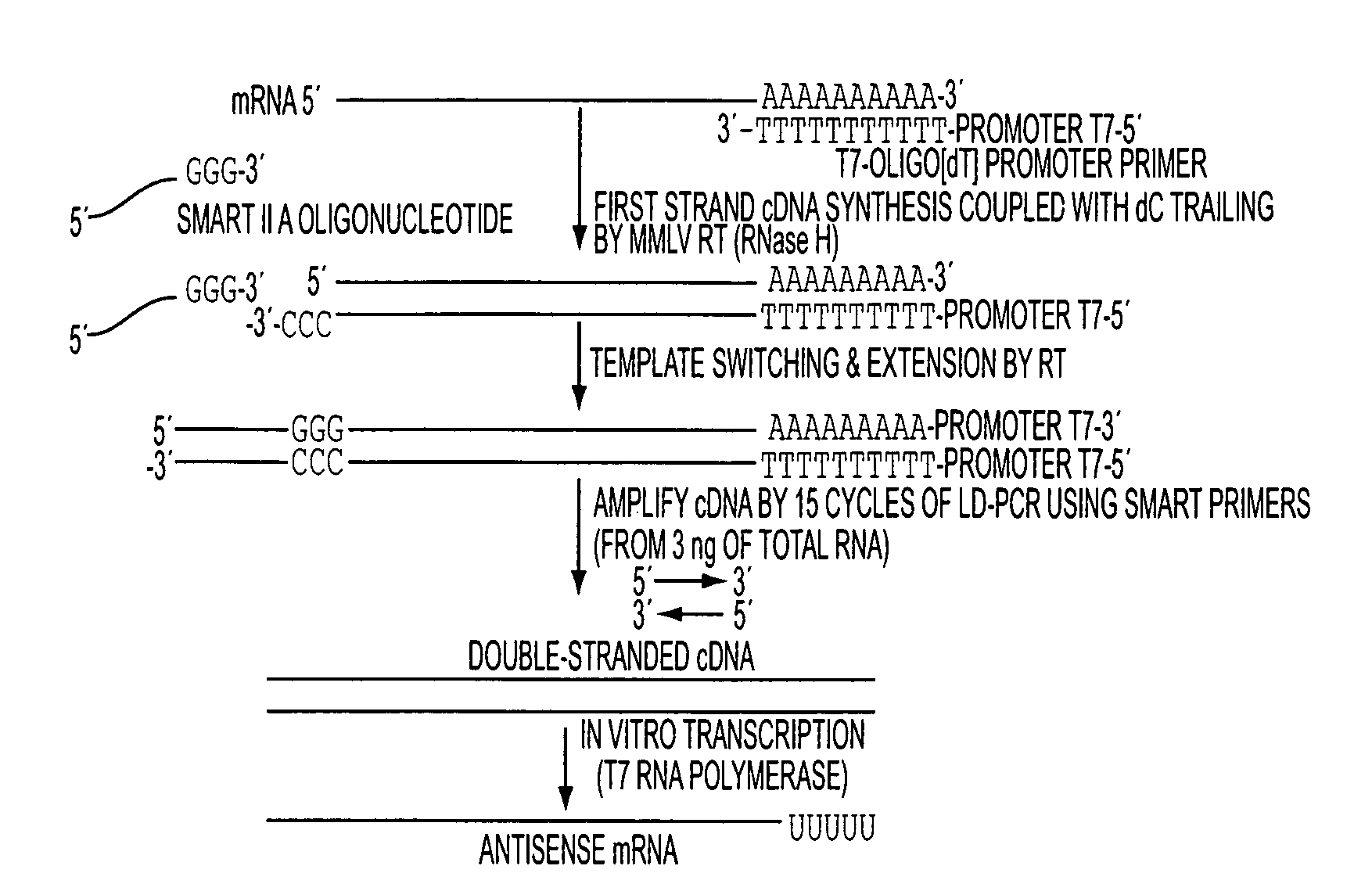
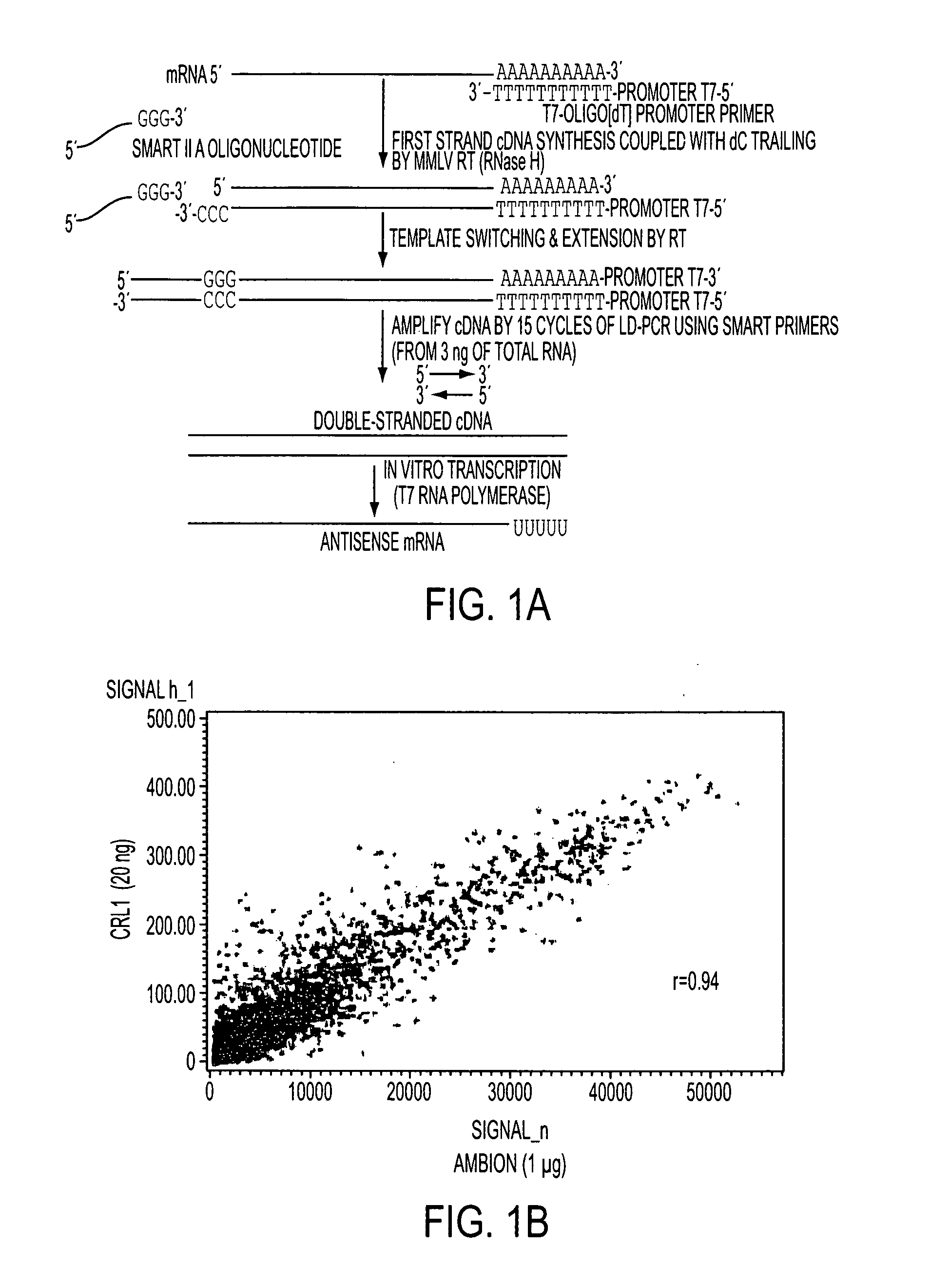
[0080]A critical step in the analysis of gene expression on small samples is the faithful amplification of mRNA molecules present in the sample. We have designed a PCR-based amplification system using the combination of SMART II A oligonucleotide (Clontech, Mountain View, Calif.) and T7-Oligo(dT) promoter primers (CRL RNA amplification protocol) (FIG. 1A). We isolated total RNA from a human cell line and 20, 3, and 1.5 ng input total RNA was amplified using the CRL amplification protocol. For each experiment, 15 μg of fragmented amplified RNA (aRNA) was hybridized to a single Affymetrix Human Genome U133 Plus 2.0 array. Nonamplified RNA from the same original sample (1 μg) was run in parallel by using the MessageAmp II aRNA Kit (Ambion, Austin, Tex.). Gene expression results from both amplified vs. nonamplified RNA samples were compared, and the correlation coefficients were found to be 0.94 (FIG. 1B), 0.93, and 0....

example 2

Validation of Microarray Data

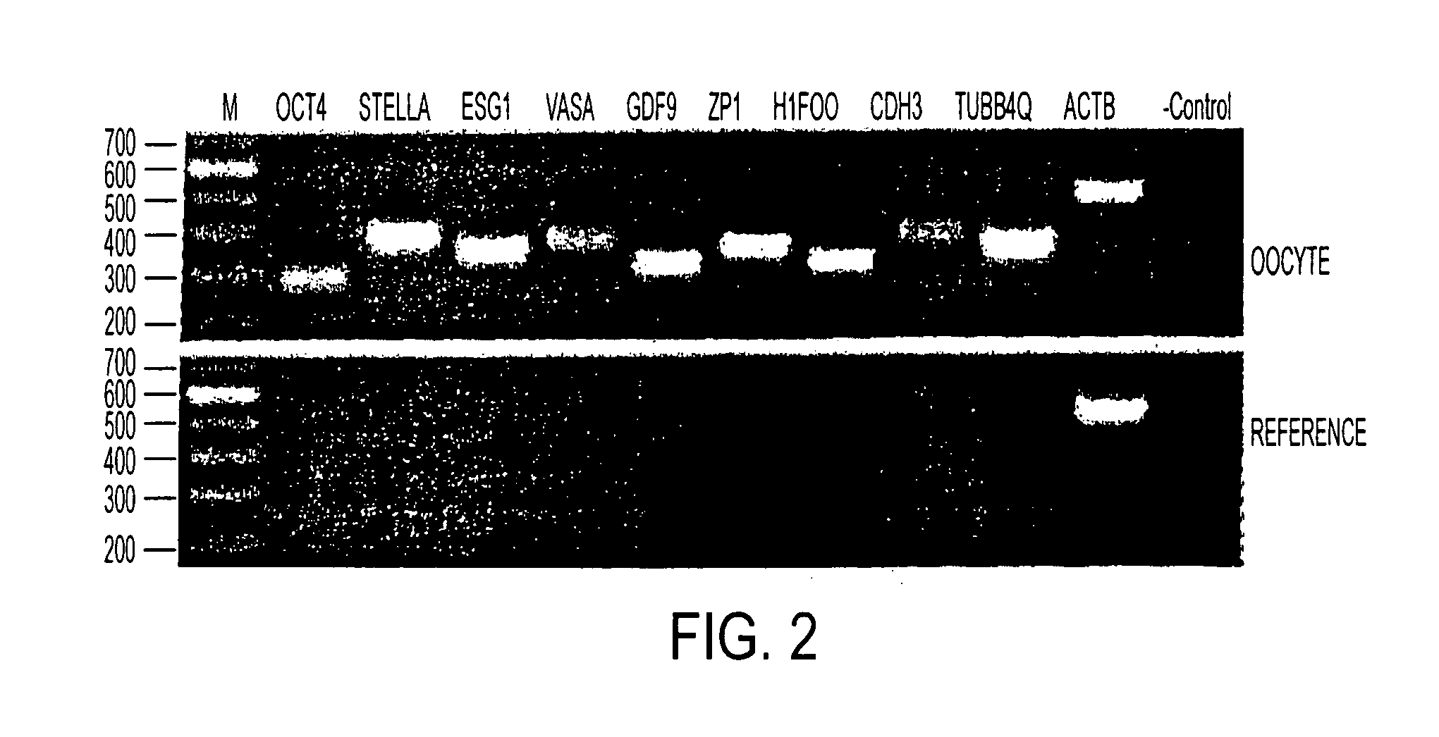
[0081]A selected list of genes was used to validate the microarray results by RT-PCR (FIG. 2). These genes were found to be present in the oocyte sample and absent in the reference RNA. FIG. 2, contains RT-PCR verification of the GeneChip array result. Loading orders of the gel were as following: M, 100 bp molecular weight standards with sizes as indicated on the left margin; OCT4, POU domain, class 5, transcription factor 1; STELLA, DPPA3, developmental pluripotency-associated 3; ESG1, embryonal stem cell-specific gene 1; VASA, DEAD box RNA helicase; GDF9, growth differentiation factor 9; ZP1, zona pellucida glycoprotein 1; H1FOO, H1 histone family, member O, oocyte-specific; CDH3, cadherin 3, type 1, P-cadherin (placental); TUBB4Q, β-tubulin; ACTB, β-actin; and negative control with no DNA template.


PUM

PropertyMeasurementUnit
Fractionaaaaaaaaaa
Electric chargeaaaaaaaaaa

Abstract

The identification of 101 genes upregulated or differentially expressed by mature human oocytes is provided herein. These genes and the corresponding gene products will facilitate a greater understanding of oogenesis, folliculogenesis, fertilization, and embryonic development. In addition these genes and the corresponding gene products can be used to effect dedifferentiation and / or transdifferentiation of desired somatic cells. The resultant dedifferentiated cells and somatic cells derived therefrom can be used in cell therapies such as in the treatment of cancer, autoimmunity, and other diseases wherein specific types of cells such as hematopoietic cells may be depleted because of the underlying disease or the treatment of the disease.Also, a core group of 66 transcripts was identified by intersecting significantly up-regulated genes of the human oocyte with those from the mouse oocyte and from human and mouse embryonic stem cells. Within the up-regulated probe sets, the top overrepresented categories were related to RNA and protein metabolism, followed by DNA metabolism and chromatin modification. This invention therefore provides a comprehensive expression baseline of genes expressed in in vivo matured human oocytes. Further understanding of the biological role of these genes will also expand knowledge on meiotic cell cycle, fertilization, chromatin remodeling, lineage commitment, pluripotency, tissue regeneration, and morphogenesis.

Description

RELATED APPLICATIONS[0001]This application claims priority to U.S. provisional Ser. No. 60 / 842,990 filed on Sep. 8, 2006. This application is incorporated by reference in its entirety herein.FIELD OF THE INVENTION[0002]The present invention relates to the identification of a set of genes which are expressed by in vivo matured human oocytes (“transcriptome” of human oocytes) and which are involved in oogenesis, folliculogenesis, fertilization, and embryonic development. These genes and the corresponding gene products are useful for dedifferentiation or transdifferentiation of somatic cells. Additionally, these genes are useful as markers of undifferentiated cell types and for assaying whether an ESC is capable of giving rise to an oocyte and for identifying pregnancy competent oocytes.[0003]The identification of genes and deduced pathways from the mature human oocyte will also facilitate a greater understanding of oogenesis, folliculogenesis, fertilization, and embryonic development....

Claims


Application Information

Patent Timeline
IPC IPC(8): A61K48/00C12N15/11C12N5/10A61P35/00A61P37/00A61P31/00C12Q1/68
CPCC12N15/8509C12N2510/00C12Q2600/158C12Q1/6886C12Q1/6883A61P31/00A61P35/00A61P37/00
Inventor CIBELLI, JOSE BERNARDOKOCABAS, ARIF MURATLIM, BINGOTU, HASAN H.
Owner MICHIGAN STATE UNIV
Learn More