
Complex shape steerable tissue visualization and manipulation catheter

a catheter and complex shape technology, applied in the field of catheters, can solve the problems of difficult direct visualization and subsequent manipulation of heart tissue, and the inability to manipulate the local tissue,

Active Publication Date: 2008-11-13
INTUITIVE SURGICAL OPERATIONS INC
View PDF107 Cites 149 Cited by
  • Summary
  • Abstract
  • Description
  • Claims
  • Application Information

AI Technical Summary

Benefits of technology

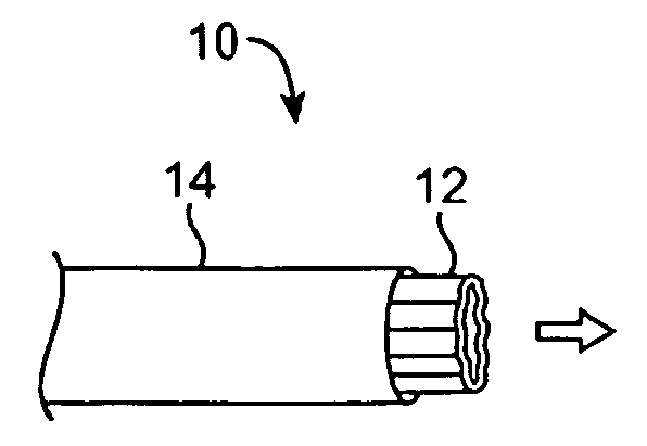
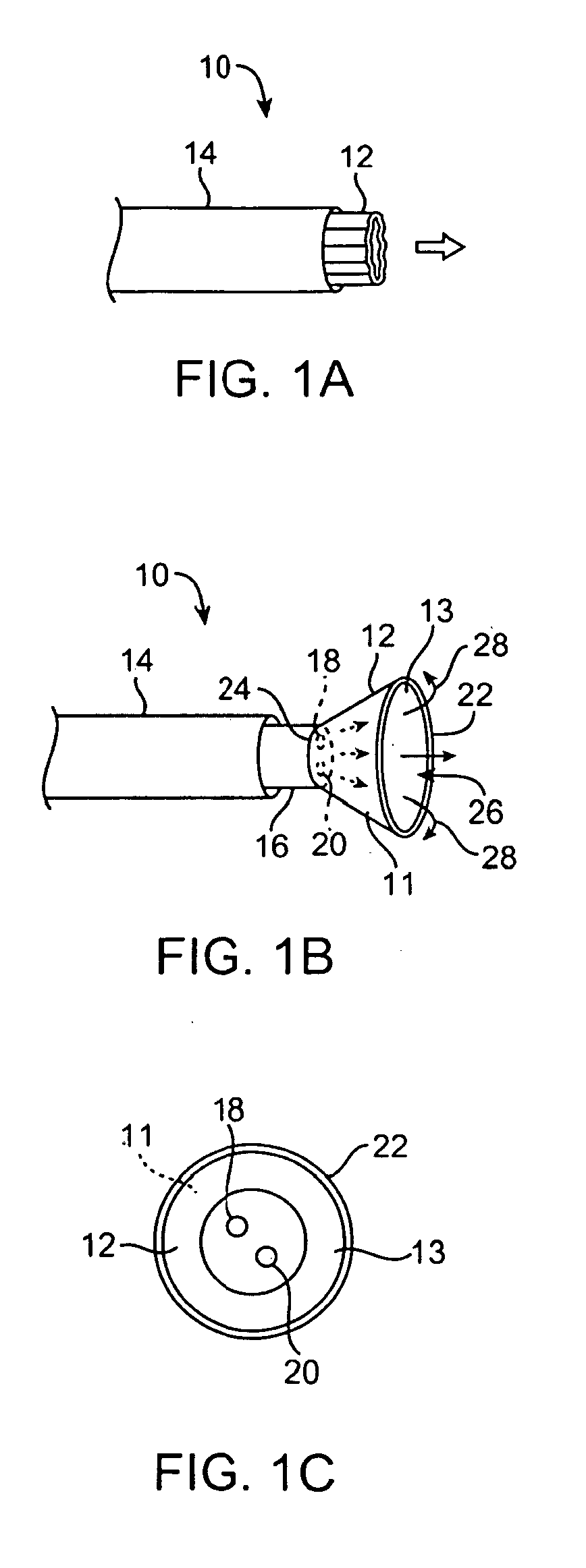
[0014]Generally, the embodiments of a tissue imaging and manipulation device depicted in the present invention meet the challenge and solve the problem of accessing regions of the body which are typically difficult to access. The design and control of the catheter shaft and the distal tip of the device as disclosed here provide a device uniquely capable of accessing a region such as the human heart, which is a region not only difficult to access, but which also has continuous blood flow. The blood flow provides a barrier to visualizing the local tissue, which in turn makes any manipulation at the local tissue nearly impossible. The unique elements that form the catheter shaft and the distal tip of the device, including the separate control of the shaft and tip and several optional modes of manipulation of either or both, provide for a device adaptable to addressing the challenges inherent in intravascular access and manipulation of heart tissue, and for accomplishing a procedure in any other difficult-to-access region in the body which is bathed in a medium that interferes with visualization.
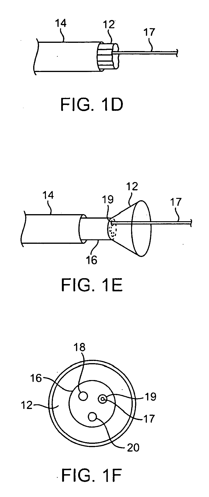
[0038]Also included are methods of accessing difficult-to-reach target tissue in a region of a body having continuous interfering blood flow by articulating the proximal steerable section within multiple planes relative to a longitudinal axis of the catheter guided by keyhole lumens, and articulating the distal steerable section within one or more planes relative to a longitudinal axis of the proximal steerable section. Accordingly, the distal hood can contact difficult to reach target tissue, for example, using complex curves generated with the proximal steerable segment so that the distal segment (the hood) can contact the target tissue perpendicularly, thus providing optimum contact of the hood with the tissue. Visualizing the target tissue within an open area through the transparent fluid can be accomplished if the visualization hood is flushed with saline or other clear fluid so that the blood is cleared providing an unobstructed visualization at the region. Processes such as ablation, or marking can occur using the distal hood of at least a portion of the target tissue within the open area that has been cleared of blood.

Problems solved by technology

The blood flow provides a barrier to visualizing the local tissue, which in turn makes any manipulation at the local tissue nearly impossible.
Blood is continuously flowing through the heart at all times, and as such presents a challenge to direct visualization and subsequent manipulation of heart tissue.

Method used


Image

  • Complex shape steerable tissue visualization and manipulation catheter
  • Complex shape steerable tissue visualization and manipulation catheter
  • Complex shape steerable tissue visualization and manipulation catheter

Examples


Embodiment Construction

[0071]The tissue-imaging and manipulation apparatus of the invention is able to provide real-time images in vivo of tissue regions within a body lumen such as a heart, which are filled with blood flowing dynamically through the region. The apparatus is also able to provide intravascular tools and instruments for performing various procedures upon the imaged tissue regions. Such an apparatus may be utilized for many procedures, e.g., facilitating transseptal access to the left atrium, cannulating the coronary sinus, diagnosis of valve regurgitation / stenosis, valvuloplasty, atrial appendage closure, arrhythmogenic focus ablation (such as for treating atrial fibrulation), among other procedures. Disclosure and information regarding tissue visualization catheters generally which can be applied to the invention are shown and described in further detail in commonly owned U.S. patent applications Ser. No. 11 / 259,498 filed Oct. 25, 2005, and published as U.S. Pat. Pub. 2006 / 0184048, which i...


PUM


Abstract

Complex steerable catheter visualization and tissue manipulation systems and their methods of use are disclosed herein. The deployment catheter is articulated using various steering mechanisms. Tissue visualization is accomplished from the visualization hood at the distal end of the deployment catheter, the hood having an ability to expand and other features to facilitate visualization and articulation at the tissue surface.

Description

CROSS-REFERENCE TO RELATED APPLICATIONS[0001]This application claims the benefit of priority to U.S. Prov. Pat. App. 60 / 916,640 filed May 8, 2007, which is incorporated herein by reference in its entirety.FIELD OF THE INVENTION[0002]The present invention relates generally to catheters having imaging and manipulation features for intravascularly accessing regions of the body.BACKGROUND OF THE INVENTION[0003]Conventional devices for accessing and visualizing interior regions of a body lumen are known. For example, ultrasound devices have been used to produce images from within a body in vivo. Ultrasound has been used both with and without contrast agents, which typically enhance ultrasound-derived images.[0004]Other conventional methods have utilized catheters or probes having position sensors deployed within the body lumen, such as the interior of a cardiac chamber. These types of positional sensors are typically used to determine the movement of a cardiac tissue surface or the elect...

Claims


Application Information

Patent Timeline
Patent Type & Authority Applications(United States)
IPC IPC(8): A61M25/01
CPCA61B1/00016A61B1/00082A61B1/00085A61B1/0051A61B1/0052A61B1/0055A61B1/015A61B1/05A61B1/3137A61M25/0147A61M25/04A61B1/008A61M25/00A61B1/018
Inventor PEH, RUEY-FENGSAADAT, VAHIDROTHE, CHRIS A.
Owner INTUITIVE SURGICAL OPERATIONS INC
Learn More