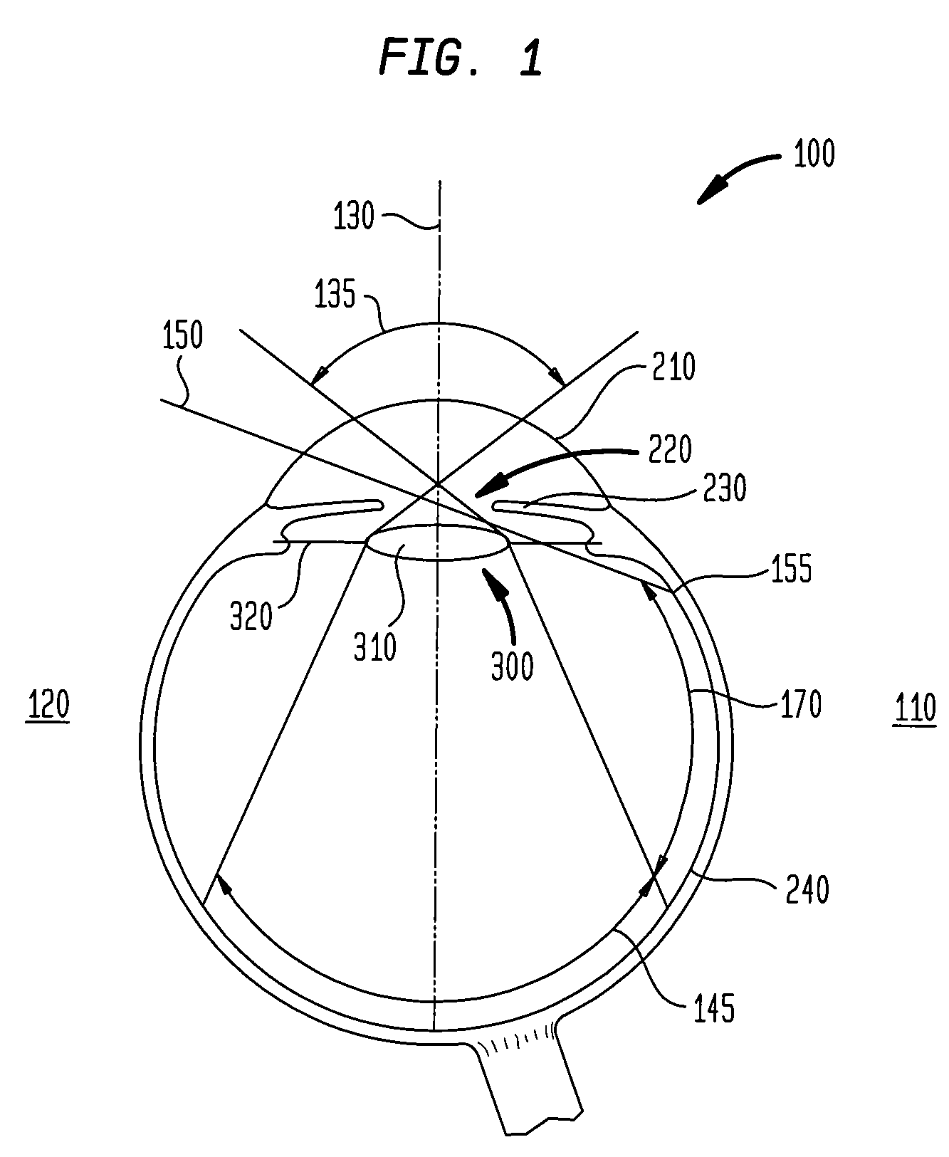
[0006]The present invention is based, in part, on the discovery that the shadows perceived by IOL patients can be caused by a double imaging effect when light enters the eye at very large visual angles. More specifically, it has been discovered that in many conventional IOLs, most of the light entering the eye is focused by both the
cornea and the IOL onto the
retina, but some of the peripheral light misses the IOL and it is hence focused only by the
cornea. This leads to the formation of a second peripheral image. Although this image can be valuable since it extends the peripheral visual field, in some IOL users it can result in the perception of a shadow-like phenomenon that can be distracting.
[0007]To reduce the potential complications of
cataract surgery, designers of modern IOLs have sought to make the optical component (the “optic”) smaller (and preferably foldable) so that it can be inserted into the
capsular bag with greater ease following the removal of the patient's natural crystalline lens. The reduced lens
diameter, and foldable
lens materials, are important factors in the success of modern IOL
surgery, since they reduce the size of the corneal incision that is required. This in turn results in a reduction in corneal aberrations from the
surgical incision, since often no suturing is required. The use of self-sealing incisions results in rapid
rehabilitation and further reductions in induced aberrations. However, a consequence of the optic
diameter choice is that the IOL optic may not always be large enough (or may be too far displaced from the iris) to receive all of the light entering the eye.
[0008]Moreover, the use of enhanced polymeric materials and other advances in IOL technology have led to a substantial reduction in capsular opacification, which has historically occurred after the implantation of an IOL in the eye, e.g., due to
cell growth. Surgical techniques have also improved along with the lens designs, and biological material that previously affected light near the edge of an IOL, and in the region surrounding the IOL, no longer does so. These improvements have resulted in a better
peripheral vision, as well as a better foveal vision, for the IOL users. Though a peripheral image is not seen as sharply as a central (axial) image,
peripheral vision can be very valuable. For example,
peripheral vision can alert IOL users to the presence of an object in their
field of view, in response to which they can turn to obtain a sharper image of the object. It is interesting to note in this regard that the
retina is a highly curved optical sensor, and hence can potentially provide better off-axis detection capabilities than comparable flat photosensors. In fact, though not widely appreciated, peripheral
retinal sensors for visual angles greater than about 60 degrees are located in the anterior portion of the eye, and are generally oriented toward the rear of the eye. In some IOL users, however, the enhanced peripheral vision can lead to, or exacerbate, the perception of peripheral visual artifacts, e.g., in the form of shadows.
[0010]The present invention generally provides
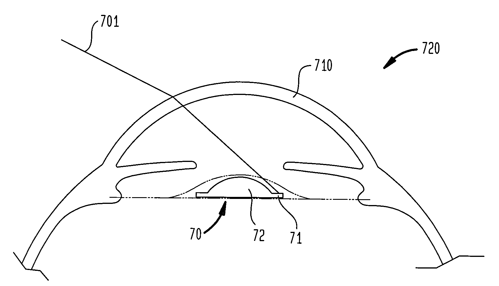
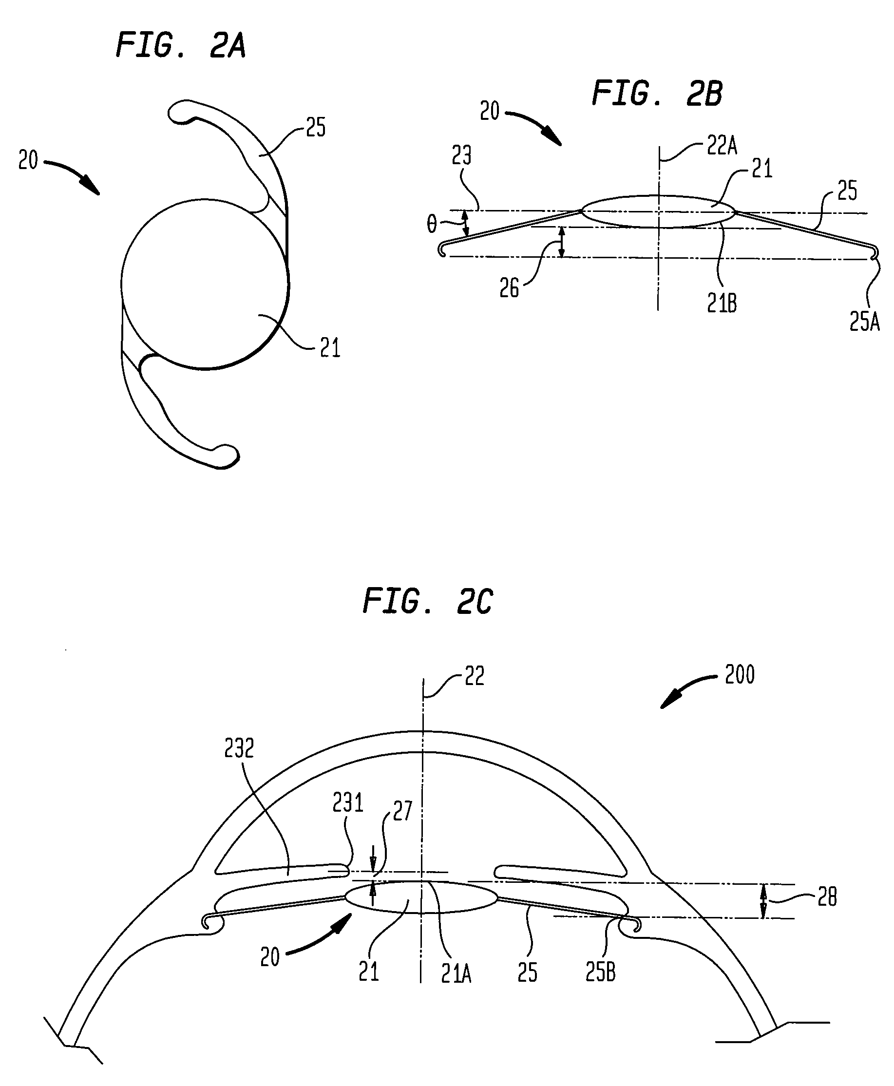
intraocular lenses (IOLs) and methods of vision correction that utilize them, which can alleviate, and preferably eliminate, the perception of dark shadows that some IOL patients occasionally report. Such IOLs can be implanted posterior or anterior to the iris of the eye. In some aspects of the present invention, the fixation members of an IOL are adapted so as to project the IOL's optic toward the iris in order to alleviate dysphotopsia. For example, an optic can be positioned sufficiently close to the iris of the eye to receive peripheral light rays entering the eye (e.g., at visual angles in a range of about 50 degrees to about 80 degrees) and to direct those rays onto the
retina so as to inhibit the formation of a secondary peripheral image or to cause a reduction of the shadow region between such a secondary image and an image formed by the IOL. For example, a fixation member can extend posteriorly from the optic to project the optic toward the iris when the IOL is appropriately implanted. In some cases, the fixation member can have arm-like extensions that extend posteriorly from the optic and form an angle relative to a
principal plane of the IOL's optic, e.g., in a range of about 5 degrees to about 45 degrees, or about 15 degrees to about 30 degrees. In many embodiments, the IOLs are preferably deformable such that their delivery to a subject's eye is facilitated. These, as well as other, aspects are disclosed in more detail herein.
[0016]Another aspect is directed to a method of inhibiting dysphotopsia in a patient having an implanted IOL by positioning an
anterior surface of the IOL's optic close enough to the iris to inhibit dysphotopsia. For instance, the
anterior surface can be positioned such that the
anterior surface would intercept peripheral light rays and would direct those rays to the retina so as to inhibit the formation of a secondary image on the retina or to reduce the extent of a
retinal dark (shadow) region between such a secondary image and an image formed by the optic. In many cases, such peripheral light rays can enter the eye at an angle in the range from about 50 degrees to about 80 degrees relative to the eye's
visual axis. By way of example, in some cases, the optic's anterior surface can be positioned an
axial distance of less than about 0.8 mm from a tip of an iris of the subject's eye.


