Neurofibrillary labels

a neurofibrillary and label technology, applied in the field of neurofibrillary labeling and detection, can solve the problems of low detection accuracy, low degree of uncertainty, and low detection accuracy of neurofibrillary tangles,

Inactive Publication Date: 2008-09-11
WISTA LAB LTD
View PDF4 Cites 18 Cited by
  • Summary
  • Abstract
  • Description
  • Claims
  • Application Information

AI Technical Summary

Benefits of technology

[0031]As described in the introduction, progression of neurofibrillary degeneration is definitive of the neuropathological staging proposed by Braak, which in turn is the best available neuropathological definition of progression of AD. The methods of the present invention can thus be used to provide an actual Braak stage result. In preferred embodiments they can be used to diagnose patients at early Braak stages (e.g. Braak stage 2) even before clinical symptoms may be readily apparent—such diagnosis can be used to direct timely treatment and advice.
[0157]The ligands described herein can suitably be detected by incorporating a positron-emitting carbon into one of the methyl groups of the compound as disclosed herein, and detecting the compound by use of positron emission tomography (PET) as is known in the art. Alternatively, or in addition, a technetium-containing chelate can be incorporated into the compound (e.g. as in the RC group of the compounds described herein), so that selective detection of extracellular tangles could be achieved. A preferred chelating group is RC=diethylenetriaminepentaacetic acid.
[0202]As shown herein, such compounds may be particularly effective when it is desired to incorporate a bulky RC group in order to facilitate detection.
[0224]The novel compounds disclosed (e.g. of formula (II)) herein are useful inter alia as synthetic ligands of neurofibrillary tangles, such as those characteristic of AD. The discovery of the minimum critical structure required for binding to these tangles thus provides for the possibility of designing high-affinity ligands which can be used to target the tangles, and can thus be used in the diagnosis, prognosis or treatment of diseases such as AD.

Problems solved by technology

Thus there exists a high degree of uncertainty, even in the best practice research setting.
However, beta-amyloid deposition is known to discriminate poorly between normal aging and Alzheimer's disease (see FIG. 2d herein), and beta-amyloid pathology does not provide a sound basis for neuropathological staging (Braak and Braak, 1991).
Therefore, FDDNP-RRT does not provide a method for in vivo neuropathological staging of Alzheimer's disease.
Although it is possible that further refinement in clinical methods with particular reference to more specific neuropsychological indicators (e.g. split attention tasks, delayed matching to sample, etc.) may improve the accuracy of clinical diagnosis, an essential problem is to develop methods for the direct measurement of underlying neuropathology during life, in particular the extent of neurofibrillary degeneration of the Alzheimer-type.
However, to date, no definitive correlation has been established between these stages, which occur at the cellular level, and possibly at different rates and probabilities in different regions in the brain, and the progression of pathology according to the defined hierarchical system of Braak and Braak which, as discussed above, is the best available definition of progression of relatively pure neurofibrillary degeneration.
Lumbar-puncture CSF measurements enable discrimination between AD and controls, and between AD and other neurological disorders, but lumbar-puncture is more invasive then nuclear medicine-based approaches, and carries a higher risk (Refs.
However, although the diagnosis of AD by monitoring global brain atrophy represents a methodology which can be made to work in a research setting, there are difficulties in defining and measuring atrophy in specific brain regions, and likewise in the measurement of global neocortical atrophy.
In any case, a diagnosis based on detectable atrophy may come too late for effective treatment.
However, although MRI and SPECT scans are useful for detecting specific patterns of perfusion defects characteristic of AD, discrimination between different neuropathological stages, or between AD and other types of dementia, is difficult.
However, SPECT changes discriminate neuropathological stages poorly (Ref 12).
Furthermore, discrimination between AD and Lewy Body dementia is difficult.

Method used


Image

  • Neurofibrillary labels
  • Neurofibrillary labels
  • Neurofibrillary labels

Examples


example 1

Aggregated Tau in Braak Staging

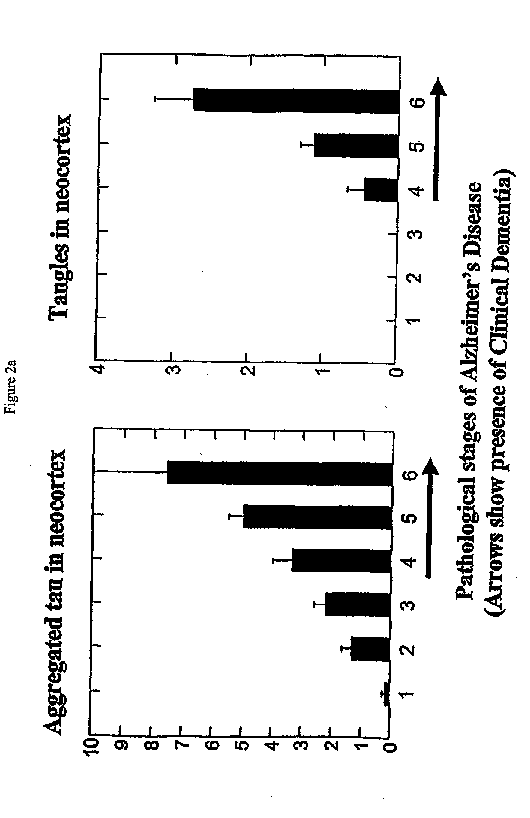
[0331]Based on immunochemical properties (Refs 26, 27, 30), it is possible to distinguish intracellular tangles from extracellular tangles. Both frequency of cases with tangles in these categories (ie probability) and their quantity (ie counts per mm2) were determined in a prospective case series and grouped into the regions known to represent stages in the progression of pathology according to the system of Braak and Braak

[0332]As shown in FIG. 25, stages 2-4 can be clearly distinguished from stage 1 on the basis of probability of extracellular tangles in E2 / Trans

and E4 / HC. Also shown are the figures for F / T / P regions (neocortical regions—frontal, temporal, parietal).

[0333]Conversely, intracellular tangles provide a poor basis for discrimination of early stages in these regions, but a good basis for discriminating stages 4 and 5 using neocortical regions. Similarly, when cases with MMSE scores greater than 21 in the 12 months prior to death were selec...

example 2

Assessment of Compounds Binding within the Aggregated Repeat Domain of PHF-Core Tau Protein

[0337]A prototype compound was obtained as one component of the crude, commercially-available preparation of thioflavin-S was separated into ˜20 distinct constituents by analytical thin-layer chromatography, and preparative chromatography. Tests showed that not all of the constituents were able to act as effective tangle ligands. Specifically, pure primulin (FIG. 5, compound 1a) was found to label tangles, but the benzothiazole thioflavin-T (FIG. 5, compound 1b) was much less effective, although it labelled amyloid preferentially.

[0338]Furthermore, compound 1a was found to displace compound 1b at tangles when the latter was introduced at 10-fold excess into crude tangle extracts.

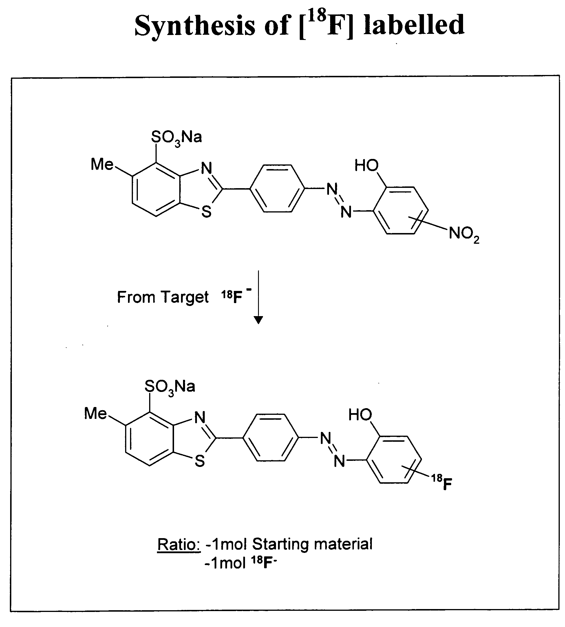
[0339]A possible difference was postulated to be the sulphonate group at position 1 (FIG. 5, compound 2 [2-(4-amino phenyl)-6-methyl-1-sulphonate benzothiazole]). However, primulin (compound 1a) was found to displace t...

example 3

Determination of Optimum Dimensions of Ligand Molecules

[0344]FIG. 14 shows three of the structures described above, along with their dimensions as indicated. For example, the C11-C1 distance and C10-C1 distance are shown for primulin, a benzothiazole analogue (denoted ‘analog’), and ‘thiazin yellow’.

[0345]FIGS. 15 and 16 illustrate the crystal structures of the ‘B’ part of the primulin structure (Soon-Beng Teo et al., 1995, Acta Crystallogr., Sect. C, 591. As can be seen from FIG. 16, which is a ‘side-on’ view, the molecule is essentially flat, although it has a slight twist. The ‘A’ part of primulin can be computed from the same molecule. From this, one can derive measures of A+B, which provide an indication of the actual length of one of the active species of the present invention.

[0346]To compute the size of the “analog” shown in FIG. 14, measurement A was used from the data of FIG. 15, and measurement B was determined from a molecule denoted N2A and shown in FIGS. 17 and 18 (Gil...


PUM


Abstract

A method for determining the Braak stage of neurofibrillary degeneration associated with a tauopathy in a subject having neurofibrillary degeneration is disclosed. The method comprises the steps of (i) administering to the subject a conjugated, chelated or detectable chemical group-associated ligand that labels aggregated paired helical filament (PHF) tau protein and is capable of crossing the blood brain barrier; (ii) determining the presence and\or amount of ligand bound to extracellular aggregated PHF tau in the medial temporal lobe of the brain of the subject, and (iii) correlating the result of the determination made in (ii) with the extent of neurofibrillary degeneration in the subject. Preferred ligands include sulphonated-benzothiazole-like compounds and diamonophenothiazines.

Description

FIELD OF THE INVENTION[0001]The present invention concerns materials, methods and models relating generally to the labelling and detection of neurofibrillary tangles. In addition, it concerns the identification and development of ligands suitable for neuropathological staging and their use in the diagnosis, prognosis or treatment of diseases such as Alzheimer's Disease (AD).BACKGROUND TO THE INVENTIONNeuropathological Staging and AD[0002]The neuropathological staging proposed by Braak (Braak, H et al. (1991), Acta. Neuropathol. 82, 239-259) provides the best available definition of progression of relatively pure neurofibrillary degeneration of the Alzheimer-type which is diagnostic of AD (Wischik et al. (2000), “Neurobiology of Alzheimer's Disease”, Eds. Dawbarn et al., The Molecular and Cellular Neurobiology Series, Bios Scientific Publishers, Oxford). This staging is shown schematically in terms of brain region in FIG. 2B, and is based on a regular regional hierarchy of neurofibri...

Claims


Application Information

Patent Timeline
Patent Type & Authority Applications(United States)
IPC IPC(8): A61K49/00C07D277/62C07D277/66C07D279/20C07D417/12G01N33/53G01N33/58G01N33/68
CPCC07D277/66C07D279/20G01N33/6896G01N33/583C07D417/12
Inventor WISCHIK, CLAUDE MICHELHARRINGTON, CHARLES ROBERTRICKARD, JANET ELIZABETHHORSLEY, DAVID
Owner WISTA LAB LTD
Why Eureka
Try Eureka