Orthovoltage radiotherapy

Active Publication Date: 2008-09-04
CARL ZEISS MEDITEC INC
View PDF99 Cites 128 Cited by
  • Summary
  • Abstract
  • Description
  • Claims
  • Application Information

AI Technical Summary

Benefits of technology

[0010]Retinal radiotherapy trials have shown stabilized or improved visual acuity without any significant toxicity. Radiation has also been shown to dry up neovascular membranes in patients and stabilize vision. However, due to limitations in the treatment of macular degeneration using radiotherapy, including localization of the region to be treated as well as specific application of the radiation to the region to be treated, macular radiotherapy often irradiates the entire retina, which is both unnecessary and possibly harmful. Moreover, the dose of radiation specifically to the macula has not been limited to multiple fractions over many days or weeks. The ability to apply a greater dose specifically to the macula in a period of time less than 24 hours will have a greater effect on the disease than was shown in previous trials.
[0078]Some embodiments further comprising a radiation source mover that moves the radiation source relative to the eye to direct the emitted radiation toward an eye structure. In some embodiments, the radiation source is configured to be stationary relative to a position of the eye during the treatment session. In some embodiments, the system further includes a system shut-off that reduces or ceases emission of radiation when an eye structure is not in a path of the collimated beam. Some embodiments further include a holder to substantially hold the eye such that an eye structure is in a path of the collimated beam.

Problems solved by technology

Macular degeneration is the leading cause of central vision loss in people over the age of fifty years.
The inflammatory process can result in direct destruction of the retina or destruction via formation of neovascular membranes which leak fluid and blood into the retina, quickly leading to scarring.
For example, the eye contains several critical structures, such as the lens and the optic nerve, that can possibly be damaged by excessive exposure to radiation.
The application of external beam therapy is limited by devices and methodologies used to apply the therapy.
These devices and methodologies are older radiation technologies used to treat conditions such as tumors anywhere in the body and were not developed specifically for ocular radiation therapy.
In addition, logistics are difficult as far as patient recruitment and administration of treatments because such treatment devices are borrowed from and displace oncologic therapies.
However, due to limitations in the treatment of macular degeneration using radiotherapy, including localization of the region to be treated as well as specific application of the radiation to the region to be treated, macular radiotherapy often irradiates the entire retina, which is both unnecessary and possibly harmful.
A major limitation of this treatment is that it requires invasive procedures involving partial removal of the vitreous fluid of the posterior chamber of the eye to place the brachytherapy probe.
In addition, the ability to fractionate the dose is limited because of the invasiveness required to deliver the therapy.
Procedures such as trabeculoplasty, trabeculotomy, canaloplasty, laser iridotomy, placement of shunts, and other procedures all suffer from a short-lived effect because of scar formation as a result of the surgical trauma.
Anti-inflammatory drugs appear to offer a palliative and / or preventative solution to the chronic scarring that occurs after these procedures; however, the drugs have to be given several times per day and are associated with their own side effect profile such as seepage into unwanted regions of the eye.
This scarring is caused by trauma from the surgery, proliferation of lens cells, and material incompatibility.
The exact cause of pterygia is not well understood, although it occurs more often in people who spend a great deal of time outdoors, especially in sunny climates, and has been linked to long-term exposure to sunlight, especially ultraviolet rays, and chronic eye irritation from dry, dusty, and windy conditions.

Method used


Image

  • Orthovoltage radiotherapy
  • Orthovoltage radiotherapy
  • Orthovoltage radiotherapy

Examples


Embodiment Construction

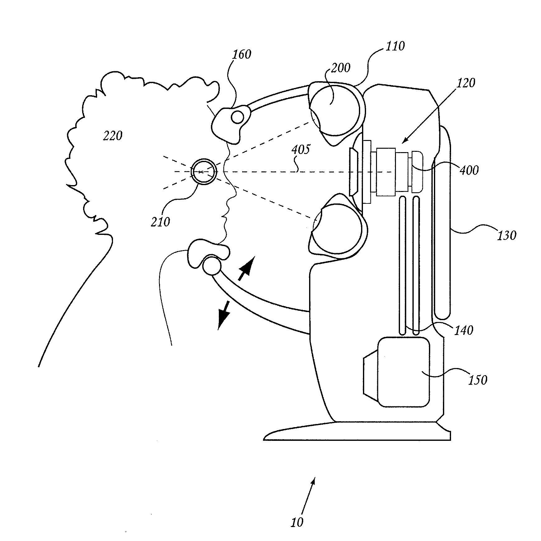
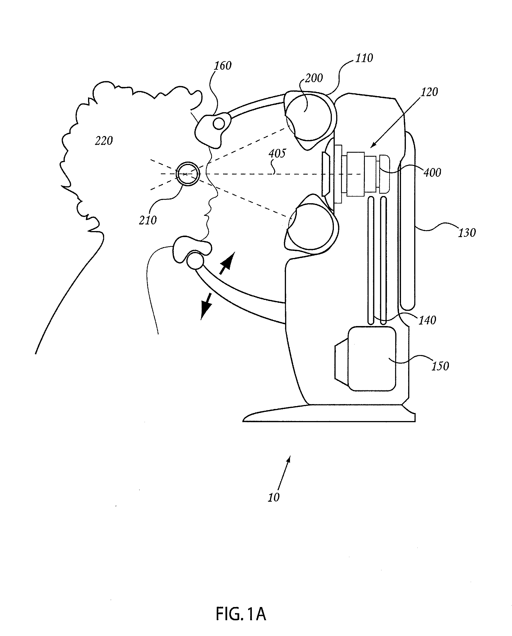
[0130]Embodiments described herein include systems and methods for treating a human eye with radiotherapy. Some embodiments described below relate to systems and methods for treating macular degeneration of the eye using radiotherapy. For example, in some embodiments, systems and methods are described for use of radiotherapy on select portions of the retina to impede or reduce neovascularization of the retina. Some embodiments described herein also relate to systems and methods for treating glaucoma or controlling wound healing using radiotherapy. For example, embodiments of systems and methods are described for use of radiotherapy on tissue in the anterior chamber following glaucoma surgery, such as trabeculoplasty, trabeculotomy, canaloplasty, and laser iridotomy, to reduce the likelihood of postoperative complications. In other embodiments, systems and methods are described to use radiotherapy to treat drusen, inflammatory deposits in the retina that are thought to lead to vision...


PUM


Abstract

A radiosurgery system is described that is configured to deliver a therapeutic dose of radiation to a target structure in a patient. In some embodiments, inflammatory ocular disorders are treated, specifically macular degeneration. In some embodiments, other disorders or tissues of a body are treated with the dose of radiation. In some embodiments, the target tissues are placed in a global coordinate system based on ocular imaging. In some embodiments, the target tissues inside the global coordinate system lead to direction of an automated positioning system that is directed based on the target tissues within the coordinate system. In some embodiments, a treatment plan is utilized in which beam energy and direction and duration of time for treatment is determined for a specific disease to be treated and / or structures to be avoided. In some embodiments, a fiducial marker is used to identify the location of the target tissues. In some embodiments, radiodynamic therapy is described in which radiosurgery is used in combination with other treatments and can be delivered concomitant with, prior to, or following other treatments.

Description

RELATED APPLICATIONS[0001]This application claims priority benefit of U.S. Provisional Application No. 60 / 933,220, filed Jun. 4, 2007; U.S. Provisional Application No. 60 / 922,741, filed Apr. 9, 2007; and U.S. Provisional Application No. 60 / 869,872, filed Dec. 13, 2006; the entirety of each of which is incorporated herein by reference.BACKGROUND[0002]1. Field of the Inventions[0003]This disclosure relates to the treatment of ocular disorders using targeted photon energy. In particular, the present disclosure relates to apparatus, systems, and methods for image-guided low energy x-ray therapy of ocular structures.[0004]2. Description of the Related Art[0005]Macular degeneration is a condition where the light-sensing cells of the macula, a near-center portion of the retina of the human eye, malfunction and slowly cease to work. Macular degeneration is the leading cause of central vision loss in people over the age of fifty years. Clinical and histologic evidence indicates that macular ...

Claims


Application Information

Patent Timeline
IPC IPC(8): A61N5/10
CPCA61N5/10A61N5/1017A61N5/1084A61N2005/105A61N2005/1091A61N2005/1097A61N5/1049A61B6/506A61B6/508A61N5/1045A61N5/1077A61N5/1064A61N5/1065A61N5/1067A61N5/1001A61N2005/1022A61N5/1048A61N2005/1059A61N2005/1051A61N2005/1019
Inventor GERTNER, MICHAELHANSEN, STEVEN D.
Owner CARL ZEISS MEDITEC INC
Why Eureka
Try Eureka