
Method and arrangement relating to x-ray imaging

a technology of x-ray imaging and method, applied in the field of tomosynthesis, can solve the problems of long processing time, difficult reconstruction time or computational cost, and many years to come, and achieve the effect of being cheap to manufactur

Inactive Publication Date: 2008-07-31
SECTRA MAMEA
View PDF6 Cites 13 Cited by
  • Summary
  • Abstract
  • Description
  • Claims
  • Application Information

AI Technical Summary

Benefits of technology

[0009]The object of the present invention is to provide an effective aid for quickly determining whether or not a tomosynthesis image acquisition was successful. The aid is quick enough to fit in the workflow of virtually any modern mammography clinic. The aid is also inexpensive to manufacture.
[0011]The present invention effectively reduces the risk of a patient needs to be recalled due to bad image quality.
[0018]Preferably, the full projection images are stored in a first memory buffer. The contents of this memory buffer will be used for reconstructing the diagnostic image volume, but that may wait. The primary focus is reconstruction of a preview image volume. Preferably, the projection images are sub-sampled, and the sub-sampled data is stored in a second buffer, which is used for reconstruction of the preview images. Preferably, the sub-sampling reduces the number of pixels by 4 or 16 times. The sub-sampled images are used for reconstructing a three-dimensional image volume, and the resolution in the image volume follows the resolution in the projection images. Preferably, the reconstruction of the preview image is performed in essentially the same way as for the diagnostic image volume, except for different parameters related to image quality and geometrical differences for the preview resolution. The primary parameter difference is that fewer layers are reconstructed, since the preview image volume shall have at least as elongated voxels as the diagnostic image volume. Thus, one octave of sub-sampling reduces the number of pixels by 2*2=4 times in the projection images, and the number of reconstructed voxels will be reduced by 2̂3=8 times. Thanks to one octave of sub-sampling, the reconstruction time will be at least 4 times, and probably close to 8 times faster. There are reasons to believe that two octaves of sub-sampling will provide enough image quality for preview, which speeds up reconstruction up to 64 times. Further speed is possible by computational approximations or sub-optimal parameters. Most tomosynthesis reconstruction algorithms are iterative (e.g. EM or Lange-Fessler 1995) and the result converges over a number of iterations. Preferably, the preview image is computed with at most half number of iterations compared to the diagnostic image volume, which doubles speed. In addition, it is possible to skip computationally expensive regularization, which simplifies computations and also tend to substantially decrease the number of iterations.

Problems solved by technology

The reconstruction time is a problem in breast imaging, due to high demands for speed, and computationally expensive reconstruction algorithms.
The long processing time arises partly due to truncation of the object at the border of projection images.
Truncation cause difficulties when using the same methods as for mainstream CT, such as filtered back-projection and direct Fourier methods.
Another problem is that it is desired to produce an image volume with many more voxels than there are pixels in the stack of projection images, which calls for regularization when using iterative methods.
Therefore, it is expected that reconstruction time or computational cost will remain a challenge for many years to come.
The long reconstruction time is a problem in normal workflow, wherein the operator of the X-ray apparatus looks at the acquired image and determines whether or not it can be used for diagnosis.
The main risk of failure is bad positioning, wherein an important part of the breast is not visible.
Other risks for reconstruction are apparatus failure, large dirt particles, silicon implants and metal pieces such as piercing.
Unfortunately, each projection image tends be characterized by a substantially worse image quality than a reconstructed volume, since it contains the combination of X-ray quantum noise and disturbing super-imposed tissue.
Normally, each projection image is very noisy, since each projection image is acquired using fraction of the total dose.

Method used


Image

  • Method and arrangement relating to x-ray imaging
  • Method and arrangement relating to x-ray imaging

Examples


Embodiment Construction

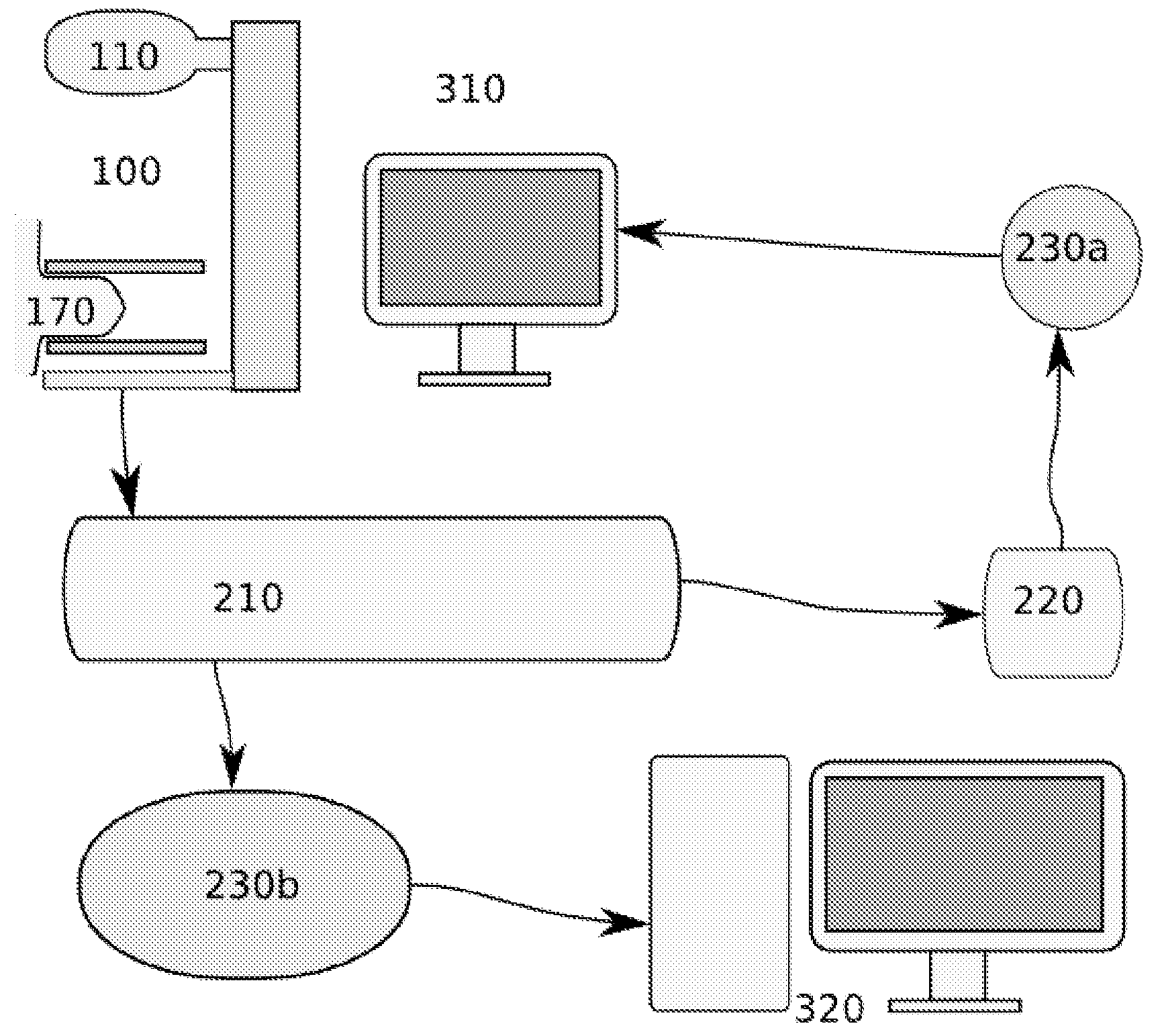
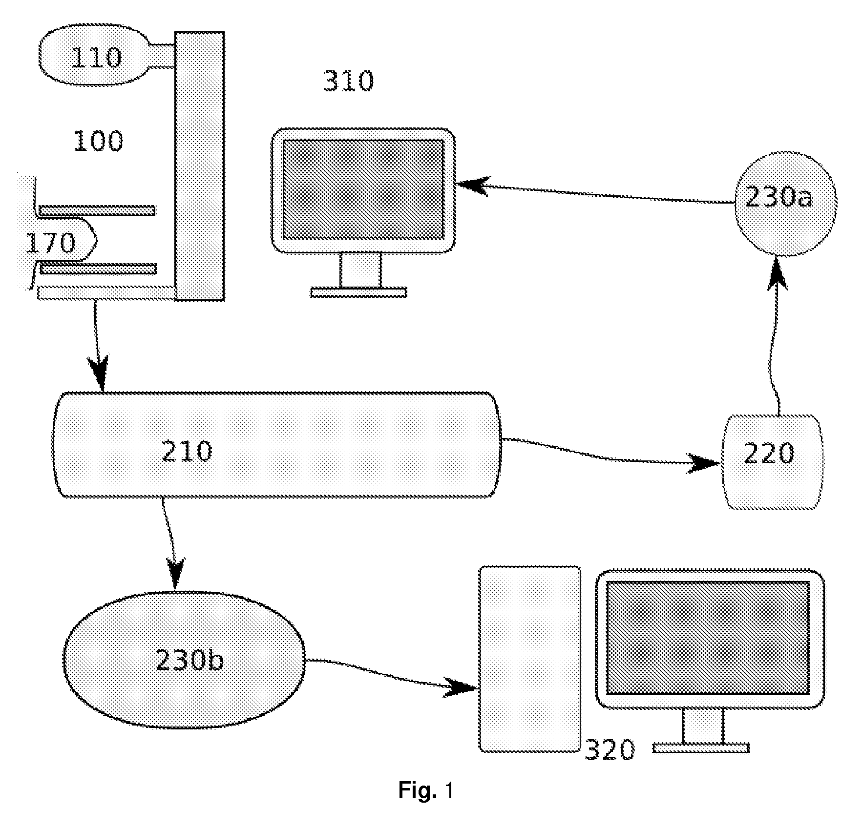
[0020]FIG. 1 shows the preferred embodiment of the present invention implemented in an X-ray acquisition device 100. The device 100 comprises an X-ray acquisition device 100, which produces a set of projection images of a compressed breast, at different angles. The full projection images are stored in a memory unit 210. From here, there are two parallel paths of processing. According to prior art, the full projection images are fed to a reconstruction means 230b, where the full projection images are combined to a three-dimensional volume, which is further used for diagnosis or exported to a Picture Archiving and Communicating System (PACS) 320. According to this embodiment of the present invention, the stored full projection images in the memory unit 210 are also fed to a preview branch, wherein the data first passes through a low resolution provider 220 for providing the projection images at a low resolution. The low resolution provider comprises a sub-sampler for conversion to low...


PUM


Abstract

To enhance image acquisition and speed up the examination during x-ray examinations, the present invention relates to an X-ray apparatus for three dimensional imaging and in particular for tomosynthesis examination, comprising a means for obtaining a set of projection images of a body part, a reconstruction means for reconstructing a three-dimensional image volume, memory for storing the projection images, and a control means. The reconstruction arrangement is arranged to reconstruct a three-dimensional image volume from data in the projection images in the memory, the reconstruction arrangement being arranged to reconstruct a first and a second image volume, wherein the second image volume is reconstructed having lower resolution than the first image volume.

Description

TECHNICAL FIELD OF THE INVENTION[0001]The present invention relates to a method and arrangement in X-ray imaging, in particular three-dimensional imaging, and more especially tomosynthesis.BACKGROUND OF THE INVENTION[0002]Tomosynthesis is used to create a three-dimensional image volume of a person's body part, e.g. her breast, or an object, using X-rays. Currently, tomosynthesis breast imaging is available only for research purposes, but an increasing number of market analysts believe that tomosynthesis breast imaging will become more widely used than conventional two-dimensional mammography.[0003]Tomosynthesis is essentially a limited form of Computed Tomography (CT). Normally, several projection images, e.g. 5 or 30, are acquired from slightly different angles, using a modified X-ray system. Each projection image is essentially a conventional 2-dimensional digital X-ray image of the examined object. The projection images are then combined using special purpose software for reconst...

Claims


Application Information

Patent Timeline
IPC IPC(8): H05G1/60
CPCA61B6/502A61B6/025
Inventor HEMMENDORFF, MAGNUSDANIELSSON, MATS
Owner SECTRA MAMEA
Learn More