Media rich imaging report generation and presentation

a report generation and media technology, applied in the field of imaging device data processing, can solve the problems of preventing users from viewing non-dicom-compliant images within the dicom platform, affecting the quality of imaging reports, and limited standards relating to the formatting of imaging modality data and its subsequent storage, so as to prevent any unauthorized access of confidential medical information

Inactive Publication Date: 2008-06-12
SONOSITE
View PDF11 Cites 39 Cited by
  • Summary
  • Abstract
  • Description
  • Claims
  • Application Information

AI Technical Summary

Benefits of technology

[0014]Embodiments of the invention use web technology in order to bring all of the information into the media rich report, which effectively mimics the display of a web page. The media rich report may identify the patient, date, time, nature of the procedure, physician observations, diagnostic findings, and the medical conclusion. The media rich report may also include images, video, audio, and textual information relating to particular measurements, calculations, annotations, and findings derived from one or more imaging procedures. The images, video, audio, and text can be embedded into the comprehensive media rich report, which can be formatted to allow for the simultaneous viewing of some or all of this information via a platform-independent interface. Once the media rich report has been generated, the report can be distributed and redistributed without the need for individual rendering of the data that comprise the report. In a preferred embodiment of the invention, the media rich report can be distributed to users via e-mail and various encryption techniques can be employed to prevent any unauthorized access of confidential medical information.

Problems solved by technology

Standards relating to the formatting of imaging modality data and its subsequent storage are limited.
Because DICOM is generally implemented in complex diagnostic environments, such as large hospitals, the systems that utilize this standard tend to be very expensive.
Even where the DICOM standard is implemented, images that are not DICOM-compliant may be embedded within a DICOM-compliant file, which may prevent the user from viewing the non-DICOM-compliant image within the DICOM platform.
Applications that enable viewing of non-DICOM-compliant image formats are limited in that the image format is unavailable for viewing simultaneously with the DICOM-compliant images.
Unfortunately, the implementation of the DICOM ultrasound application can be cost prohibitive for small medical practices due to the significant costs associated with acquiring, implementing, administering, and maintaining the DICOM computing environment.
Because the DICOM standard is limited to facilitating communication between imaging devices and archival devices, the DICOM standard does not provide a user with the ability to easily retrieve the archived information or subsequently utilize that information in any meaningful way.
And although the DICOM standard is a fixture in large hospital enterprises, its use in small medical practices is practically nonexistent.
Once raw data is acquired by the imaging modality and exported to an archive or portable media, the organization of that data becomes problematic.
Generally, medical practices will not only save the imaging modality data in a proprietary format, but will do so using a haphazard approach to the organization of this data within the archival system.
Because custom applications are normally required to efficiently retrieve and utilize the imaging modality data, the costs associated with software development, deployment, and maintenance of these systems can be substantial.
The physician may have purchased or been provided with the software by the manufacturer, or the physician may not have access to that application at all, which would prevent any subsequent review of the video.
Even if the physician has access to the proprietary program that will allow him to view the video of the ultrasound, the video alone may not be as diagnostically useful as a comprehensive presentation of audio data, video data, image data, patient data, and / or report data.
In order for the physician to access such a diverse range of data, multiple software applications may be necessary and the simultaneous display of such data may not be possible.
The lack of a standard platform for reviewing this information may prevent other interested parties from reviewing information relating to one or more imaging procedures.
This lack of information flow may have detrimental consequences to the medical decision-making process by preventing the medical knowledge base from expanding and potentially preventing the patient from making an informed decision regarding his / her healthcare.

Method used


Image

  • Media rich imaging report generation and presentation
  • Media rich imaging report generation and presentation
  • Media rich imaging report generation and presentation

Examples


Embodiment Construction

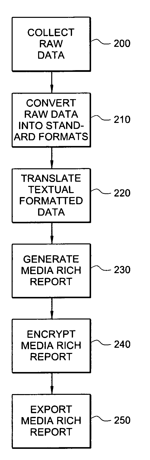
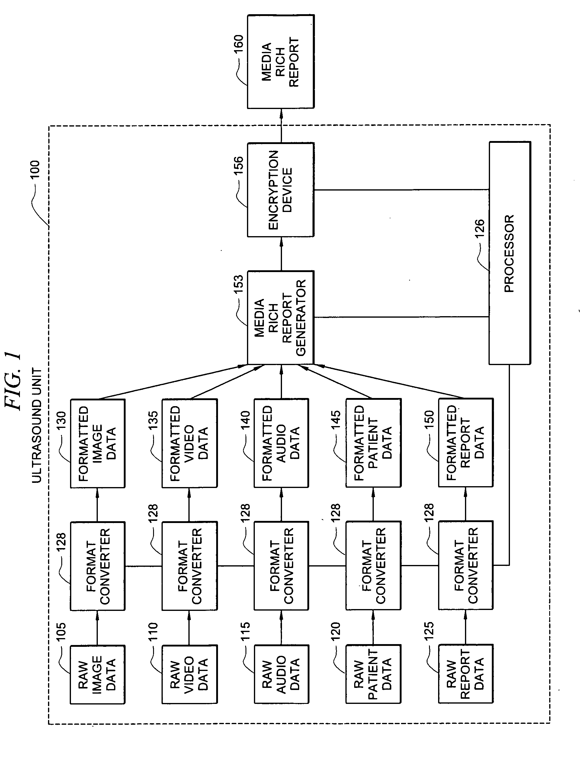
[0020]FIG. 1 is a functional block diagram of an embodiment of the invention showing a system for processing the raw data by ultrasound unit 100, converting the raw data into one or more standard formats, and generating a media rich report 160. The specification will generally use ultrasound as an example of an imaging device that can generate a wide variety of relevant data types (e.g., audio, video, text) to aid in understanding the concepts of the present invention. For example, with respect to an ultrasound procedure performed to monitor a developing fetus, the movement of the fetus (video) and its heartbeat (audio) may be of interest to both the patient and the treating physician. However, it should be appreciated that embodiments of the invention may be utilized with respect to various imaging devices, such as X-ray machines, computer tomography (CT) machines, magnetic resonance imaging (MRI) machines, endoscopic ultrasonography units, nuclear medicine imaging machines, and vi...


PUM


Abstract

A system and method that collects and converts raw data and other relevant information related to imaging procedures in order to provide medical personnel, patients, and other authorized parties with a comprehensive media rich report that can be accessed using a platform-independent interface. The invention facilitates the collection and conversion of audio, video, image, and textual data from various sources in order to produce a single comprehensive report that provides the user with maximum utility in evaluating the information derived from one or more the imaging procedures.

Description

CROSS-REFERENCE TO RELATED APPLICATIONS[0001]This application is related to concurrently filed, co-pending, and commonly assigned U.S. patent application Ser. No. 065744-P032US-10610770 entitled “ORGANIZATION OF DISSIMILAR DATA FOR PLATFORM-INDEPENDENT DIAGNOSTIC IMAGING PRESENTATION,” the disclosure of which application is hereby incorporated herein by reference.TECHNICAL FIELD[0002]The present invention relates generally to the processing of imaging device data and more particularly to the processing of imaging device data to generate a media rich report.BACKGROUND OF THE INVENTION[0003]An imaging device is any device that acquires information and transforms the information into a form suitable for visual perception and / or electronic use, such as an imaging modality unit that may include related peripheral equipment. An imaging modality unit is a medical system that is used to display an image that accurately represents anatomy. Prevalent imaging modalities used today include ultr...

Claims


Application Information

Patent Timeline
Patent Type & Authority Applications(United States)
IPC IPC(8): G06F17/00
CPCG06F19/3487G06F19/321G16H15/00G16H30/40G16H30/20
Inventor CATALLO, LEO R.HENDRICH, JONATHAN P.
Owner SONOSITE
Why Eureka
Try Eureka