
Hemoglobin A1c Determination Method, Enzyme to be Used therefor, and Production Method Thereof

a technology of hemoglobin and enzyme, applied in the field of hemoglobin a1c determination method, enzyme to be used therefor, and production method thereof, can solve the problems of low processing speed, low accuracy, and dedicated devices

Active Publication Date: 2008-05-15
ASAHI KASEI PHARMA
View PDF6 Cites 23 Cited by
  • Summary
  • Abstract
  • Description
  • Claims
  • Application Information

AI Technical Summary

Benefits of technology

The present invention provides a method for specifically determining the glycated β-chain N-terminal of glycated hemoglobin or a fragment thereof without a separation operation. This is achieved by using a protease that cleaves a glycated amino acid or a glycated peptide in the β-chain N-terminal without substantially cleaving a glycated amino acid or a glycated peptide in the α-chain N-terminal of glycated hemoglobin. The inventors have also discovered that a specific ketoamine oxidase can be used to react with the glycated amino acid or peptide without reacting with another amino acid or peptide. This allows for the specific determination of the glycated β-chain N-terminal without the need for separation.

Problems solved by technology

However, the electrophoresis and chromatography methods require expensive dedicated devices, and processing speeds thereof are low, so that those methods are inappropriate for clinical examinations to process many samples.
However, the method is performed using an antigen-antibody reaction, so that it is problematic that the accuracy is not always good due to reproducibility and effects of coexisting substances.
However, there is no description about specificity to cleave a glycated amino acid and / or a glycated peptide from a glycated β-chain N-terminal without substantially cleaving a glycated amino acid or a glycated peptide from a glycated α-chain N-terminal from glycated hemoglobin or a fragment thereof.
However, the glycation rate of a glycated hemoglobin product existing in nature is low (about 5%).
Therefore, the yield in separating hemoglobin where an α-chain N-terminal has been glycated and hemoglobin where a β-chain N-terminal has been glycated was extremely low, and it was difficult to detect the activity of the protease using those substances as substrates because hemoglobin is red.
In general, a step for cleaving FV from a hemoglobin β-chain N-terminal glycated peptide using a protease has disadvantages in that the reaction hardly proceeds in general and must be performed using a large amount of enzymes for a long time, so that, in order to overcome such disadvantages, a ketoamine oxidase that reacts also with a glycated peptide cleaved from a hemoglobin β-chain N-terminal glycated peptide by a protease has been required as a ketoamine oxidase to be used in determining hemoglobin A1c.
However, it has not been known which amino acid residue in the primary structure of the enzyme contributes to reduction of a reaction with FK or FZK.
However, centrifugation was performed as an operation, and there is no description that the determination can be performed without a separation operation.
However, this method have the following disadvantages: it requires two stages of protease reactions; it is difficult to strictly control the protease reaction for cleaving the carboxyl group side of leucine at the third position from a hemoglobin β-chain N-terminal in the first stage; and a reaction of the step for cleaving an α-glycated amino acid from a hemoglobin β-chain N-terminal glycated peptide in the second stage hardly proceeds in general and must be performed using a large amount of enzymes for a long time.
Moreover, in the method shown in examples, a cumbersome separation operation (ultrafiltration) was performed twice, and there is no description that the method of the present application enables specific determination of a glycated β-chain N-terminal of glycated hemoglobin without a separation operation.
In addition, there is no description that the suggested method can be performed without a separation operation.
Although the above-described determination methods relating to glycated hemoglobin were known, there have not been known a method and a reagent kit for specifically determining a glycated β-chain N-terminal of glycated hemoglobin using enzymes without a separation operation.

Method used


Image

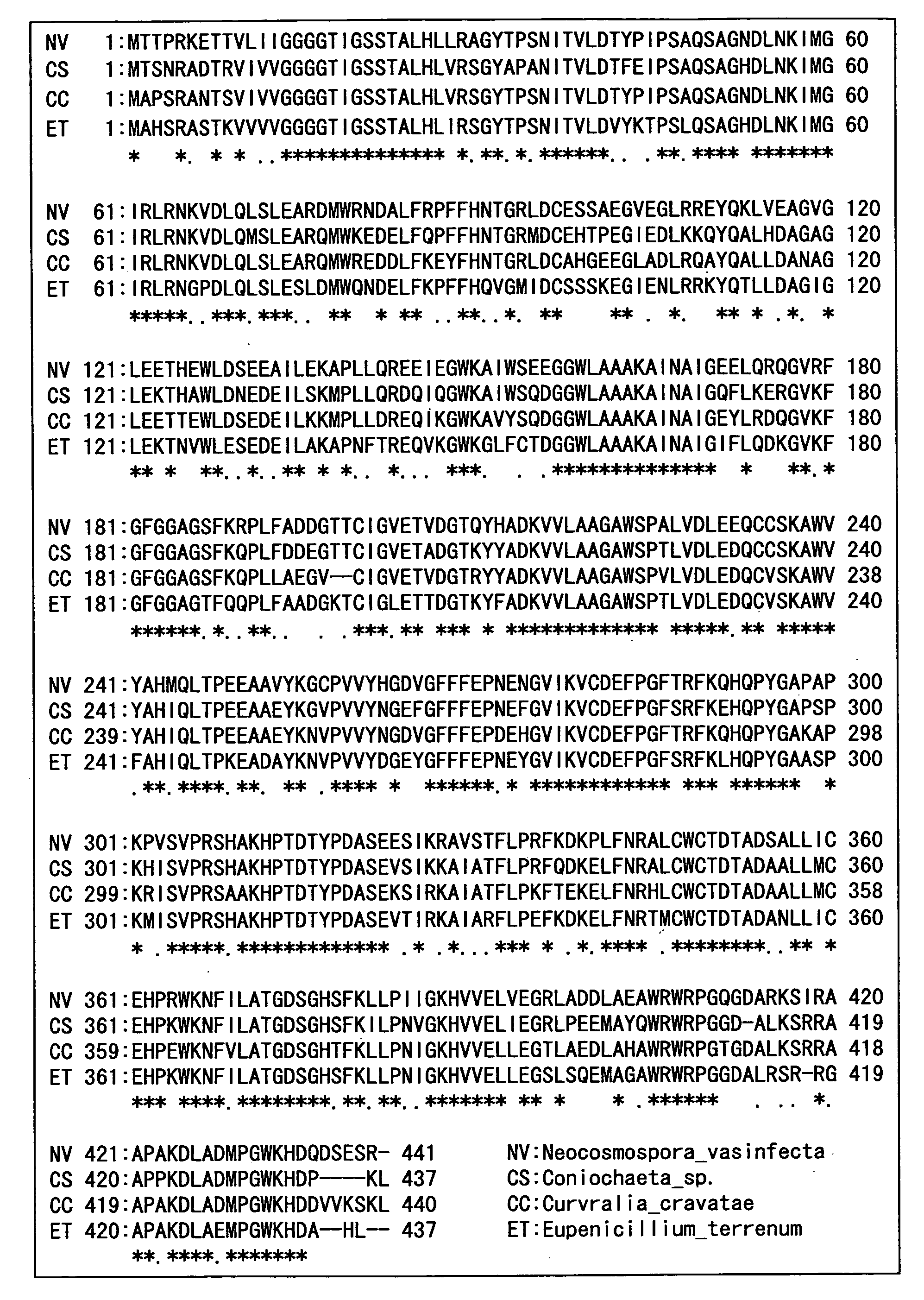
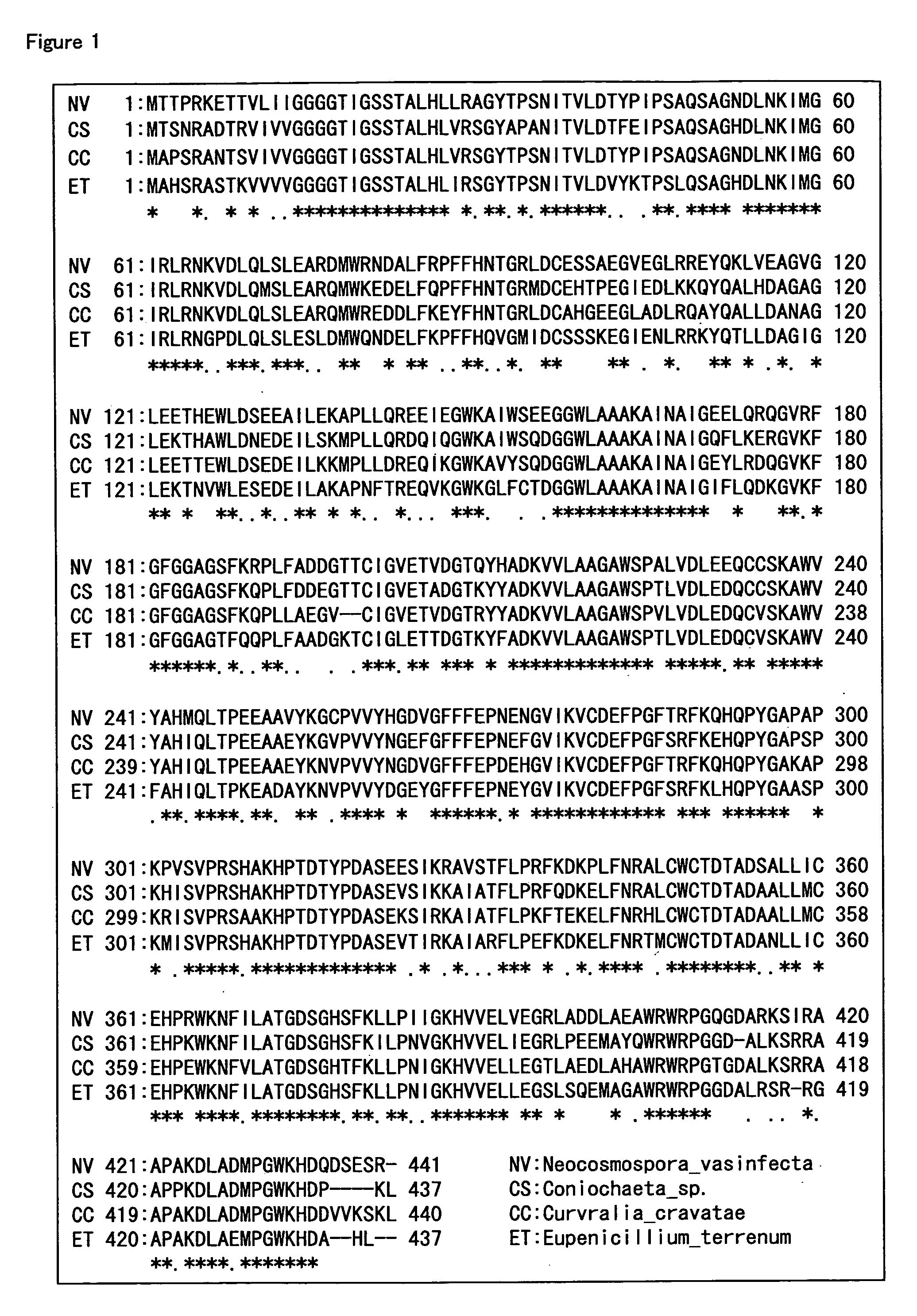
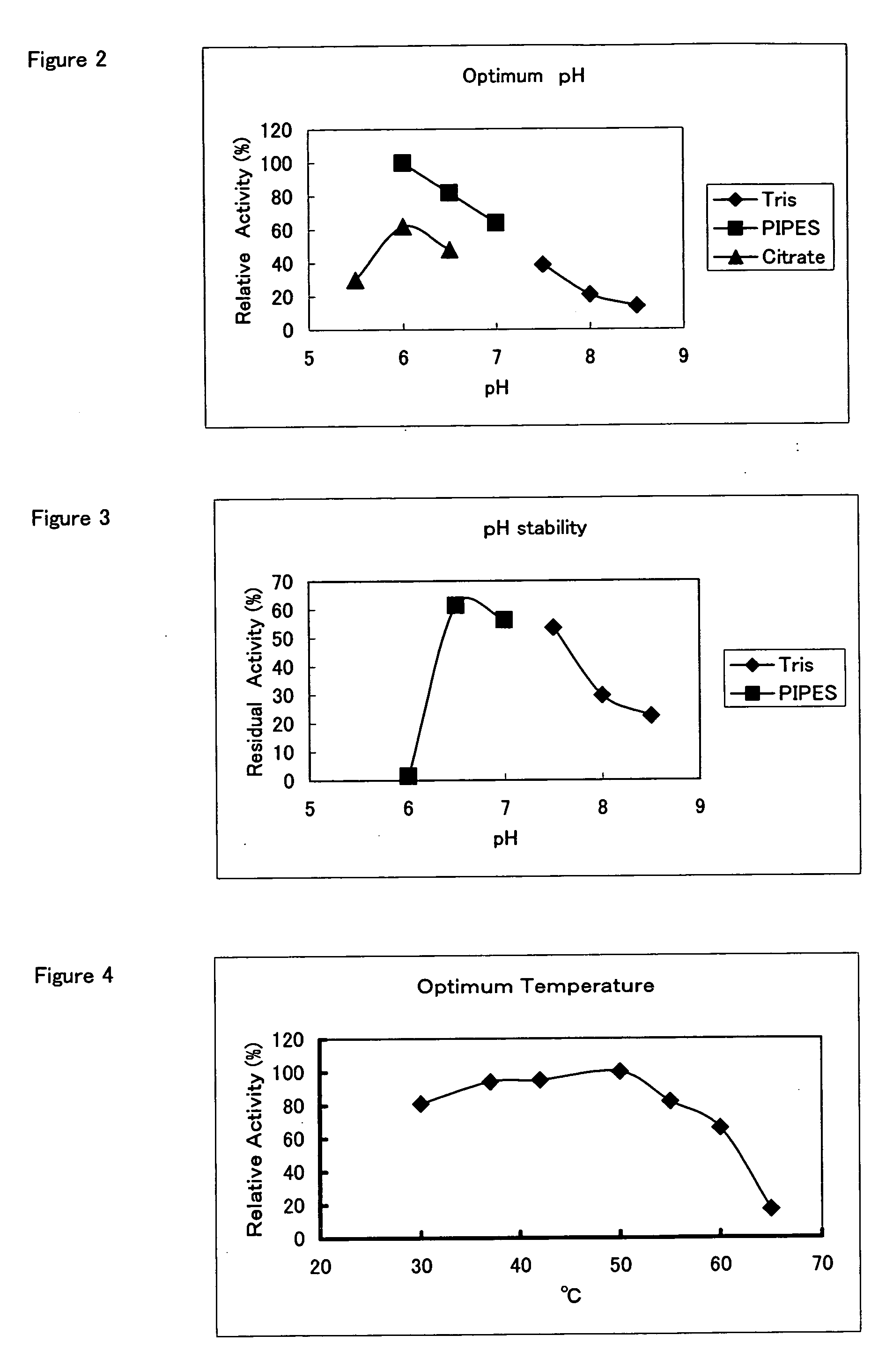
  • Hemoglobin A1c Determination Method, Enzyme to be Used therefor, and Production Method Thereof
  • Hemoglobin A1c Determination Method, Enzyme to be Used therefor, and Production Method Thereof
  • Hemoglobin A1c Determination Method, Enzyme to be Used therefor, and Production Method Thereof

Examples


example 1

Screening of Protease for Hemoglobin A1c Determination (pH 7.5)

[0200]A screening method is shown in which a protease that cleaves a glycated peptide from N-terminal-glycated pentapeptide of a hemoglobin β-chain without substantially cleaving a glycated amino acid or glycated peptide from N-terminal-glycated pentapeptide of a hemoglobin α-chain. Each 10 μl of a sample for screening was placed in 3 wells of a 96-well plate, respectively. Then, the first well was supplemented with 50 μl of an α-solution, the second well was supplemented with 50 μl of a β-solution, and the third well was supplemented with 50 μl of a control solution. After the plate was incubated at 37° C. for 60 minutes, 50 μl of distilled water and 50 μl of a developing solution were added, and the whole was left at room temperature for 30 minutes. Then, the absorbance at a measuring wavelength of 550 nm and a reference wavelength of 595 nm were determined using a Microplate Reader (model 550, manufactured by Bio-Rad)...

example 2

Screening of Protease for Hemoglobin A1c Determination (pH 3.5)

[0208]A screening method is shown in which a protease that cleaves a glycated peptide from N-terminal-glycated pentapeptide of a hemoglobin β-chain without substantially cleaving a glycated amino acid or glycated peptide from N-terminal-glycated pentapeptide of a hemoglobin α-chain. Each 10 μl of a sample for screening was placed in 3 wells of a 96-well plate, respectively. Then, the first well was supplemented with 50 μl of an α-solution, the second well was supplemented with 50 μl of a β-solution, and the third well was supplemented with 50 μl of a control solution. After the plate was incubated at 37° C. for 60 minutes, 50 μl of pH-regulating solution and 50 μl of a developing solution were added, and the whole was left at room temperature for 30 minutes. Then, the absorbance at a measuring wavelength of 550 nm and a reference wavelength of 595 nm were determined using a Microplate Reader (model 550, manufactured by B...

example 3

Screening of Protease for Hemoglobin A1c Determination Derived from Microorganisms

[0217]Various bacteria were cultured with shaking in Difco Lactobacilli MRS Broth (manufactured by Becton, Dickinson and Company), a MM medium (2.5% mannose, 2.5% beer yeast extract, pH7.0), and a YPG medium (2% glucose, 1% polypeptone, 2% yeast extract, 0.1% potassium dihydrogen phosphate, 0.05% magnesium sulfate heptahydrate, pH 7.0) at a temperature of from 28 to 30° C. for 1 to 7 days. Then, cells were removed by centrifugation from the culture solutions to obtain culture supernatants. A sample was prepared by diluting the obtained culture supernatants 10-fold with a 10 mM potassium phosphate buffer (pH7.5), and the determination was performed in a similar manner as Example 1. Table 7 shows the results from the bacteria cultured in MRS Broth, Table 8 shows the results from the bacterium cultured in the MM medium, and Table 9 shows the results from the bacterium cultured in the YPG medium (note that...


PUM

PropertyMeasurementUnit
pHaaaaaaaaaa
pHaaaaaaaaaa
pHaaaaaaaaaa

Abstract

There is provided a method for specifically determining a glycated β-chain N-terminal of glycated hemoglobin using enzymes without a separation operation, and a determination reagent kit therefor.A protease that cleaves a glycated amino acid and / or a glycated peptide from a glycated β-chain N-terminal without substantially cleaving a glycated amino acid or a glycated peptide from a glycated α-chain N-terminal of glycated hemoglobin or a fragment thereof is screened. The method of specifically determining a glycated β-chain N-terminal of glycated hemoglobin and the determination reagent kit are provided by using the protease obtained by the screening method.According to the present invention, a glycated β-chain N-terminal of glycated hemoglobin can specifically be determined without a separation operation.

Description

TECHNICAL FIELD[0001]The present invention relates to a method of specifically determining a glycated β-chain N-terminal of glycated hemoglobin using enzymes without a separation operation, a determination reagent kit, a protease that may be used for the specific determination, a production method thereof, a screening method thereof, and a ketoamine oxidase that may be used for the specific determination.BACKGROUND ART[0002]Determining glycated proteins is very important in diagnosing and controlling diabetes. In particular, recent studies on hemoglobin A1c have proved that risk of occurrence and progression of complications is significantly lowered by controlling the level to 7% or less, so that the level is often used as an index that is essential in clinical fields. As a quantification method for hemoglobin A1c, there are generally known electrophoresis, ion-exchange chromatography, affinity chromatography, immunization, and enzymatic methods. However, the electrophoresis and chr...

Claims


Application Information

Patent Timeline
Patent Type & Authority Applications(United States)
IPC IPC(8): G01N33/53C12N9/50C12N5/02C12N5/04C12Q1/37C12N9/06C07H21/04C12N15/00C12N1/21C12N9/02C12N15/53G01N33/66G01N33/72
CPCC12N9/0004C12N9/50C12Q1/37G01N2333/95C12R1/07G01N33/66G01N33/723C12R1/01C12N9/52C12N9/54C12R2001/07C12N1/205C12R2001/01
Inventor MATSUOKA, TAKESHIKOGA, SHINJIKOUZUMA, TAKUJIYOSHIOKA, ISSEINISHIMURA, ATSUHISAITOU, HOMAREKUMAZAWA, TOSHIHIKOKUROYANAGI, TAKASHI
Owner ASAHI KASEI PHARMA
Learn More