
Method Of Producing Vascular Endothelial Cells From Primate Embryonic Stem Cells

Inactive Publication Date: 2008-01-31
MITSUBISHI TANABE PHARMA CORP
View PDF1 Cites 13 Cited by
  • Summary
  • Abstract
  • Description
  • Claims
  • Application Information

AI Technical Summary

Benefits of technology

[0008] In a second aspect, the present invention relates to the provision of a method for producing early developmental vascular endothelial cells, capable of achieving at least any one of the provision of early developmental vascular endothelial cells having excellent properties that make it possible to have at least any one of long-term culture, subculture, in vitro proliferation, a graft survival at a high efficiency, obtainment of a high compatibility with an individual, development of vascular endothelial cells in an individual, treatment of a condition and / or a disease in which a therapeutic effect can be expected by ameliorating local blood flow, and the like; the supply of the early developmental vascular endothelial cells in a large amount; the provision of a material for the treatment of a condition and / or a disease in which a therapeutic effect can be expected by ameliorating local blood flow; and the like.
[0031] According to the method for constructing a vascular structure of the present invention, an excellent effect that a vascular structure showing a graft survival at a high efficiency and a high compatibility with an individual can be constructed is exhibited. According to the method for constructing a vascular structure of the present invention, an excellent effect that a material for the treatment of a condition and / or a disease in which a therapeutic effect can be expected by ameliorating local blood flow can be provided is exhibited.

Problems solved by technology

However, as described above, the angiogenesis therapy using angiogenesis factor has a defect that the angiogenesis medicine has disadvantages in safety, since according to a random double blind control clinical trial when bFGF is used as an angiogenesis factor for a coronary artery disease, a symptom is likely to be ameliorated by the angiogenesis medicine in a short term but not in a long term.

Method used


Image

  • Method Of Producing Vascular Endothelial Cells From Primate Embryonic Stem Cells
  • Method Of Producing Vascular Endothelial Cells From Primate Embryonic Stem Cells
  • Method Of Producing Vascular Endothelial Cells From Primate Embryonic Stem Cells

Examples


example 1

(1) Differentiation from Monkey Embryonic Stem Cells

[0156] As monkey embryonic stem cells, CMK-6 cell strain, which is cynomolgus monkey (Macaca fascicularis) embryonic stem cells, [Hirofumi Suemori et al., Developmental Dynamics, 222, 273-279 (2001)] was used.

[0157] A cell dissociation buffer {manufactured by GIBCO} was added to the monkey embryonic stem cells maintained on a dish containing a medium for maintaining undifferentiated cells {composition per 200 ml: 163 ml of Dulbecco's modified minimum essential medium (DMEM) / F12, 30 ml of fetal bovine serum (final concentration: 15% by weight), 2 ml of L-glutamine (final concentration: 2 mM), 2 ml of penicillin (100 U / ml)-streptomycin (100 μg / ml), 2 ml of MEM non-essential amino acid solution (manufactured by GIBCO), 1 ml of 2-mercaptoethanol (final concentration: 0.1 mM)}. The monkey embryonic stem cells were incubated at 37° C. for 10 minutes. Thereafter, the embryonic stem cells were collected by tapping the dish and then detac...

example 2

[0170] The VE cadherin-positive cell population obtained in the above item (2) of Example 1 (5×10⁴ cells) were sown in each well of a 6-well dish {manufactured by BD Biosciences} coated with collagen IV or fibronectin, and 20 ml of a culture medium {α-MEM culture medium (manufactured by GIBCO) containing 5×10⁻⁵ M 2-mercaptoethanol and 10% by weight serum) in the presence of 50 ng / ml human recombinant VEGF (trade name) {manufactured by PEPROTECH} was added thereto. The cells were cultured at 37° C. in the presence of 5% by volume of CO2. Here, the culture medium mentioned above was exchanged once in every two days.

[0171] At a stage where the VE cadherin-positive cells reached confluence, the VE cadherin-positive cells were detached from the dish with a 0.25% by weight trypsin solution {manufactured by GIBCO}. The resulting cells were diluted with the culture medium mentioned above so as to have a dilution fold of 1:2 to 1:3, and the resulting cells were further sown in each well of ...

example 3

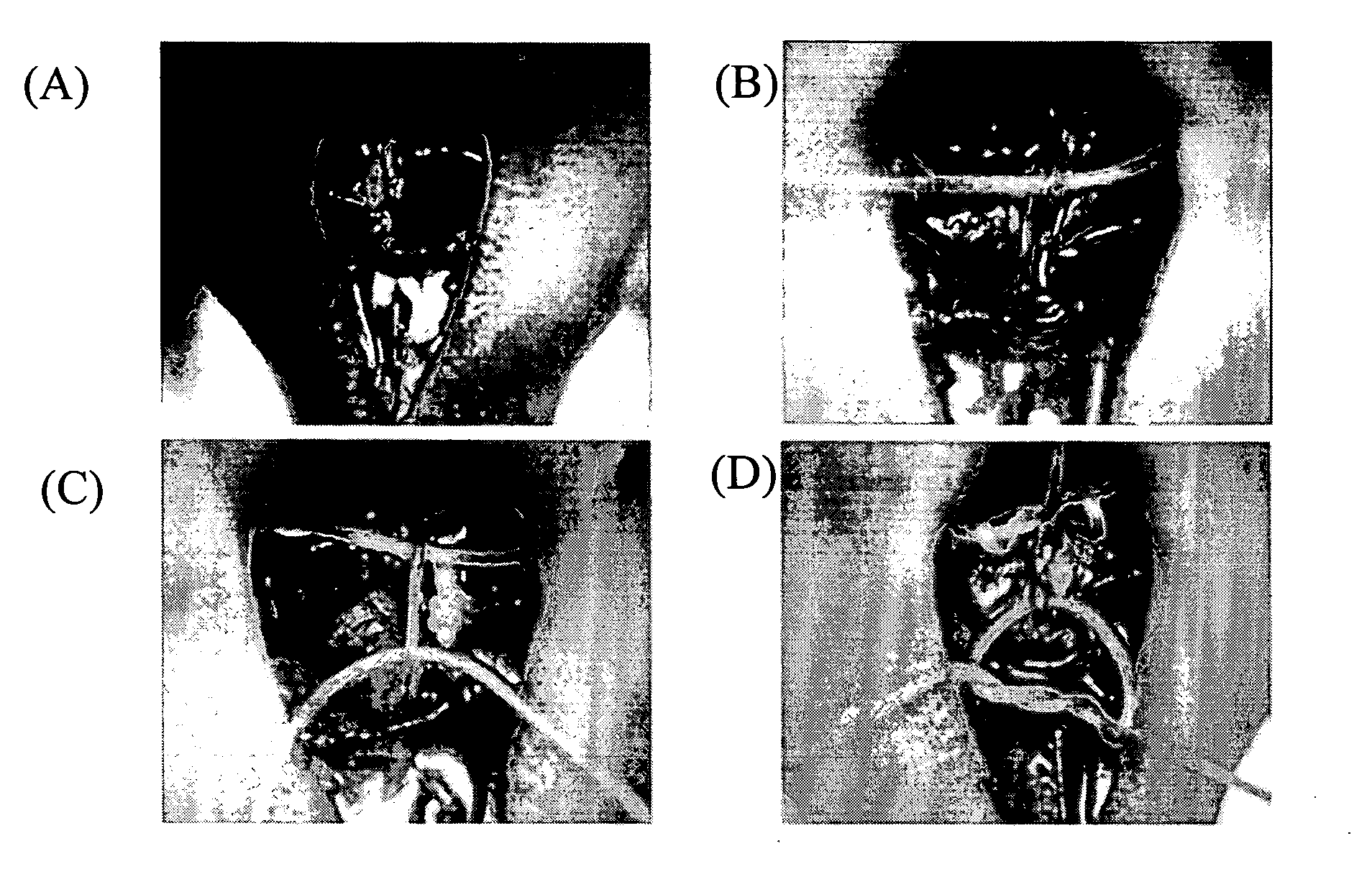
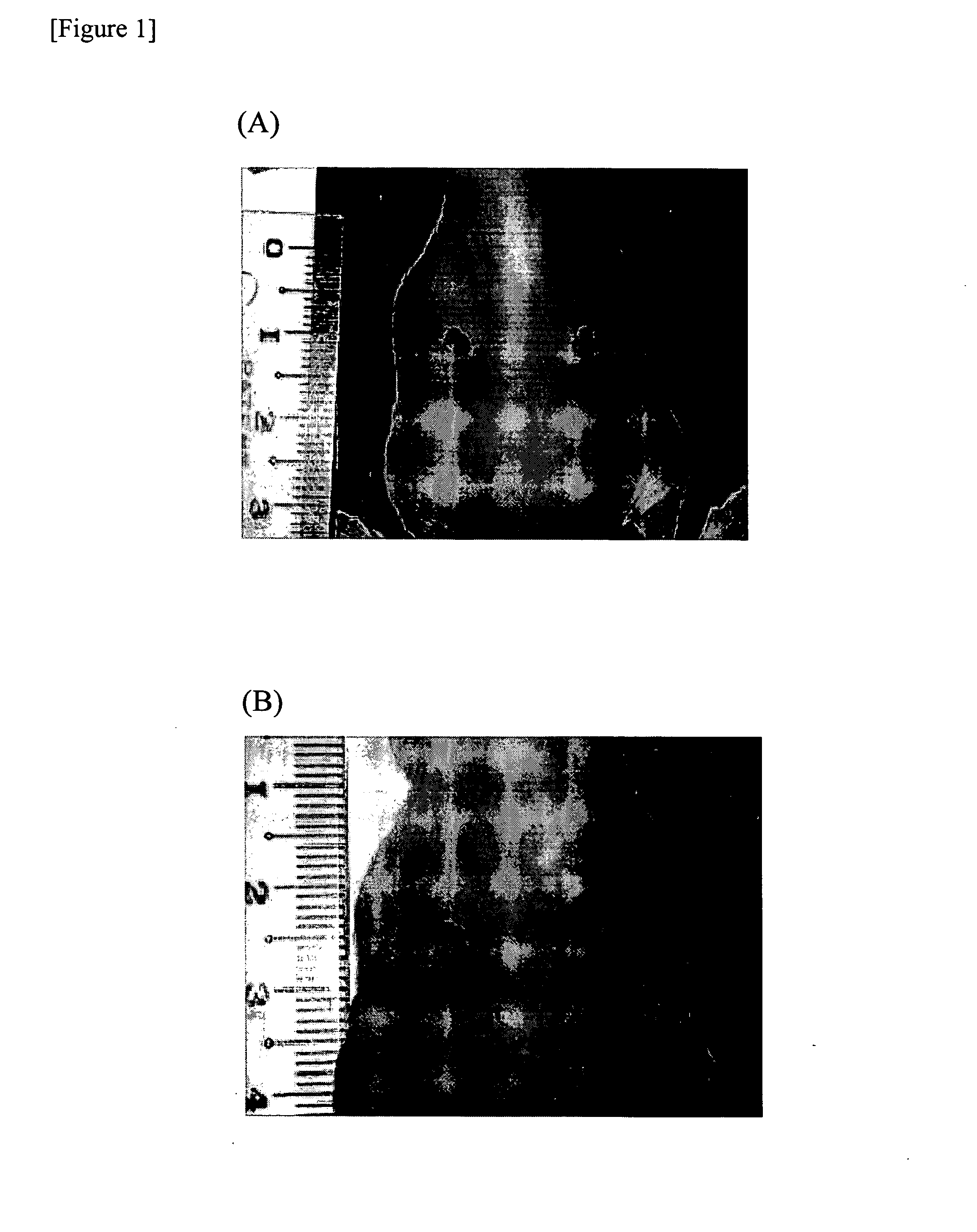
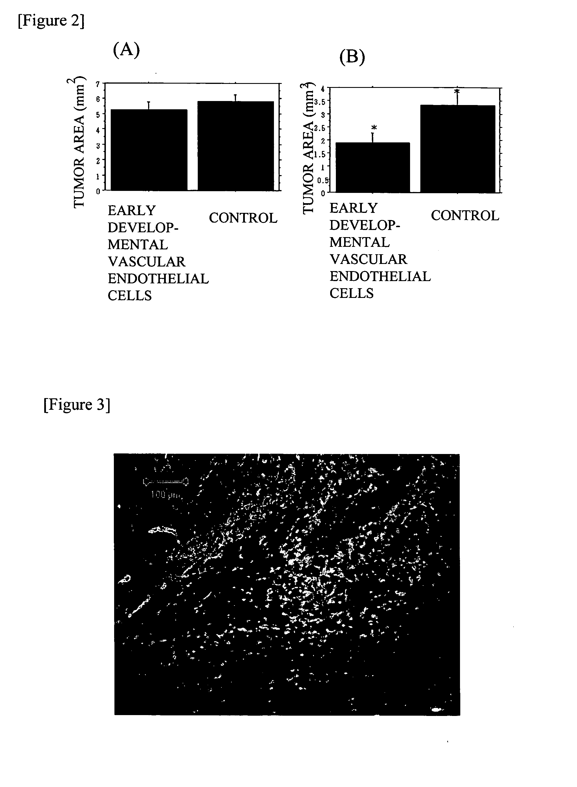
Transplantation of Vascular Endothelial Marker-Positive Cells into Skin Ulcer Model

[0176] Skin ulcer was generated on both sides of back side of 8- to 11-week-old KSN nude mice (Japan SLC, Inc.) with a product under the trade name of KAI STERILE DERMAL BIOPSY PUNCH (manufactured by kai industries co., ltd., size: 4.00 mm, Commercial Product No. BP-40F).

[0177] In addition, the VE cadherin-positive cells proliferated by subculture obtained in Example 2 (5×10⁵ cells / 50 μl phosphate buffered saline) was labeled with a product under the trade name of Vybrant CM-DiI cell-labeling solution {manufactured by Molecular Probes}, in accordance with the manual of Molecular Probes attached to the manufactured article.

[0178] The labeled VE cadherin-positive cells were subcutaneously injected to a skin ulcer on one side of the KSN nude mice mentioned above. In addition, as a control, only 50 μl of phosphate buffered saline (manufactured by GIBCO) was injected on the opposite side of the skin ulc...


PUM


Abstract

To provide a method for differentiating primate embryonic stem cells into vascular endothelial cells, and a technique using the method. A method for differentiating primate embryonic stem cells into early developmental vascular endothelial cells comprising differentiating primate embryonic stem cells into vascular endothelial cell parker-positive cell population; the early developmental vascular endothelial cells; a method for producing the vascular endothelial cells; a method for amplifying the vascular endothelial cells; a vascular lesion-therapeutic agent containing the early developmental vascular endothelial cells as an active ingredient; a method for revascularization comprising supplying the early developmental vascular endothelial cells; a method for transplantation comprising transplanting the early developmental vascular endothelial cells; and a method for treatment comprising administering the early developmental vascular endothelial cells in a therapeutically effective dose.

Description

TECHNICAL FIELD [0001] The present invention relates to a method for differentiating primate embryonic stem cells into vascular endothelial cells, and a technique using the method. More specifically, the present invention relates to a method for differentiating primate embryonic stem cells into vascular endothelial cells (for example, early developmental vascular endothelial cells, and the like), a method for producing the vascular endothelial cells (for example, early developmental vascular endothelial cells, and the like), a method for amplifying the early developmental vascular endothelial cells, early developmental vascular endothelial cells obtained by the method for production, a vascular lesion-therapeutic agent, a method for revascularization, use of the early developmental vascular endothelial cells, a method for treating a condition and / or a disease in which a therapeutic effect can be expected by ameliorating local blood flow, a method for constructing a vascular structur...

Claims


Application Information

Patent Timeline
IPC IPC(8): A61K35/12A61P9/00C12N5/06A61L27/00C12N5/071
CPCA61K35/12C12N2506/02C12N5/0691C12N5/069A61P9/00
Inventor NAKAO, KAZUWAITOH, HIROSHIYAMASHITA, JUNSONE, MASAKATSUYAMAHARA, KENICHIKONDO, YASUSHISUZUKI, YUTAKA
Owner MITSUBISHI TANABE PHARMA CORP
Learn More