
Hydrophilic light absorbing compositions for determination of physiological function in critically ill patients

a composition and light absorbing technology, applied in the field of optical probes for physiological function monitoring, can solve the problems of inability to correlate the single value returned several hours after sampling with other important physiologic events, inability to obtain new or repeat data, and inability to accurately predict the effect of the patient's physiological function, so as to achieve simple, efficient and effective monitoring of organ function.

Inactive Publication Date: 2007-11-22
MALLINCKRODT INC
View PDF3 Cites 4 Cited by
  • Summary
  • Abstract
  • Description
  • Claims
  • Application Information

AI Technical Summary

Benefits of technology

Provides a real-time, accurate, and repeatable measure of renal excretion rate, independent of age or muscle mass, enabling precise monitoring of organ function without radioactivity, enhancing clinical decision-making in critically ill patients.

Problems solved by technology

In addition, a single value returned several hours after sampling is difficult to correlate with other important physiologic events such as blood pressure, cardiac output, state of hydration and other specific clinical events (e.g., hemorrhage, bacteremia, ventilator settings and others).
New or repeat data are equally cumbersome to obtain.
Frequently, dosing of medication is adjusted for renal function and thus can be equally as inaccurate, equally delayed, and as difficult to reassess as the values upon which they are based.
However, these markers have several undesirable properties such as the use of radioactivity or ex-vivo handling of blood and urine samples.
The main drawback of high molecular weight polypeptides is that they are immunogenic.
In addition, large polymers with narrow molecular weight distribution are difficult to prepare, especially in large quantities.
This represents a substantial improvement over any currently available or widely practiced method, since currently, no reliable, continuous, repeatable bedside method for the assessment of specific renal function by optical methods exists.

Method used


Image

  • Hydrophilic light absorbing compositions for determination of physiological function in critically ill patients
  • Hydrophilic light absorbing compositions for determination of physiological function in critically ill patients
  • Hydrophilic light absorbing compositions for determination of physiological function in critically ill patients

Examples


example 1

Synthesis of Indole Disulfonate

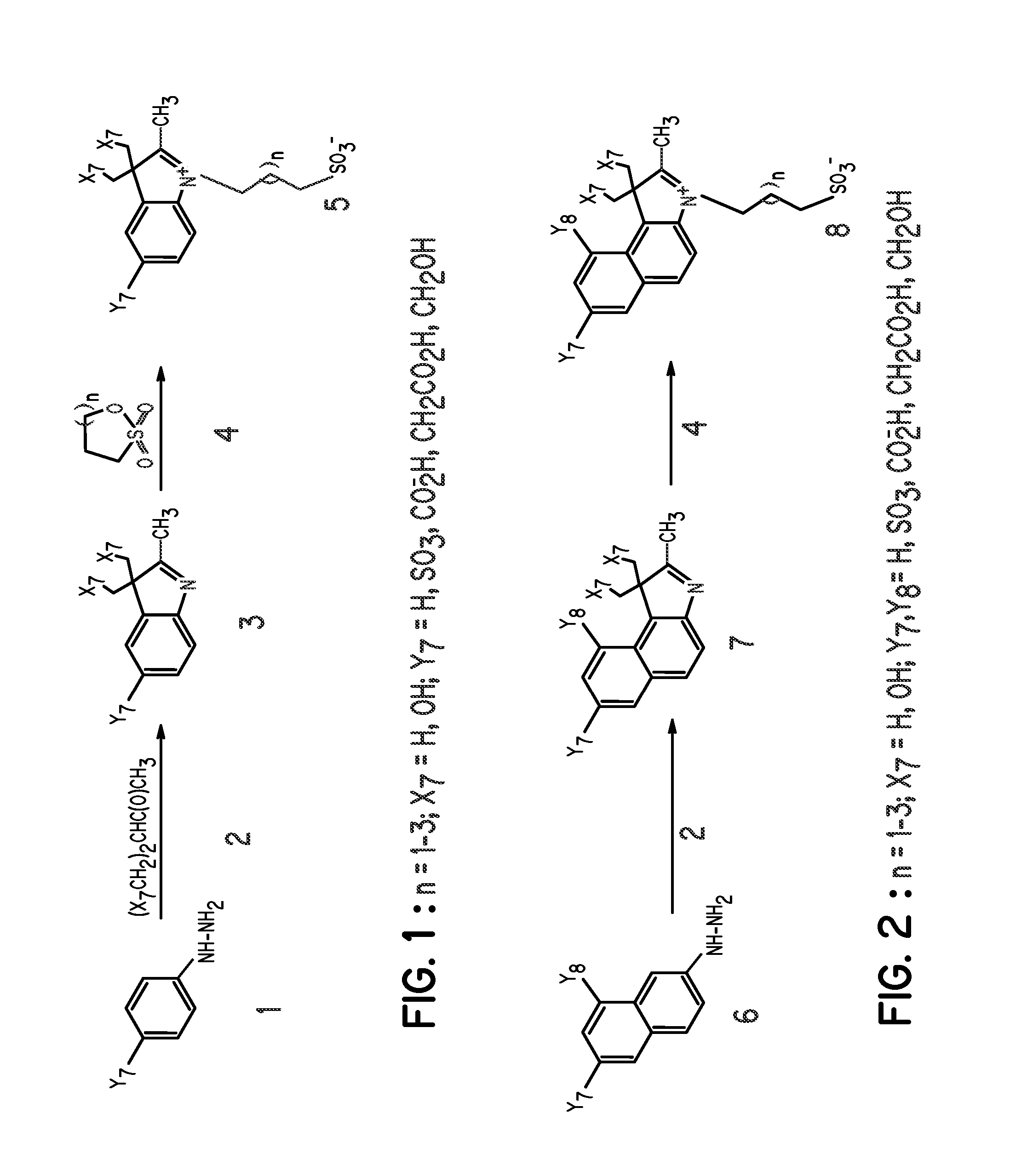
FIG. 1, Compound 5, Y7═SO3−; X7═H; n=1

[0062] A mixture of 3-methyl-2-butanone (25.2 mL), and p-hydrazinobenzenesulfonic acid (15 g) in acetic acid (45 mL) was heated at 110° C. for 3 hours. After reaction, the mixture was allowed to cool to room temperature and ethyl acetate (100 mL) was added to precipitate the product, which was filtered and washed with ethyl acetate (100 mL). The intermediate compound, 2,3,3-trimethylindolenium-5-sulfonate (FIG. 1, compound 3) was obtained as a pink powder in 80% yield. A portion of compound 3 (9.2 g) in methanol (115 mL) was carefully added to a solution of KOH in isopropanol (100 mL). A yellow potassium salt of the sulfonate was obtained in 85% yield after vacuum-drying for 12 hours. A portion of the 2,3,3-trimethylindolenium-5-sulfonate potassium salt (4 g) and 1,3-propanesultone (2.1 g) was heated in dichlorobenzene (40 mL) at 110° C. for 12 hours. The mixture was allowed to cool to room temperature and the re...

example 2

Synthesis of Indole Disulfonate

FIG. 1, Compound 5, Y7═SO3−; X7═H; n=2

[0064] This compound was prepared by the same procedure described in Example 1, except that 1,4-butanesultone was used in place of 1,3-propanesultone.

example 3

Synthesis of Benzoindole Disulfonate

FIG. 2, Compound 8, Y7, Y8═SO3−; X7═H; n=2

[0065] This compound was prepared by the same procedure described in Example 1, except that hydrazinonaphthalenedisulfonic acid was used in place of hydrazinobenzenesulfonic acid.

[0066] Other compounds prepared by a similar method include polyhydroxyindoles such as:


PUM

PropertyMeasurementUnit
wavelengthaaaaaaaaaa
volumesaaaaaaaaaa

Abstract

Highly hydrophilic indole and benzoindole derivatives that absorb and fluoresce in the visible region of light are disclosed. These compounds are useful for physiological and organ function monitoring. Particularly, the molecules of the invention are useful for optical diagnosis of renal and cardiac diseases and for estimation of blood volume in vivo.

Description

RELATED APPLICATIONS [0001] This application is the second of two Continuations, each filed Jul. 16, 2007, and each a Continuation of pending U.S. patent application Ser. No. 10 / 744,334, filed on Dec. 23, 2003, which is a Continuation-In-Part of U.S. patent application Ser. No. 09 / 688,943, filed on Oct. 16, 2000, now U.S. Pat. No. 6,669,926, the disclosures of each hereby incorporated by reference in its entirety.TECHNICAL FIELD [0002] Novel optical probes for use in physiological function monitoring, particularly indole and benzoindole compounds. BACKGROUND OF THE INVENTION [0003] Dynamic monitoring of physiological functions of patients at the bedside is highly desirable in order to minimize the risk of acute renal failure brought about by various clinical, physiological, and pathological conditions (Rabito et al., Renal function in patients at risk with contrast material-induced acute renal failure: Noninvasive real-time monitoring, Radiology 1993, 186, 851; Tilney and Lazarus, A...

Claims


Application Information

Patent Timeline
Patent Type & Authority Applications(United States)
IPC IPC(8): A61K49/20A61K31/19C07D235/04C07D277/84C07D293/10C07D263/60A61K31/4184A61K31/403C07D405/14A61K49/00C07D209/08C07D209/12C07D209/58C07D209/60
CPCA61K31/403A61K49/0021A61K49/0032C07D209/60C07D209/12C07D209/58C07D209/08A61P13/12A61P43/00
Inventor ACHILEFU, SAMUELRAJAGOPALAN, RAGHAVANDORSHOW, RICHARD B.BUGAJ, JOSEPH E.PERIASAMY, MUTHUNADAR P.
Owner MALLINCKRODT INC
Learn More