Enhanced Planar Single Photon Emission Imaging

a single photon emission and imaging technology, applied in the field of nuclear medicine imaging, can solve the problems of long acquisition time, large patient discomfort, and small overall number of patients who can be imaged in a given time, and achieve the effect of enhancing the image quality of a planar nuclear emission imag

Inactive Publication Date: 2007-09-20
ULTRASPECT
View PDF12 Cites 24 Cited by
  • Summary
  • Abstract
  • Description
  • Claims
  • Application Information

AI Technical Summary

Benefits of technology

[0030] There is thus provided, in accordance with some preferred embodiments of the present invention, a method of enhancing image quality of a planar nuclear emission image acquired by a gamma camera, comprising the steps of:

Problems solved by technology

SPECT is considered to be a very useful technique and a good tool for obtaining diagnostic information, however it requires the collection of large number of emitted photon (large statistics) and this means that in order to obtain the required number of photons, a long acquisition time is necessary.
Long acquisition time means that the patient is subjected to a relatively long period of discomfort and furthermore, making the overall number of patients who can be imaged in a given time relatively small—a feature that many medical institutes and hospitals regard as extremely unfavorable and undesirable.
The main limitations to the quality of SPE images comes from:
Resolution is thus limited by sensitivity, and vice versa.
B) Limited number of registered gamma ray photons. a) The constraints imposed on the sensitivity of the collimator by geometrical resolution needs (typically only 1, out of every 10000 emitted photons, is collected), b) the low doses of radiopharmaceutical that can be administered to patients due to hazards associated to exposure to gamma radiation, c) limited acquisition time.
Compton scattering within the patient, in the collimator or in the detector may cause a gamma ray photon to be registered in an erroneous place.

Method used


Image

  • Enhanced Planar Single Photon Emission Imaging
  • Enhanced Planar Single Photon Emission Imaging
  • Enhanced Planar Single Photon Emission Imaging

Examples


Embodiment Construction

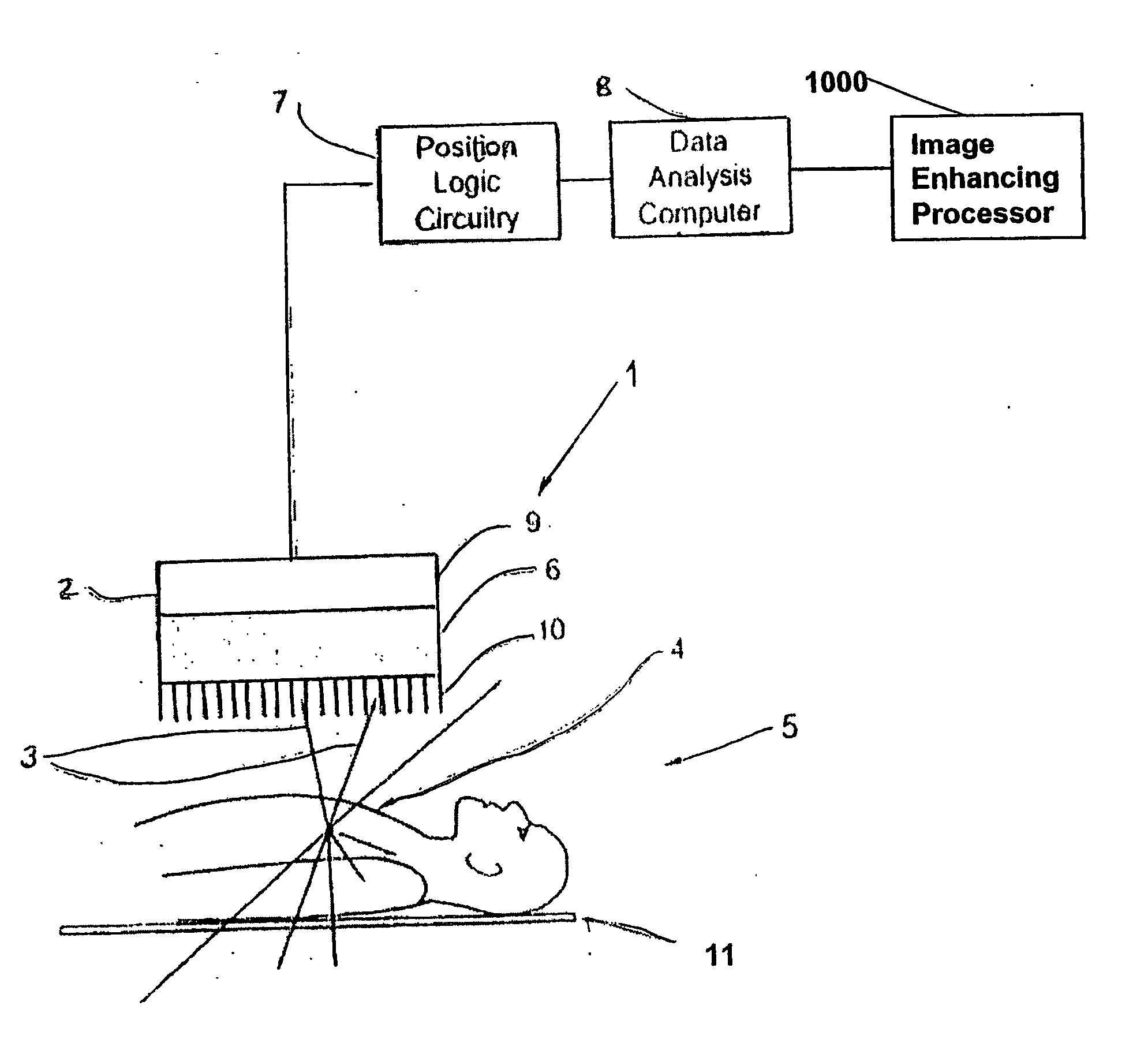
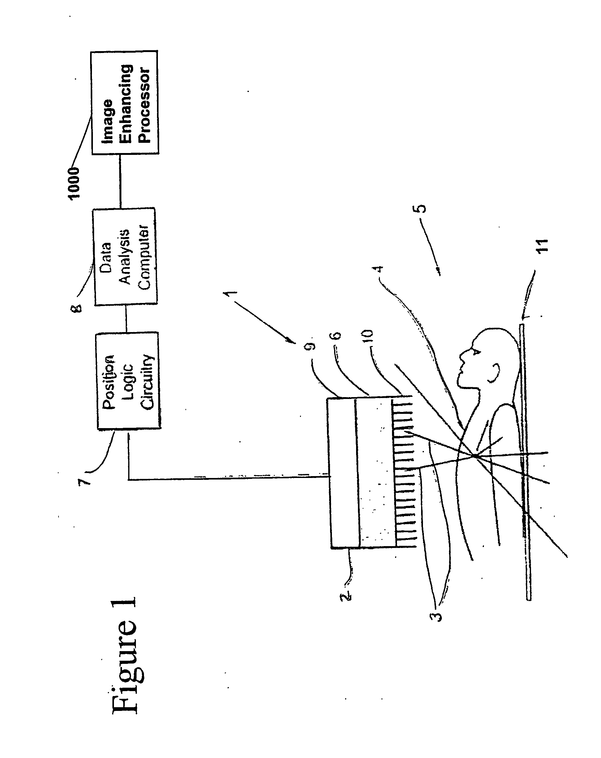
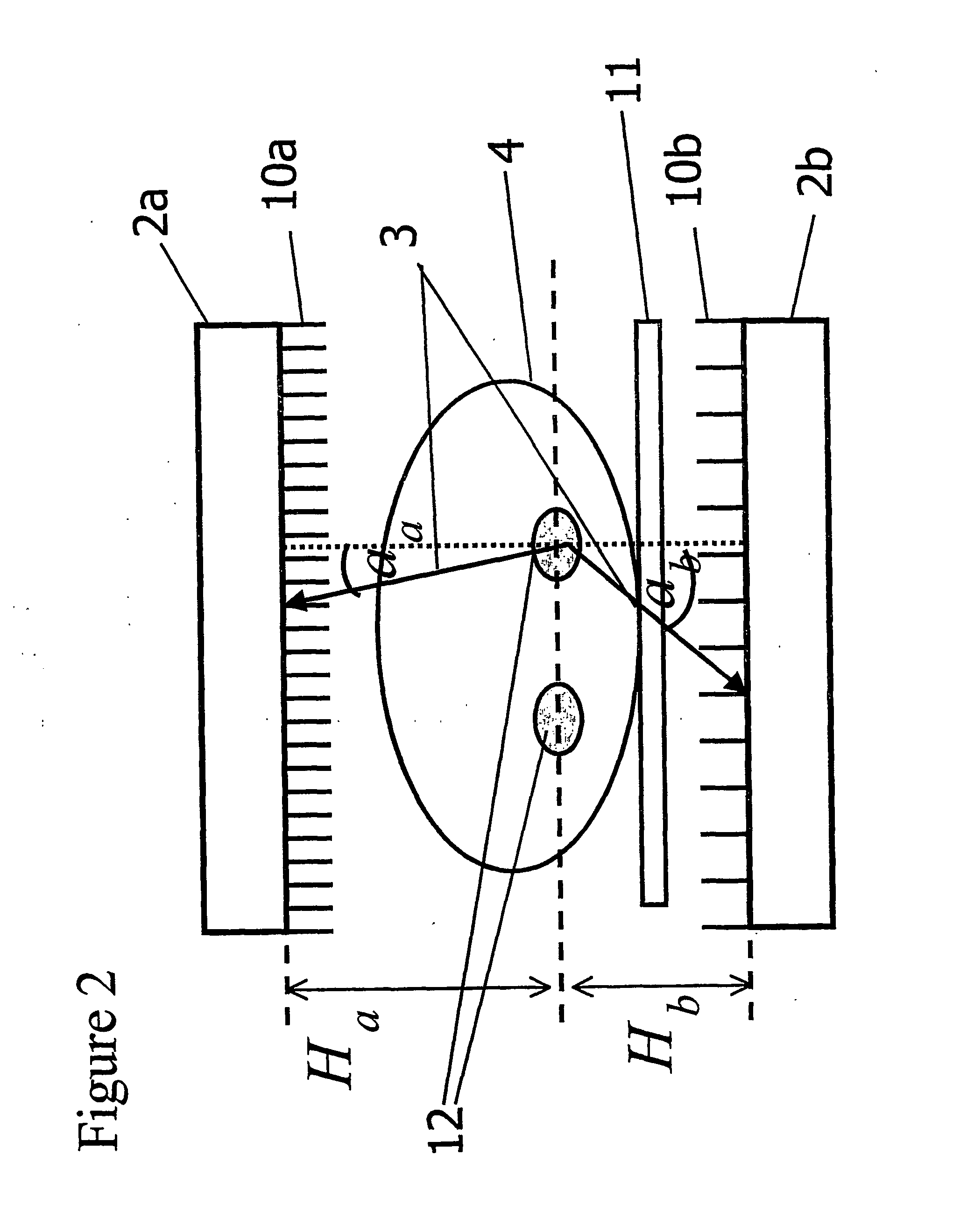
[0139] The general object of the present invention, which will be described subsequently in greater detail, is to provide a novel technique for acquisition and reconstruction of planar single photon emission images.

[0140] Standard planar single photon emission imaging merely collects data from a single angular position, and displays the resulting 2D projection of the imaged body. Thus, it fails to take into account any particulars of the imaging system

[0141] The technique of the present invention, conversely, makes use of available information regarding the physical conditions on which emitted photons are collected by the gamma camera, for example: a) a detailed description of the collimator used, if any; b) a detailed account of the positioning of the camera with respect to the patient's body; c) distance from the patient's body to the camera; d) attenuation map of the patient's body, if available; e) energy of the gamma rays; f) Compton scattering within the patient or the detec...


PUM


Abstract

An apparatus and method for obtaining a planar image of a portion (4) of a body (5) and enhancing image quality of at least one specific organ or volume of interest located within the portion (4) of the body (5), administered with radiopharmaceutical substance radiating gamma rays, by using single photon emission imaging, for determination of functional information thereon, comprising: (a) acquiring at least one projection data of the portion (4), by means of a gamma camera detector (2); (b) determining the effective distance between the detector (2) and the specific organ of interest; (c) calculating weight values taking into account acceptance angles of the gamma camera detector (2) and the effective distance; and (d) obtaining a two dimensional image of a spatial distribution of the pharmaceutical substance within the portion (4) by mathematically analyzing the data in conjunction with weight values.

Description

FIELD OF THE INVENTION [0001] The present invention relates to a nuclear medicine imaging. More particularly it relates to enhancing planar single photon emission imaging. BACKGROUND OF THE INVENTION [0002] Single photon emission (SPE) imaging is a known medical imaging technique. It involves injecting radiopharmaceutical substance into a patient's body and evaluating the distribution of the radiopharmaceutical substance, which is indicated by the distribution of gamma rays emitted from within the patient's body. [0003] A radiation-detecting system, often referred to as Gamma camera, detects those gamma rays. Gamma camera detects gamma rays emitted from the radiopharmaceuticals substances, and the data acquired is analyzed to form an image representing the distribution of concentrations of the radiopharmaceutical substance within a specific body area. [0004] Several modalities of SPE imaging are in use: [0005] One of them is Single Photon Emission Computerized Tomography (SPECT). In...

Claims


Application Information

Patent Timeline
IPC IPC(8): G06T5/00G01BG01TG01T1/161G01T1/164
CPCG01T1/1642A61B6/4258A61B6/037G01T1/1647
Inventor GAL, YANIVOAKNIN, JACOBSHWARTZ, SHOULAMITOHANA, ISRAEL
Owner ULTRASPECT
Why Eureka
Try Eureka