Specific binding sites in collagen for integrins and use thereof
a technology of integrins and specific binding sites, applied in the field of computer-aided molecular modeling and cell signaling interaction of extracellular matrix proteins with receptors, can solve the problems of reducing the apparent affinity of i domains, and the precise molecular mechanisms that lead to these activities are not understood
- Summary
- Abstract
- Description
- Claims
- Application Information
AI Technical Summary
Benefits of technology
Problems solved by technology
Method used
Image
Examples
example 1
Recombinant I Domains
[0040] Recombinant I domains of integrin α1 and α2 subunits were generated and isolated as (Xu, Y et al., 2000; Rich, R. L. et al., 1999). Purified recombinant proteins were examined by SDS-polyacrylamide gel electrophoresis (PAGE) followed by staining with Coomassie blue.
example 2
Purification of Recombinant Procollagen
[0041] Frozen yeast cells expressing recombinant type I and III procollagens were obtained from FibroGen, Inc (San Francisco, Calif.). The yeast cells expressed both genes encoding human collagen and prolyl 4-hydroxylase enabling formation of hydroxyproline residues and thermally stable triple helical collagen. The cells were thawed in an ambient-temperature water bath and resuspended in a Start buffer (0.1 M Tris, 0.4 M NaCl, 25 mM EDTA, 1 mM phenylmethylsulfonyl fluoride (PMSF), 1 mM pepstatin, pH 7.5). The cells were lysed using a French press, and the lysate was centrifuged at 30,000×g for 30 min at 4° C. The supernatant was filtered through a 0.45-mm membrane, and the pH of the filtrate was adjusted to 7.5. An affinity column was prepared by coupling a recombinant collagen-binding MSCRAMM from Staphylococcus aureus (Patti et al., 1992) to CNBr-activated Sepharose 4B (Amersham Biosciences). The supernatant was applied to the column and in...
example 3
Surface Plasmon Resonance (SPR) Measurements
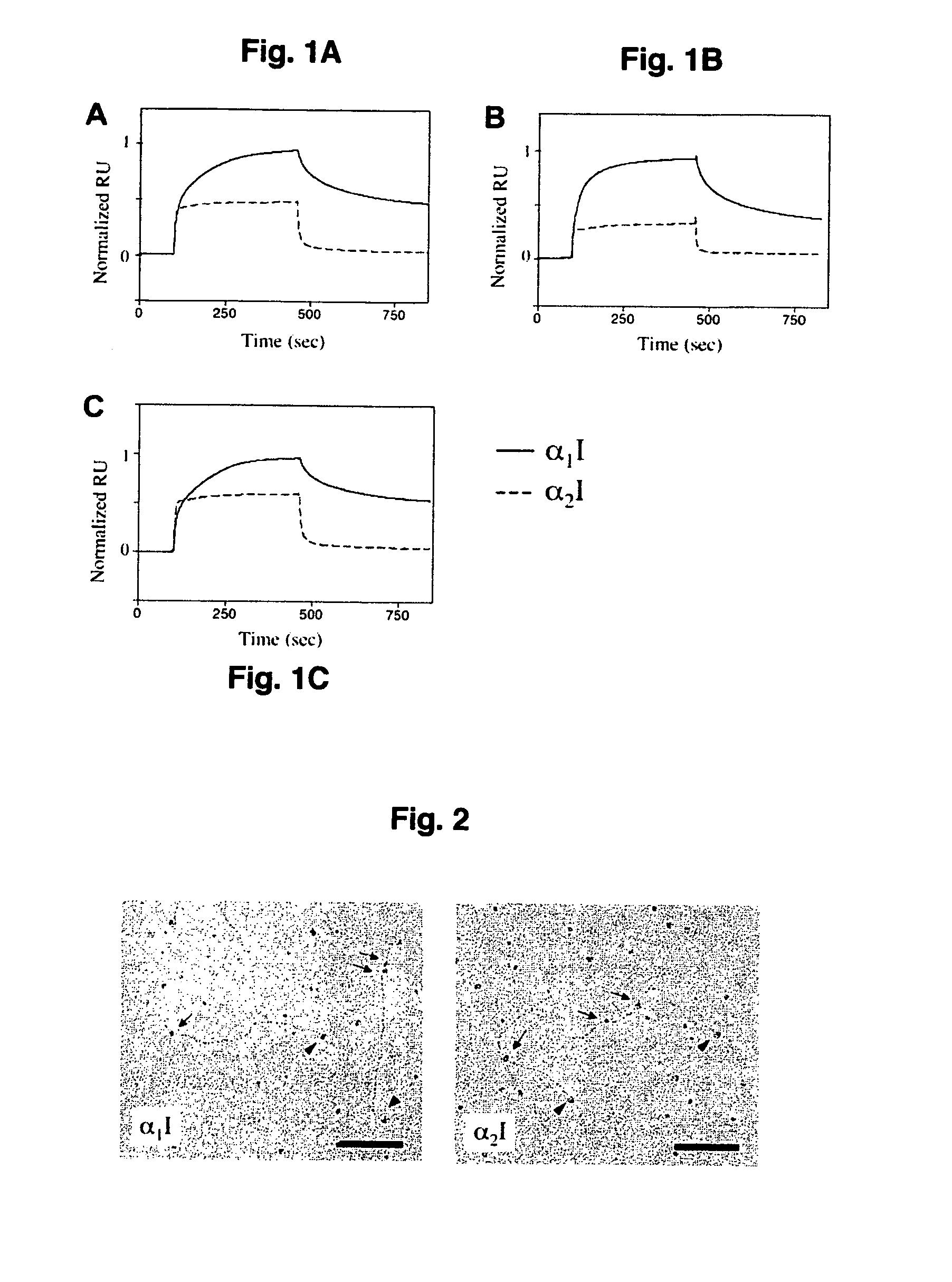
[0042] For the analyses of interactions between recombinant I domains and fibrillar collagens, SPR measurements were carried out at ambient temperature using the BIAcore 3000 system (BIAcore, Uppsala, Sweden) as described previously (Rich et al., 1999) with following modifications. First, purified recombinant human procollagens I and III (described above), or bovine mature type II collagen (Sigma) were immobilized on the flow cells of a CM5 chip resulting in 200-700 response units of immobilized protein. Different concentration of the α1I and α2I proteins in HBS buffer (25 mM HEPES, 150 mM NaCl, pH 7.4) containing 5 mM β-mercaptoethanol, 1 mM MgCl2, and 0.05% octyl-D-glucopyranoside were passed over the immobilized collagen at 30 μl / min for 4 min. Regeneration of the collagen surfaces was achieved with 20 μl of HBS containing 0.01% SDS.
[0043] Binding of α1I and α2I to a reference flow cell, which had been activated and deactivated witho...
PUM
| Property | Measurement | Unit |
|---|---|---|
| pH | aaaaa | aaaaa |
| v/v | aaaaa | aaaaa |
| path length | aaaaa | aaaaa |
Abstract
Description
Claims
Application Information
 - R&D
- Intellectual Property
- Life Sciences
- Materials
- Tech Scout
- Unparalleled Data Quality
- Higher Quality Content
- 60% Fewer Hallucinations
Browse by: Latest US Patents, China's latest patents, Technical Efficacy Thesaurus, Application Domain, Technology Topic, Popular Technical Reports.
© 2025 PatSnap. All rights reserved.Legal|Privacy policy|Modern Slavery Act Transparency Statement|Sitemap|About US| Contact US: help@patsnap.com


