
Method for the pre-conditioning/fixation and treatment of diseases with heat activation/release with thermo-activated drugs and gene products

a technology of heat activation/release and gene products, which is applied in the direction of biocide, therapy, genetic material ingredients, etc., can solve the problems of ineffective cold spots in the treatment zone, limited area, and limited use of previous treatments, and achieve the effect of accurate treatment of diseased tissu

Inactive Publication Date: 2007-04-05
MEDIFOCUS
View PDF11 Cites 49 Cited by
  • Summary
  • Abstract
  • Description
  • Claims
  • Application Information

AI Technical Summary

Benefits of technology

[0017] The present invention relates to an apparatus and a method for administering focused energy to a target site in a subject using either a single energy applicator or multiple energy applicators to supply heat prior to, concurrently with and / or after delivery of a liposome and / or a nanoparticle, for example a thermosensitive liposome or nanoparticle, and / or a thermo-activated drug, gene and / or viral vector. Embodiment of the invention relate to a multi-modality method of treatment using a localized, focused and / or regional heating apparatus, which supplies heat to a defined treatment area, for example an area of a patient's body. The apparatus is used to heat or pretreat a specific site, and to trigger the release of active agents from liposomes and / or activate thermo-activated drugs at the body site. The heating apparatus includes one or more variable and adjustable probes and one or more delivery ports to heat the specific treatment site and to activate liposomes or drugs to release an active agent at the specific treatment site. Each probe may optionally include one or more temperature sensors to permit the regulation of temperature at the specific treatment site and in the surrounding tissue. The use of the apparatus and method allow for the heat conditioning of a specific treatment site and the activation of thermosensitive liposomes and / or thermo-activated drugs at the specific treatment site, allowing for a more accurate treatment of diseased tissue without damaging healthy tissue.
[0018] Furthermore, the apparatus according to the invention addresses the shortcoming mentioned above in that the apparatus permits the user to see in real time the amount of heat generated at a specific location and the actual location where the high heat is generated; thus, the user can effectively target the heat and avoid significant cell death in surrounding tissue.
[0020] A further embodiment of the method according to the present invention is to pre-condition or condition the targeted tissue prior to the delivery of the thermosensitive liposomes and / or thermo-activated drugs. The step of introducing the heat initially enhances the release of active agents from liposomes, particularly thermosensitive liposomes, and / or activates thermo-activated drugs in the targeted tissue.
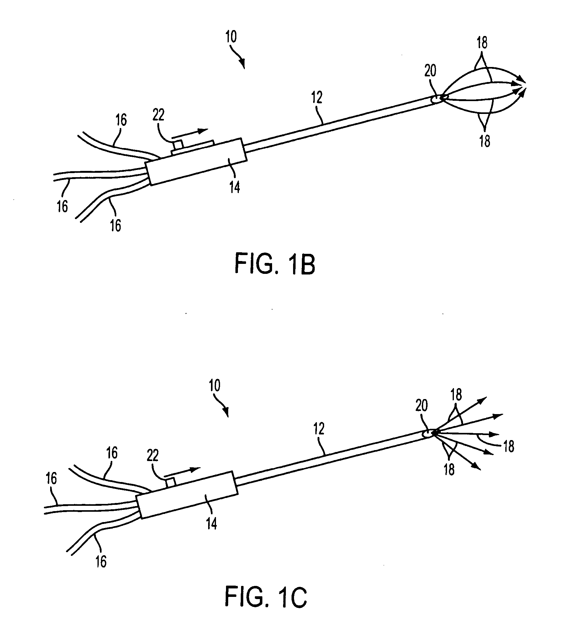
[0027] The apparatus of the present invention can include a catheter, at least one probe, a control unit, and conduits for transferring liquids and gases, for transferring energy, and for transferring temperature readings. There can be a plurality of probes, each including at least one of a temperature sensor, a fluid or gas port, and an energy emitter. In one embodiment, each probe includes a plurality of temperature sensors, fluid or gas ports, and energy emitters. Some embodiments may have probes that include energy emitters alone or energy emitters with temperature sensors and fluid ports or temperature sensors. The temperature sensors can be used to read the temperature in different sections of the treatment zone to allow for the heating of the treatment zone and to control the heating process to ensure that the treatment zone remains at the desired temperature and that the surrounding healthy tissue around the treatment zone does not become overly heated. The fluid and gas ports can be used for direct injection of a thermosensitive liposome and / or thermo-activated drug into the treatment zone. These ports may also be used to deliver cooling liquid or gas into the treatment zone. The energy emitters can deliver an energy source to the treatment zone that heats the diseased tissue in the treatment zone to the desired temperature. The desired temperature can be below the normal ablation temperature so that surrounding tissue is not damaged by the heating of the diseased tissue, but is sufficient to release active agents the liposomes and / or activate thermo-activated drugs introduced into the treatment zone. By using a lower heat level in combination with the liposomes or drugs, the apparatus of the invention can provide for the precise treatment of diseased tissue without significantly harming the surrounding healthy tissue. The design of the device allows for a user to fix and control the size and shape of a treatment zone, which allows for the more directed treatment of only the diseased tissue, and can kill the diseased tissue at the margins of the treatment area, while not damaging the surrounding healthy tissue.

Problems solved by technology

The previous uses of these treatments were limited in focus to small effective treatment regions.
There may be ineffective cold spots throughout the treatment zone due to the non-homogeneous nature of these previous heating methods.
The use of modalities such as RFA can indeed effectively heat a small defined area of tissue, but this area is limited to tissue in close proximity to a deployed heating antenna.
Prior heat treating apparatuses have resulted in unsatisfactory tumor control, generally limited to the immediate center of the treatment site.
Hepatic tumors are either primary or secondary (e.g., metastatic liver cancer (MLC)) and are a substantial medical problem both in the United States and worldwide.
Generally, chemotherapy and radiation therapy are ineffective for treatment of hepatic tumors and certain localized tumors where the above heating modalities are used.
Unfortunately, less than 20% of patients of primary or secondary liver tumors are eligible for surgical resection due to tumor size.
This is also the case where solid tumors have advanced in size, so that it may not be possible to remove the tumor from the organ without compromising the well being of the patient.
The outlook is less favorable for patients with unresectable hepatic tumors.
Thus, all of the devices have similar limitations in the ability to effectively treat larger lesions, especially viable cancer cells in the margins of the lesion.
It is desirable to attempt to create tumor-free margins beyond the imaged tumor lesion of 1 cm or greater; however, RFA is often limited in its ability to produce such consistent margins especially for tumors with a maximum diameter greater than 3 cm.
As a result, known RFA devices are only effective in limited areas that can be heated to high enough temperatures; generally greater than 50° C., and sometimes to temperatures greater than 80° C., in order to ablate the viable cancer.
The high temperature makes it difficult to prevent damage to surrounding non-cancerous tissues.
That is, it is difficult to heat the cancer cells at the margins to greater than 50° C. to kill the diseased tissue and at the same time avoid damaging the surrounding non-cancerous tissue.
With regard to the treatment of prostate tumors, these systems may be less invasive than surgery, cyroblation, or seed implants, but the use of HIFU has also been associated with adverse effects, such as incontinence, recto-urethral fistulas, edema, and chronic necrotic debris and infection.
In addition, due to the time taken to treat using the pinpoint focus of HIFU and limitations on the size of the treatment zone, complete ablation and thus control of the tumor will be very difficult to achieve with these known systems.
HIFU has also been used for other localized cancers; however, it has only achieved marginal success due to difficulty of use, limited size of ablation area, and difficulty of focusing and directing the energy to exactly where it is required.
These shortcomings include the limited size of the effective targeted area and potential adverse effects caused by the high intensity heat.
The inability to see in real time the amount of heat generated and the actual location of where the greatest amount of heat is generated can lead to significant cell death of the adjacent healthy cells.
In addition, heat is distributed non-uniformly within the targeted treatment zone and does not extend effectively to the margins of the lesions or tumors.
Microwave ablation probes have been used to deliver heat to lesions and tumors, but this technology is invasive.
To some extent, MA may be limited by the fact that the regions around blood vessels can sometimes act as heat sinks; these heat sinks can result in cool spots that fail to achieve sufficiently high temperature to kill the lesion or tumor.
Another potential limitation of MA is that it can take more time to heat a very confined area of lesion or tumor tissue.
As a result, the delivery of cytotoxic drugs can damage normal cells and vital organs.
Furthermore, due to certain physical and physiological limitations, higher and more effective doses of anti-cancer agents are generally not achievable.
However, for many localized lesions within organs such as the liver, prostate, lung, esophageal and breast, complete tumor control (including the tumor margins) has not been significantly improved, nor has there been a dramatic increase in survival rates.
Even with thermotherapy, it can be difficult to heat a significant portion of the prostate gland while sparing healthy tissues therein as well as in the surrounding tissues (such as the urethral and rectal walls).
Thus, cancer cells in the margins may not be effectively treated.
Recent treatment of BPH includes transurethral microwave thermotherapy in which microwave energy is employed to elevate the temperature of tissue surrounding the prostatic urethra above about 45° C., thereby thermally damaging the tumorous prostate tissue.

Method used


Image

  • Method for the pre-conditioning/fixation and treatment of diseases with heat activation/release with thermo-activated drugs and gene products
  • Method for the pre-conditioning/fixation and treatment of diseases with heat activation/release with thermo-activated drugs and gene products
  • Method for the pre-conditioning/fixation and treatment of diseases with heat activation/release with thermo-activated drugs and gene products

Examples


example 1

Modeling of Heating

[0111] A. Phantom Study

[0112] To validate the computer models, a study in phantoms made of Agar (5%), NaCl (0.25%), Albumin (0.5%), and water was performed. These phantoms reproduce thermal and electrical tissue properties, and coagulate at 75° C. Phantoms were created with 2.5 cm diameter cylindrical holes approximating esophageal dimensions. The setup is shown in FIG. 13. After ablation, the phantom was sliced to determine dimensions of the coagulated regions.

[0113] A 90° C. electrode temperature was maintained for 2 minutes to obtain larger coagulation zones (clinically, 80-85° C. and 1 min are used). FIGS. 14 and 15 show coagulation zones (i.e., zones >75° C.) from these experiments. On average, the 75° C. boundary was ˜2 mm in diameter (FIG. 15).

[0114] B. Computer Models

[0115] Geometrically accurate models of the Stretta catheter needle (FIG. 16a, left) were created. The needle was inserted into tissue inside the simulated esophageal lumen (FIG. 16). The...

example 2

Testing Protocol

[0118] Three dosage levels are used that comprise a thermosensitive liposome prepared from DPPC, MSPC, and DSPE in a molar ratio of about 90:10:4. Doxorubicin is encapsulated within the liposomes. The three dose levels are about 20, 30, 40 mg / m2 doxorubicin. The thermosensitive liposome is infused over 30 minutes; the RF heating procedure begins approximately 15 minutes after start of the infusion.

[0119] The RF heating procedure is designed to create local tissue temperatures above 40° C. to a depth of at least 3 cm measured from the deepest portion of the tumor. Temperature is maintained below locally ablative temperatures. Under endoscopic guidance, the catheter is placed in the center of greatest tumor mass. As many of the catheter heating prongs as possible are deployed directly into the tumor.

[0120] This procedure is repeated at approximately 21 day intervals or as tolerated. Therapy continues unless: 1) there is a complete pathological response, 2) there are...


PUM

PropertyMeasurementUnit
temperatureaaaaaaaaaa

Abstract

A method of treating cancer by introducing heat into cancerous tissue and delivering a liposome containing an active agent or a thermo-activated drug, gene or virus to said tissue. The heat delivered is sufficient to release the active agent or activate the thermo-activated drug, gene or virus. The cancer can be esophageal cancer. The liposome containing an active agent or a thermo-activated drug, gene or virus can be a thermosensitive liposome. The active agent can be an anti-neoplastic agent, for example doxorubicin.

Description

CROSS REFERENCE TO RELATED APPLICATIONS [0001] This application is a continuation-in-part of U.S. application Ser. No. 11 / 280,199 which claims the benefit of priority of U.S. Provisional Application No. 60 / 664,699, filed on Mar. 24, 2005, the subject matter of which is incorporated herein by reference.BACKGROUND OF THE INVENTION [0002] 1. Field of the Invention [0003] The present invention generally relates to an apparatus and a method for administering a focused energy treatment to a limited, defined area of a patient's body. The energy treatment can be delivered by the use of one or more energy applicators. The energy applicators can be used to trigger the release of active agents such as pharmaceutically active agents, nutritional agents, diagnostic agents, cosmetic agents, imaging agents, polynucleotides, and the like from liposomes and / or nanoparticles, in particular thermosensitive liposomes or nanoparticles, for the treatment of cancerous, precancerous, and benign lesions, as...

Claims


Application Information

Patent Timeline
Patent Type & Authority Applications(United States)
IPC IPC(8): A61N1/39A61K48/00A61K31/704A61K9/127
CPCA61B18/1477A61B18/18A61B2018/143A61B2218/002A61K41/0052A61N7/02
Inventor MON, JOHN
Owner MEDIFOCUS
Learn More