Kits for Prenatal Testing
a technology for prenatal testing and kits, applied in the field of kits for prenatal testing, can solve the problems of insufficient accuracy of prenatal tests, time-consuming analysis, and prone to errors
- Summary
- Abstract
- Description
- Claims
- Application Information
AI Technical Summary
Benefits of technology
Problems solved by technology
Method used
Image
Examples
example 1
A Silicon Device Multiplexing 14 Three-Stage Array Duplexes
[0204]FIGS. 11A-11E show an exemplary size-based separation module of the invention, characterized as follows:
[0205] Dimensions: 90 mm×34 mm×1 mm
[0206] Array design: 3 stages, gap size=18, 12 and 8 μm for the first, second and third stage, respectively. Bifurcation ratio=1 / 10. Duplex; single bypass channel
[0207] Device design: multiplexing 14 array duplexes; flow resistors for flow stability
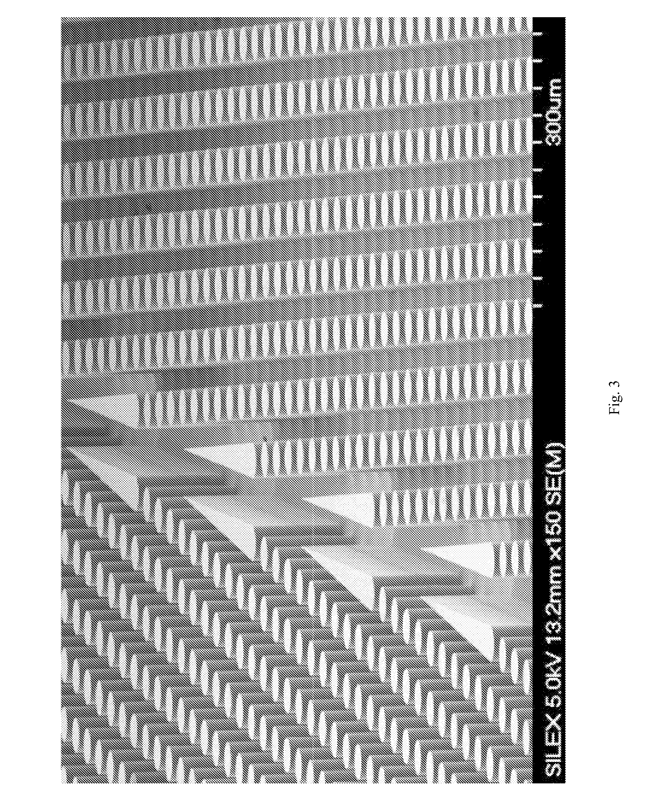
[0208] Device fabrication: The arrays and channels were fabricated in silicon using standard photolithography and deep silicon reactive etching techniques. The etch depth is 150 μm. Through holes for fluid access are made using KOH wet etching. The silicon substrate was sealed on the etched face to form enclosed fluidic channels using a blood compatible pressure sensitive adhesive (9795, 3M, St Paul, Minn.).
[0209] Device packaging: The device was mechanically mated to a plastic manifold with external fluidic reservoirs to deliver bl...
example 2
A Silicon Device Multiplexing 14 Single-Stage Array Duplexes
[0214]FIGS. 13A-13D shows an exemplary device of the invention, characterized as follows.
[0215] Dimensions: 90 mm×34 mm×1 mm
[0216] Array design: 1 stage, gap size=24 μm. Bifurcation ratio=1 / 60. Duplex; double bypass channel
[0217] Device design: multiplexing 14 array duplexes; flow resistors for flow stability
[0218] Device fabrication: The arrays and channels were fabricated in silicon using standard photolithography and deep silicon reactive etching techniques. The etch depth is 150 μm. Through holes for fluid access are made using KOH wet etching. The silicon substrate was sealed on the etched face to form enclosed fluidic channels using a blood compatible pressure sensitive adhesive (9795, 3M, St Paul, Minn.).
[0219] Device packaging: The device was mechanically mated to a plastic manifold with external fluidic reservoirs to deliver blood and buffer to the device and extract the generated fractions.
[0220] Device ope...
example 3
Separation of Fetal Cord Blood
[0224]FIGS. 14A-14D shows a schematic of the device used to separate nucleated cells from fetal cord blood.
[0225] Dimensions: 100 mm×28 mm×1 mm
[0226] Array design: 3 stages, gap size=18, 12 and 8μm for the first, second and third stage, respectively.
Bifurcation ratio=1 / 10. Duplex; single bypass channel.
[0227] Device design: multiplexing 10 array duplexes; flow resistors for flow stability.
[0228] Device fabrication: The arrays and channels were fabricated in silicon using standard photolithography and deep silicon reactive etching techniques. The etch depth is 140 μm. Through holes for fluid access are made using KOH wet etching. The silicon substrate was sealed on the etched face to form enclosed fluidic channels using a blood compatible pressure sensitive adhesive (9795, 3M, St Paul, Minn.).
[0229] Device packaging: The device was mechanically mated to a plastic manifold with external fluidic reservoirs to deliver blood and buffer to the device ...
PUM
| Property | Measurement | Unit |
|---|---|---|
| volumes | aaaaa | aaaaa |
| volumes | aaaaa | aaaaa |
| volumes | aaaaa | aaaaa |
Abstract
Description
Claims
Application Information
 - R&D
- Intellectual Property
- Life Sciences
- Materials
- Tech Scout
- Unparalleled Data Quality
- Higher Quality Content
- 60% Fewer Hallucinations
Browse by: Latest US Patents, China's latest patents, Technical Efficacy Thesaurus, Application Domain, Technology Topic, Popular Technical Reports.
© 2025 PatSnap. All rights reserved.Legal|Privacy policy|Modern Slavery Act Transparency Statement|Sitemap|About US| Contact US: help@patsnap.com


