
Method and apparatus for detection of rare cells

a rare cell and detection method technology, applied in biochemistry apparatus and processes, instruments, optical elements, etc., can solve the problems of difficult fluorescent imager focus and difficulty in locating fluorescent dye-tagged cells in fluorescent images

Inactive Publication Date: 2007-02-22
BATTELLE MEMORIAL INST
View PDF71 Cites 75 Cited by
  • Summary
  • Abstract
  • Description
  • Claims
  • Application Information

AI Technical Summary

Benefits of technology

[0013] Numerous advantages and benefits of the present invention will become apparent to those of ordinary skill in the art upon reading the following detailed description of the preferred embodiments.

Problems solved by technology

This makes it difficult to focus the fluorescent imager.
In view of these difficulties, locating fluorescent dye-tagged cells in the fluorescent images is challenging.

Method used


Image

  • Method and apparatus for detection of rare cells
  • Method and apparatus for detection of rare cells
  • Method and apparatus for detection of rare cells

Examples


Embodiment Construction

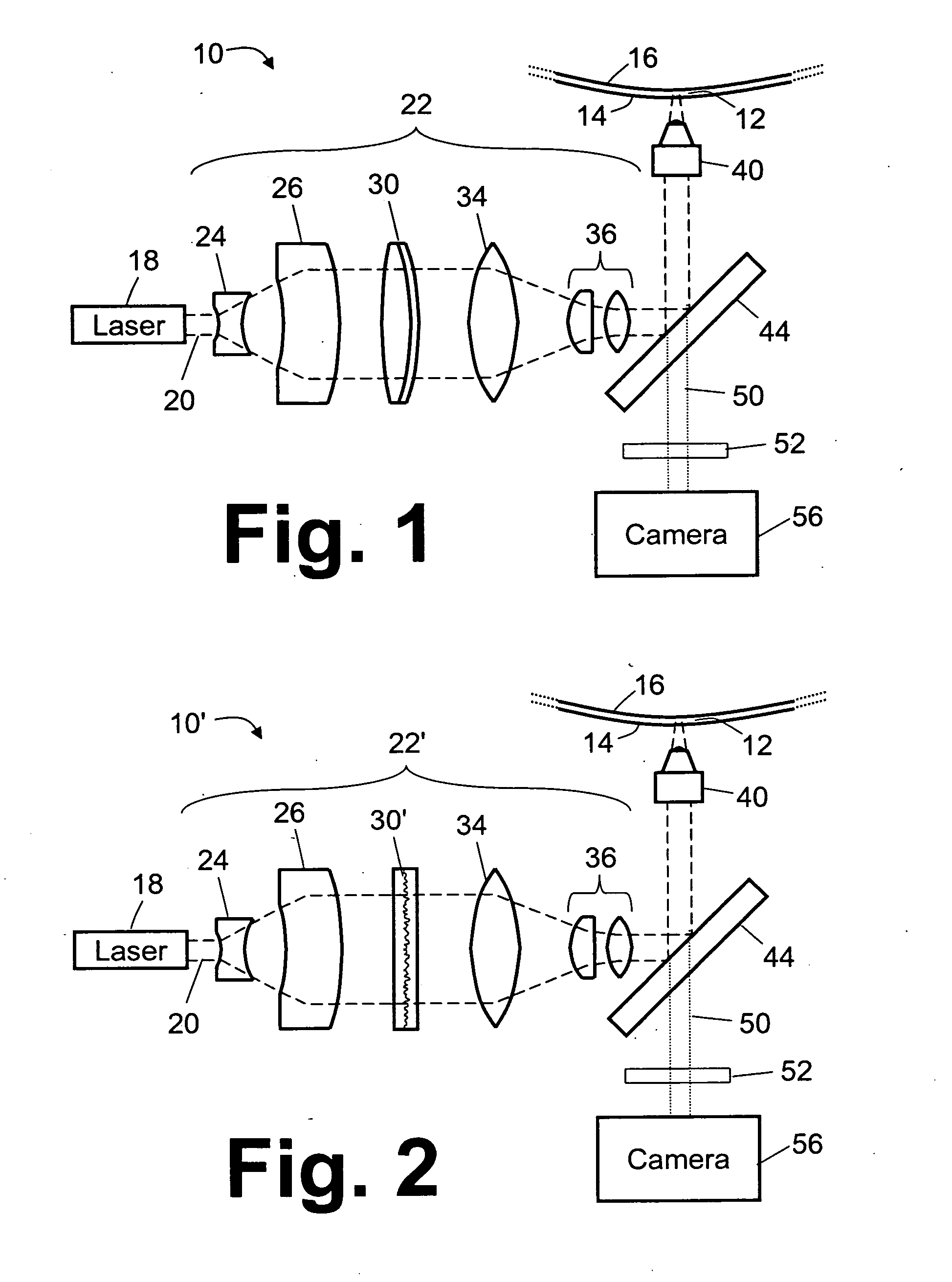
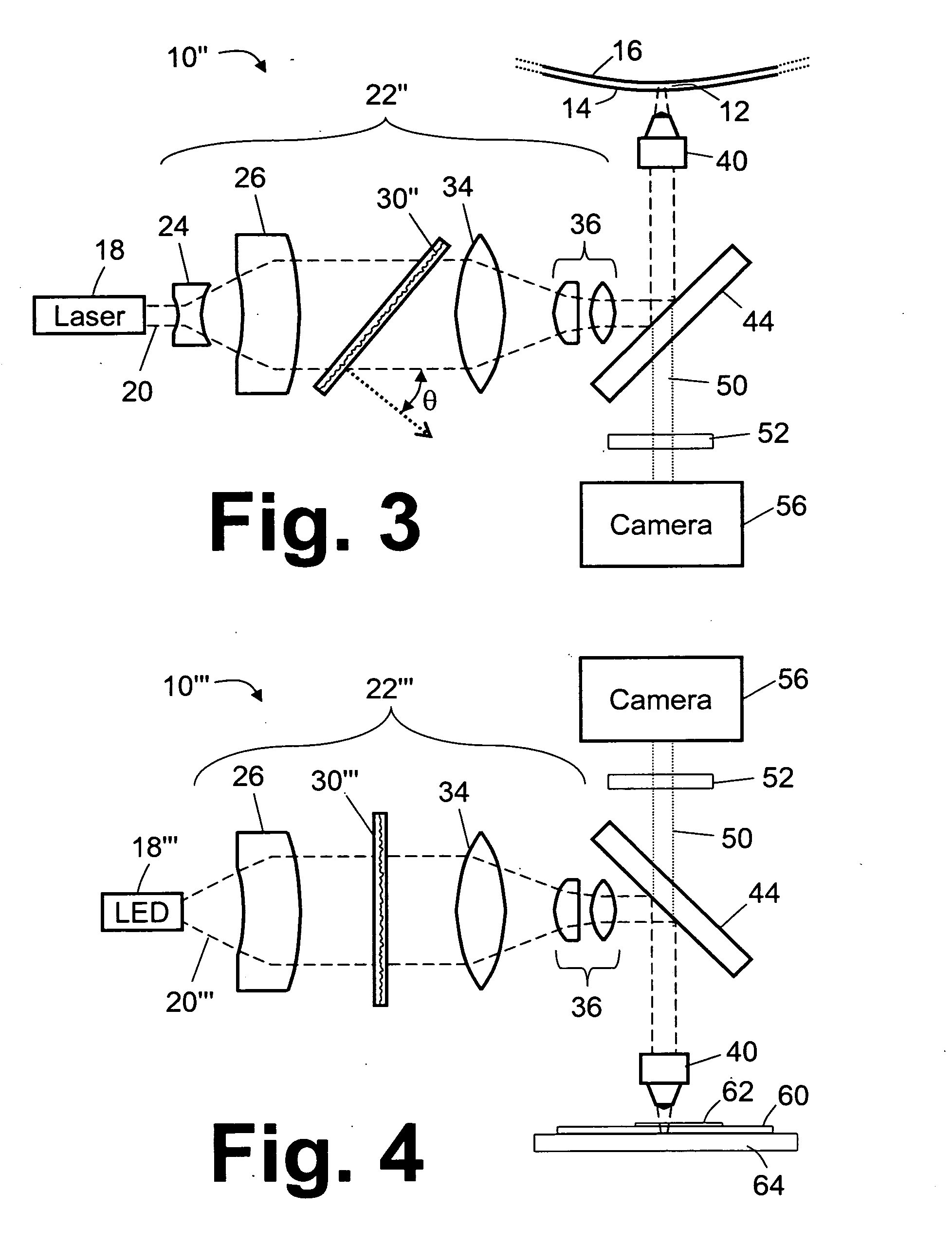
[0038] With reference to FIG. 1, a microscope system 10 images a microscope field of view coinciding with a buffy coat sample disposed in a generally planar portion of an annular gap 12 between a light-transmissive test tube wall 14 and a float wall 16 of a float disposed in the test tube. Suitable methods and apparatuses for acquiring and preparing such buffy coat samples are disclosed, for example, in U.S. Publ. Appl. No. 2004 / 0067162 A1 and U.S. Publ. Appl. No. 2004 / 0067536 A1.

[0039] The microscope field of view is generally planar in spite of the curvatures of the test tube and the float, because the microscope field of view is typically much smaller in size than the radii of curvature of the test tube wall 14 and the float wall 16. Although the field of view is substantially planar, the buffy coat sample disposed between the light-transmissive test tube wall 14 and the float wall 16 may have a thickness that is substantially greater than the depth of view of the microscope sys...


PUM


Abstract

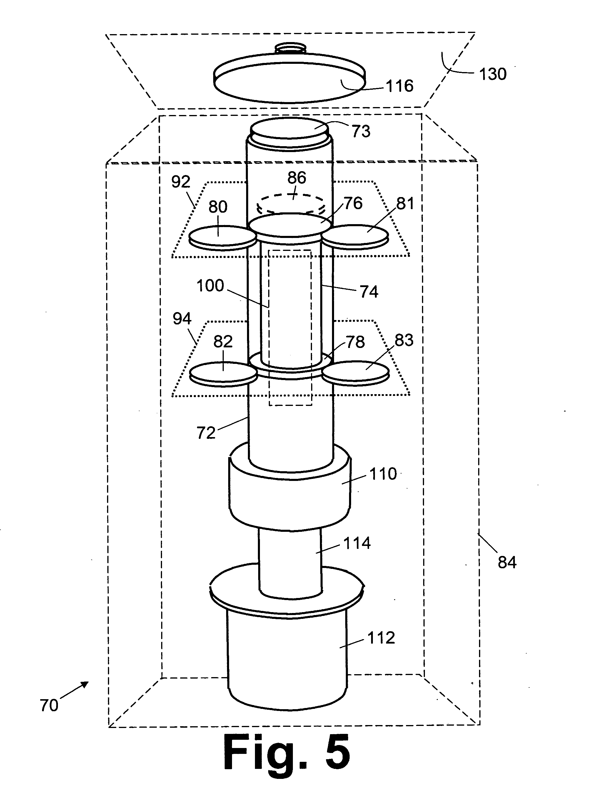
Fluorescent dye-tagged rare cells are disposed in a biological fluid layer contained in an annular gap (12) between an inside test tube wall (14) and a float wall (16). One or more analysis images are acquired at one or more focus depths using a microscope system (10, 10′, 10″, 10′″). Each acquired analysis image is image processed each to identify candidate rare cell images. The test tube (72) is selectively rotated. The test tube and the microscope system are selectively relatively translated along a test tube axis (75). The acquiring and image processing are repeated for a plurality of fields of view accessed by the selective rotating and selective relative translating and substantially spanning the annular gap.

Description

[0001] The present application is a divisional application of U.S. patent application Ser. No. 11 / 261,105, filed Oct. 27, 2005. [0002] This application claims the benefit of U.S. Provisional Application No. 60 / 631,025, filed Nov. 24, 2004, which is incorporated by reference herein in its entirety. This application also claims the benefit of U.S. Provisional Application No. 60 / 631,026, filed Nov. 24, 2004, which is incorporated by reference herein in its entirety. This application also claims the benefit of U.S. Provisional Application No. 60 / 631,027, filed Nov. 24, 2004, which is incorporated by reference herein in its entirety.BACKGROUND [0003] The following relates to the biological assay arts. It is described with particular reference to example embodiments that relate to detection of rare cells, such as epithelial cells, in the buffy coat of a centrifuged blood sample. However, the following relates more generally to identifying fluorescent dye-tagged cells in a biological fluid...

Claims


Application Information

Patent Timeline
Patent Type & Authority Applications(United States)
IPC IPC(8): C12Q1/00G06T9/00
CPCB01L9/06B01L2200/025G01N33/5094G02B21/002G02B21/06G02B21/248G02B21/365G02B27/095G02B27/48
Inventor WELLER, ALBERT E. III
Owner BATTELLE MEMORIAL INST
Learn More