
Devices for component removal during blood collection, and uses thereof

a technology for blood collection and components, applied in the field of blood collection devices and methods, can solve the problems of insufficient specificity of clean-up methods, increased processing errors, and increased risk of operator exposure to potentially infectious blood components

Active Publication Date: 2007-01-25
BECTON DICKINSON & CO
View PDF25 Cites 49 Cited by
  • Summary
  • Abstract
  • Description
  • Claims
  • Application Information

AI Technical Summary

Benefits of technology

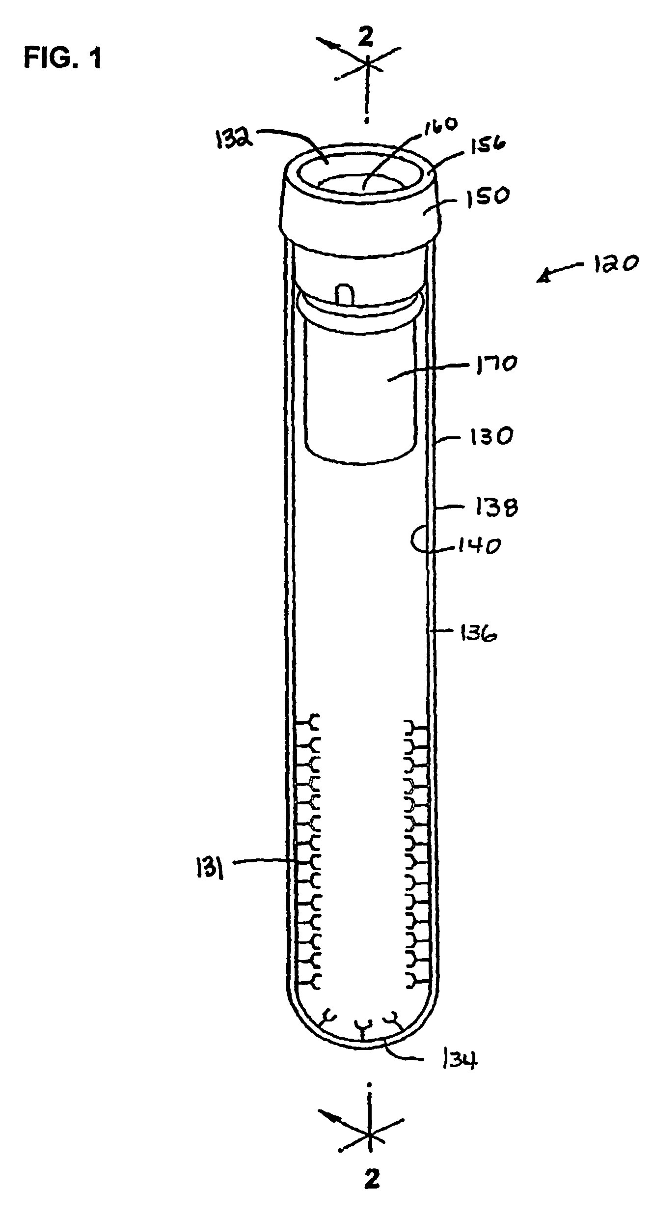
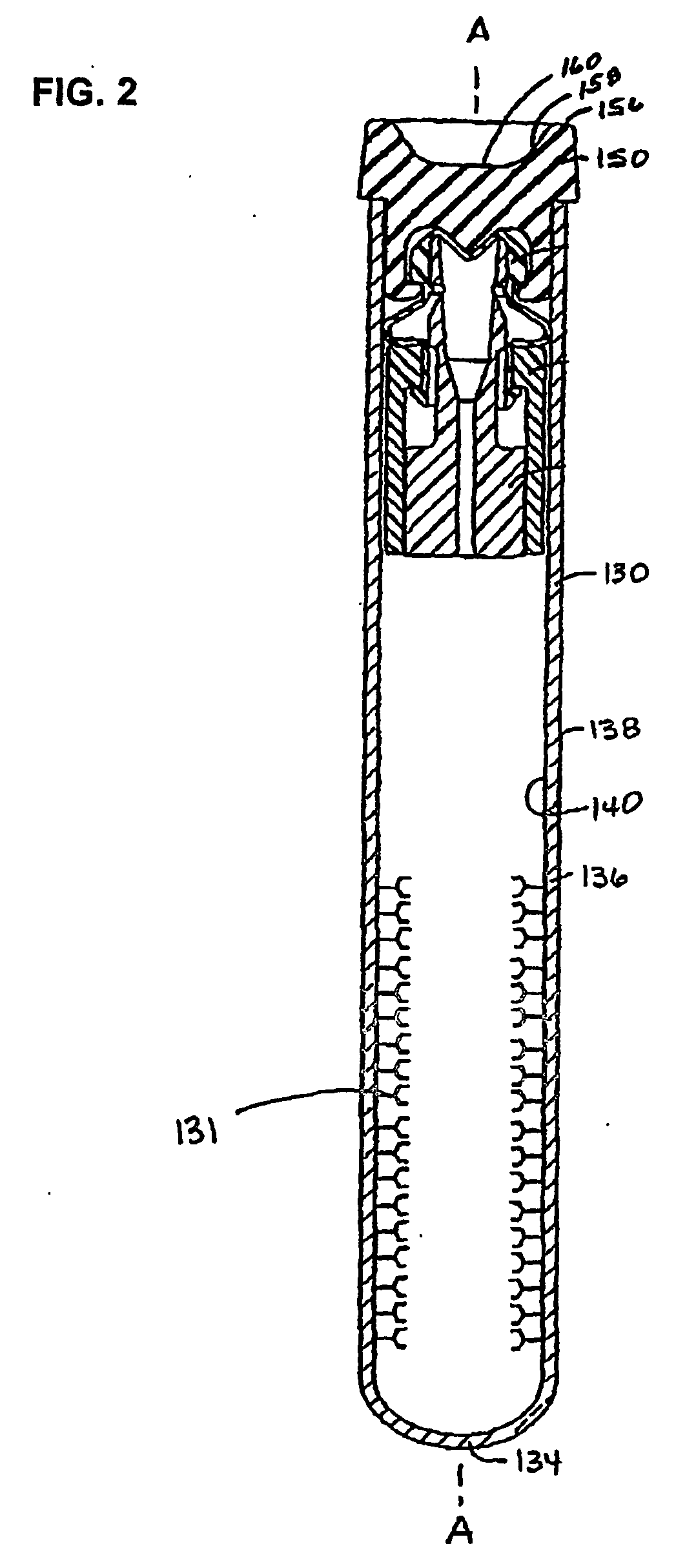
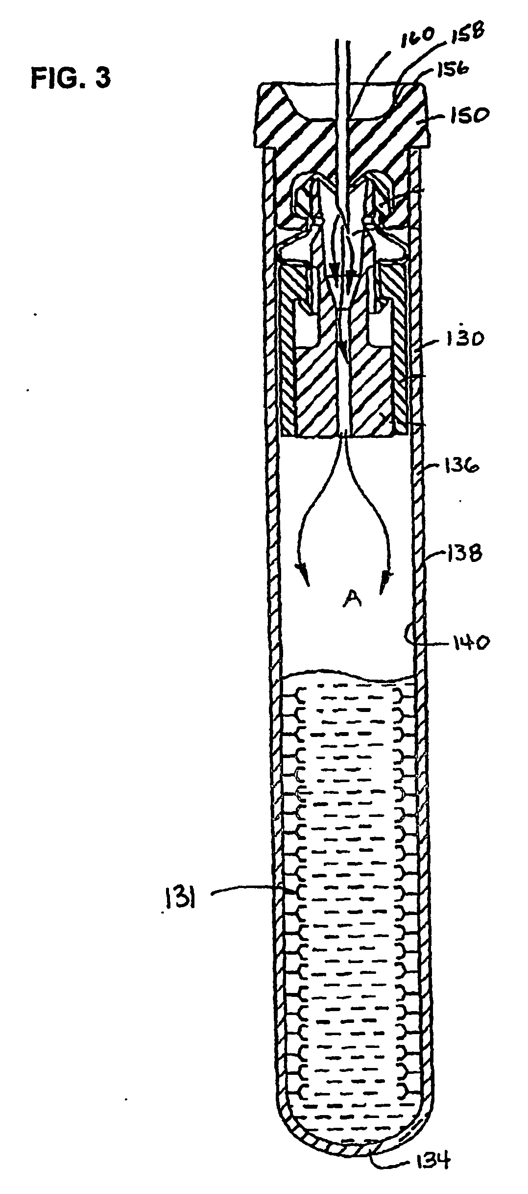
[0056] The present invention provides distinct advantages over current methods, these advantages being achieved by exposing the affinity molecules to the blood during and / or immediately after the blood collection process. As used herein, “during the time of collection” is used to mean that the affinity molecules are exposed to the blood during the blood collection process. The exposure of the blood to the affinity molecules need not be brief or instantaneous in time, so long as the exposure of the blood to the affinity molecules occurs prior to any subsequent processing or analysis of the blood sample. Thus, the affinity molecules must be situated in such a manner as to come into contact with the blood during the time of collection. As such, the affinity molecules may be situated to where they come into contact with the blood either before or after the blood collects in the reservoir, so long as the affinity molecules contact the blood during the time of collection. The purpose of contacting the affinity molecules with the blood during the time of collection is to reduce the number of handling and processing steps.

Problems solved by technology

However, plasma proteins or other components present in high concentrations, for example albumin, can hinder the separation and obscure the identification of these lower abundance proteins and, at times, make it difficult or impossible to determine their identity on 2D gels or by any other downstream testing of blood samples.
Currently, many different methods exist to remove albumin and other high abundance components from blood samples; however, most of these clean-up methods are not specific enough and may actually remove many of the desirable proteins or components.
Current albumin clean-up methods, for example, require transferring the sample into another container, thus requiring additional handling steps, and increasing the risk of processing errors, sample contamination, and extra risk of operator exposure to possibly infectious blood components.
Furthermore, current methods do not use any kind of specifically directed affinity molecule targeting the target components in the clean-up process, and may thus remove components of interest.

Method used


Image

  • Devices for component removal during blood collection, and uses thereof
  • Devices for component removal during blood collection, and uses thereof
  • Devices for component removal during blood collection, and uses thereof

Examples


example 1

Coating Evacuated Polystyrene Tubes with High Affinity Antibodies

[0066] High affinity antibodies (˜10−8 M-˜10−12 M) directed toward, for example, albumin (“albumin antibodies”) can be purchased from various suppliers. Polystyrene tubes are sterilized, and the albumin antibodies are covalently linked to the tubes as outlined in Tijssen, P., Practice and Theory of Enzyme ImmunoAssays, in Burdon, R H and Knippenberg, P H, (Eds.), Laboratory Techniques in Biochemistry and Molecular Biology, Elsevier Science Publishers, Vol. 15: 297-329 (1985), the entirety of which is hereby incorporated by reference. Briefly, the tubes are pretreated with 0.2% (v / v) glutaraldehyde in 100 mM sodium phosphate buffer, pH 5.0, for 4 hours at room temperature. The tubes are then washed twice with the same buffer and an antibody solution, which is prepared by adding 2-10 ug / mL in 100 mM sodium phosphate buffer, pH 8.0, is poured into the tubes and allowed to incubate for 3 hours at 37° C. After incubation, ...

example 2

Collection of Blood In Coated Tubes

[0067] Blood is drawn from a patient and collected in the coated evacuated tubes as prepared in Example 1. The blood is allowed to incubate in the tube at room temperature for approximately 5-90 minutes (depending on the affinity and concentration the antibody used in coating the tubes), while the tubes are gently rocked or otherwise mixed to allow binding of albumin to the antibodies on the sides of the tubes. The tubes can be incubated overnight at room temperature.

example 3

Testing the Ability of the Coated Tubes to Remove Albumin or Other Interfering Proteins

[0068] The blood collected in Example 2 is then centrifuged at 1100 G for about 10 minutes to separate the components. After component separation, the proteins in the albumin-depleted plasma is subjected to 2D gel, as described in Celis, J E, and Bravo, R, (Eds.), Two-Dimensional gel Electrophoresis of Proteins, Academic Press (1984), the entirety of which is hereby incorporated by reference. 2D gel electrophoresis will separate the remaining proteins in the sample, based on molecular weight and isoelectric point. The separated proteins are then isolated and purified using the Montage In-Gel Digest96 Kit™ (Millipore Corp.) according to the manufacturer's protocol. Proteins of interest are identified using Matrix-assisted laser desorption / ionisation-time of flight mass spectrometry (MALDI-TOF MS) on an Axima CFR MALDI-TOF Mass Spectrometer (Kratos Analytical, Inc.), as described in Worrall, T A, e...


PUM

PropertyMeasurementUnit
Timeaaaaaaaaaa
Affinityaaaaaaaaaa

Abstract

The invention relates to a device and method for collecting blood whereby certain target components are isolated or removed from the blood sample at the time of collecting the blood, as well as methods for using such devices.

Description

BACKGROUND OF THE INVENTION [0001] 1. Field of the Invention [0002] The present invention relates to devices and methods for collecting blood whereby certain target components are isolated or removed from the blood sample at the time of collecting the blood, as well as methods of using such devices. [0003] 2. Background of the Invention [0004] Today with the growth of proteomics, large-scale isolation and identification of proteins is becoming the focus of research, drug development and diagnostics. Protein separation methods are well-established in the molecular and biochemical arts; however, with the growth of proteomics, new technologies have arisen which require different, more stringent protein separation techniques. For example, exploring the plasma proteome requires the separation and identification of low abundance proteins and the determination of their characteristics using electrophoresis in a 2-dimensional (2D) gel system. However, plasma proteins or other components pre...

Claims


Application Information

Patent Timeline
IPC IPC(8): C12Q1/68G01N33/53C12M1/34B01L3/14G01N33/543
CPCB01L3/50215B01L3/5082B01L2200/16Y10T436/25375G01N33/5306G01N33/54346G01N33/54386G01N33/5304G01N33/543
Inventor ROSS, JULIEHAYWOOD, BRUCE
Owner BECTON DICKINSON & CO
Learn More