
Methods of using bone morphogenic proteins as biomarkers for determining cartilage degeneration and aging

a technology of bone morphogenic proteins and biomarkers, which is applied in the direction of biochemistry apparatus and processes, instruments, material analysis, etc., can solve the problems of increased susceptibility to disease and injury, decreased anti-inflammatory agent dose in patients, and gradual decline in function

Inactive Publication Date: 2007-01-18
RUSH PRESBYTERIAL ST LUKES MEDICAL CENT +1
View PDF6 Cites 7 Cited by
  • Summary
  • Abstract
  • Description
  • Claims
  • Application Information

AI Technical Summary

Benefits of technology

[0023] In another aspect, the invention provides methods for determining the effective dose of an anti-inflammatory agent in a subject by administering to a subject a dose of an anti-inflammatory agent, obtaining a tissue, body fluid or cell sample from the subject, determining OP-1 protein concentration or OP-1 mRNA concentration in the sample, determining the concentration of protein or mRNA encoded by a second gene whose expres

Problems solved by technology

However, in general, as any biological tissue or organ ages, function gradually declines and susceptibility to disease and injury increases (Buckwalter et al.
These methods, however, possess disadvantages in that blood rheumatoid factor is not specific to patients with RA.
Rheumatoid factor assay kits based on such methods also have poor accuracy and reproducibility.
Assays for erythrocyte sedimentation rate (ESR) or C-reactive protein (CRP) are useful for determining the activity of inflammatory diseases such as RA but are not suitable for use in their diagnosis.
Detection of anti-nuclear antibodies or LE cells may be used to detect RA but it is difficult to accurately diagnose RA because these methods are not specific for RA and because such antibodies or cells are frequently detectable in other collagen diseases.
In addition, these methods do not specifically detect the cartilage degradation which is associated with RA.

Method used


Image

  • Methods of using bone morphogenic proteins as biomarkers for determining cartilage degeneration and aging
  • Methods of using bone morphogenic proteins as biomarkers for determining cartilage degeneration and aging
  • Methods of using bone morphogenic proteins as biomarkers for determining cartilage degeneration and aging

Examples


example 1

OP-1 Protein and mRNA Levels Decrease with Increased Age

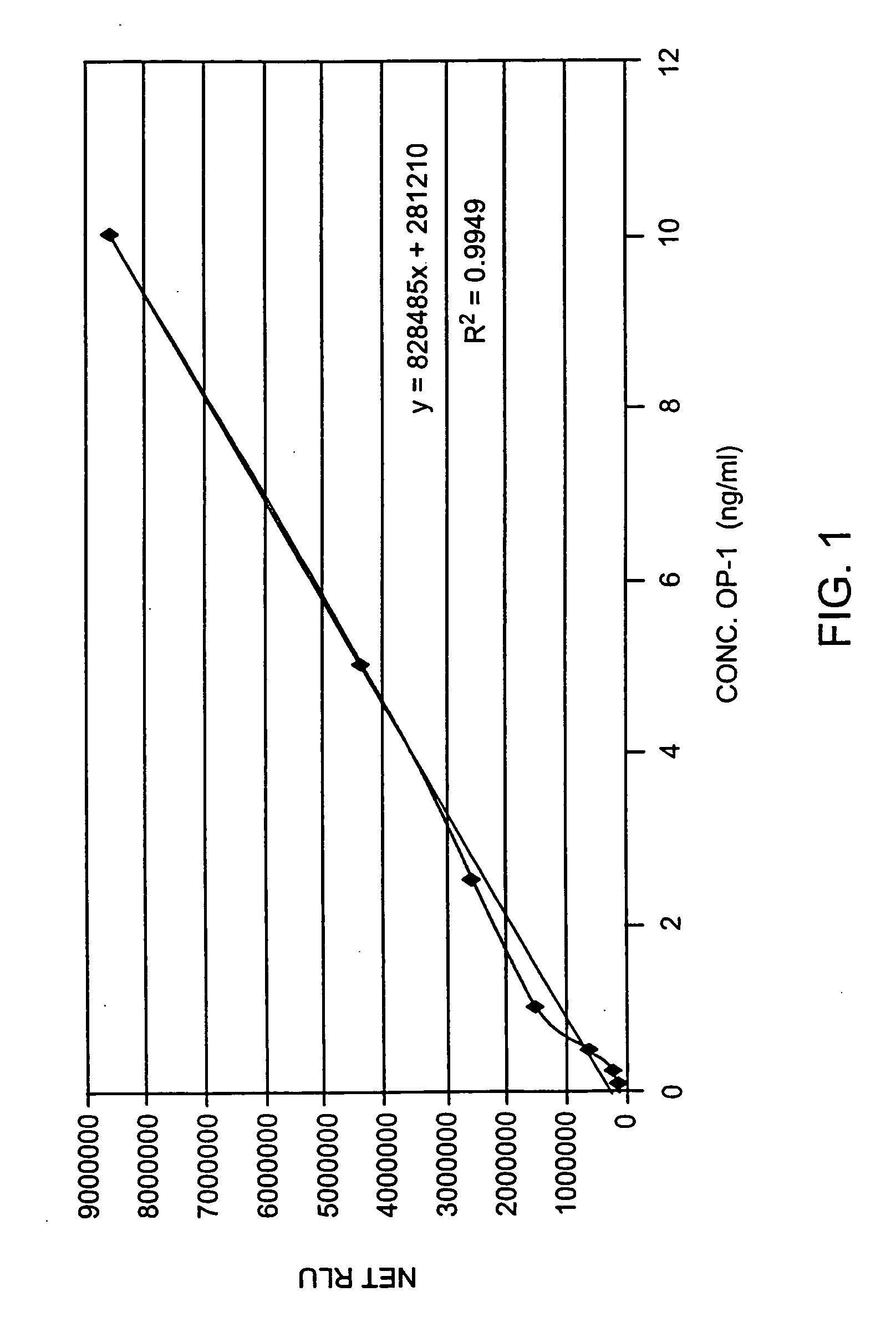
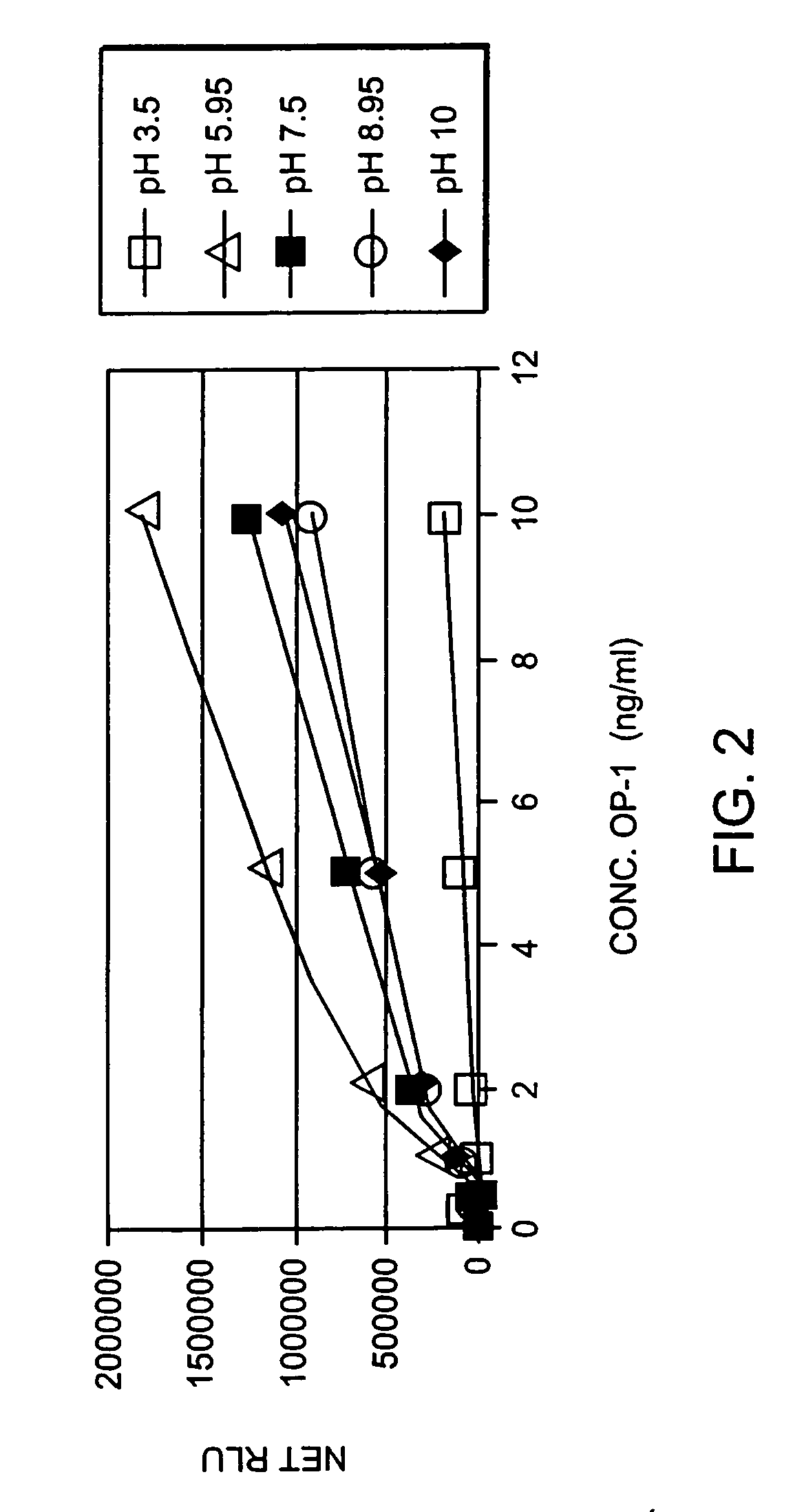
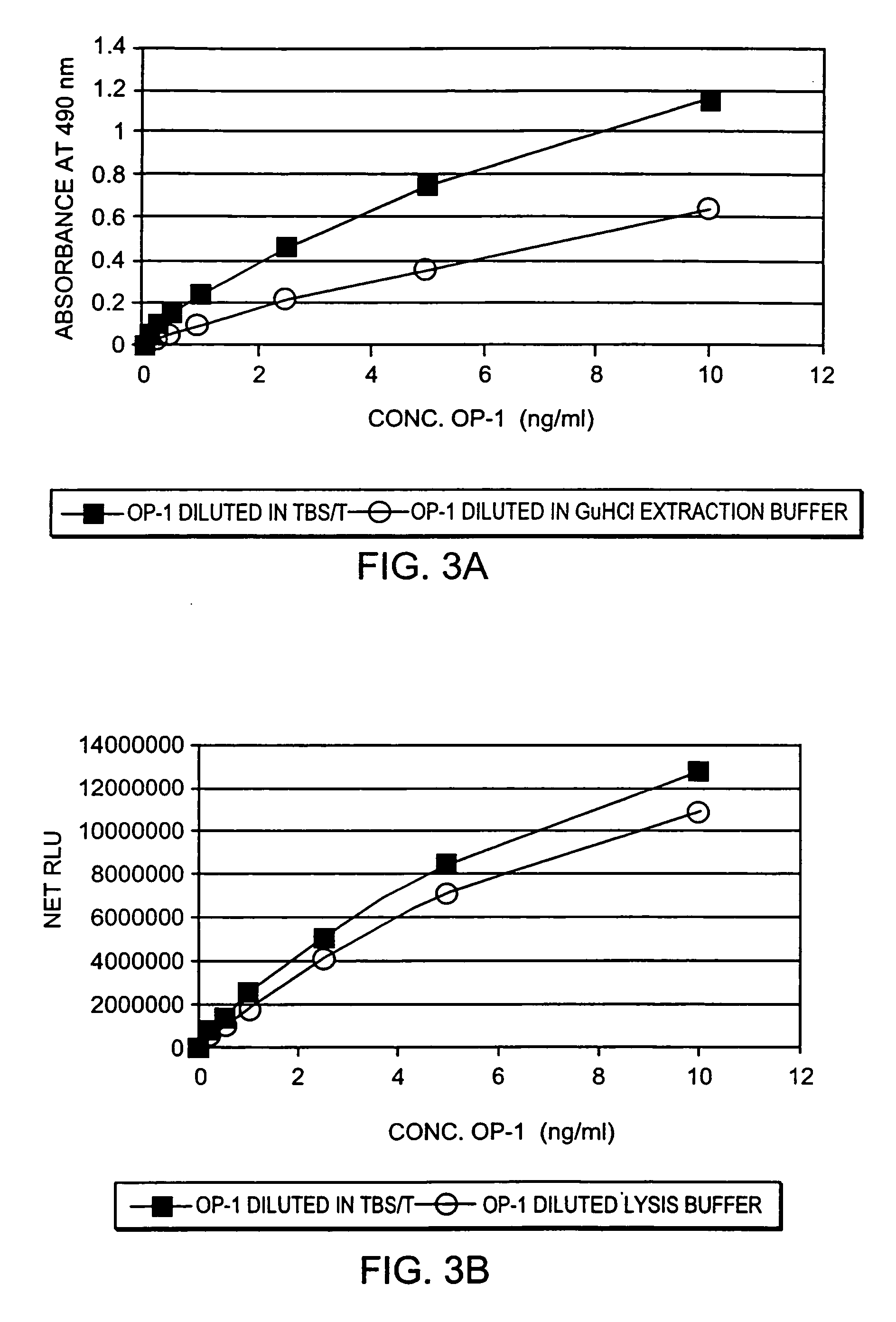
[0060] The changes in endogenous OP-1 (protein and mRNA) expression with aging of human articular cartilage were studied. In order to assess quantitatively the concentration of total endogenous OP-1 protein in cartilage extracts, a sandwich enzyme-linked immunosorbent assay (ELISA) was developed and compared with Western Blot and reverse transcription polymerase chain reaction (RT-PCR) measurements. Results indicate that there is a correlation between a decrease in total and mature OP-1 protein and OP-1 mRNA with increased age.

Materials and Methods

Reagents

[0061] Human recombinant pro- and mature-OP-1, BMP-6, anti-pro (R2854) and anti-mature (1B12) OP-1 antibodies were obtained from Stryker Biotech (Hopkinton, Mass.). Two other, anti-OP-1 antibodies (#SC-9305 and #MAB354) were purchased from Santa Cruz Biotechnology, Inc. (Santa Cruz, Calif.) and R&D Systems (Minneapolis, Minn.), respectively. Electrophoresis grade reagent...

example 2

OP-1 Protein and mRNA Levels in Rheumatoid Arthritis and Osteoarthritis

[0077] The above-described OP-1 sandwich ELISA was used to determine whether OP-1 protein could be detected in synovial fluid, whether quantitative approaches could be adapted for the assessment of OP-1 protein in synovial fluid, and whether there are differences in the levels of OP-1 protein between normal donors and patients with rheumatoid arthritis (RA) and osteoarthritis (OA). The results suggest that synovial fluid OP-1 is a useful diagnostic and prognostic marker for both RA and OA.

[0078] Synovial fluid was aspirated from subjects with RA and OA as well as from normal joints of human organ donors according to standard methods. Cartilage specimens from 74 joints (13 normal, 25 RA, 29 OA and 7 other inflammatory diseases) were also obtained. Synovial fluid and cartilage was analyzed by Western Blot with anti-pro and anti-mature OP-1 antibodies and the concentration of OP-1 protein was measured using the OP...

example 3

OP-1 Protein and mRNA Levels Decrease in Osteoarthritis

[0086] Human normal cartilage derived from normal newborn and normal adult donors with no documented history of joint disease were obtained according to standard procedures. Osteoarthritis cartilages (OA) were removed from patients diagnosed with OA who underwent knee arthroplasty. Three samples of each type were tested. RT-PCR of OP-1 and GADPH mRNA was performed as described in Example 1. Levels of OP-1 mRNA in normal newborn and normal adult cartilage were similar, whereas OP-1 mRNA expression in OA cartilage was up-regulated two to three-fold (Table 2).

TABLE 2OP-1 mRNA Expression In Human Articular CartilageType of CartilageOP-1 / GADPH# specimensNormal newborn cartilage0.518 ± 0.066N = 3Normal adult cartilage0.567 ± 0.067N = 3OA cartilage1.148 ± 0.234N = 3P

[0087] OP-1 protein was extracted from tissues with 1 M GuHCl in the presence of protease inhibitors, lyophilized, and analyzed by Western Blot under non-reduced condit...


PUM


Abstract

Methods are provided for determining cartilage degeneration, regeneration, or aging in a joint tissue in a patient by measuring levels of osteogenic protein-1 (OP-1) protein and / or mRNA in synovial fluid or joint tissue. The methods according to the invention are useful for detecting, diagnosing, predicting, determining a predisposition for, or monitoring joint tissue degeneration, regeneration, or aging in a patient including inflammatory joint disease or age-related disorders.

Description

RELATED APPLICATIONS [0001] This application is related to, and claims the benefit of U.S. Ser. No. 60 / 348,111, filed Nov. 9, 2001 and U.S. Ser. No. 60 / 270,528, filed Feb. 21, 2001, the contents of which are herein incorporated by reference.FIELD OF THE INVENTION [0002] The invention relates generally to uses of OP-1 and other bone morphogenic proteins as biomarkers of tissue integrity or deterioration, and more particularly to methods for diagnosing and / or monitoring cartilage degeneration associated with inflammatory disease and age. BACKGROUND OF THE INVENTION [0003] A number of factors can cause or contribute to cartilage degeneration in mammals, including trauma and inflammatory disease. Damage to cells resulting from the effects of inflammatory response has been implicated as the cause of reduced cartilage function or loss of cartilage function in diseases of the joints (e.g., rheumatoid arthritis (RA) and osteoarthritis (OA)). In addition, autoimmune diseases such as systemic...

Claims


Application Information

Patent Timeline
IPC IPC(8): C12Q1/68G01N33/50G01N33/543G01N33/53G01N33/564G01N33/68
CPCG01N33/564G01N2800/105G01N2333/51G01N33/6893
Inventor CHUBINSKAYA, SUSANNARUEGER, DAVID C.KUETTNER, KLAUS E.
Owner RUSH PRESBYTERIAL ST LUKES MEDICAL CENT
Learn More