
Interventional medical closure device

a medical closure and medical technology, applied in the field of interventional medical closure devices, can solve the problems of prolonging the time, affecting the healing effect of patients, and affecting the healing effect of patients, and achieve the effect of reducing the trauma caused upon creation and being simple yet effectiv

Active Publication Date: 2006-12-21
VIVASURE MEDICAL LTD
View PDF36 Cites 80 Cited by
  • Summary
  • Abstract
  • Description
  • Claims
  • Application Information

AI Technical Summary

Benefits of technology

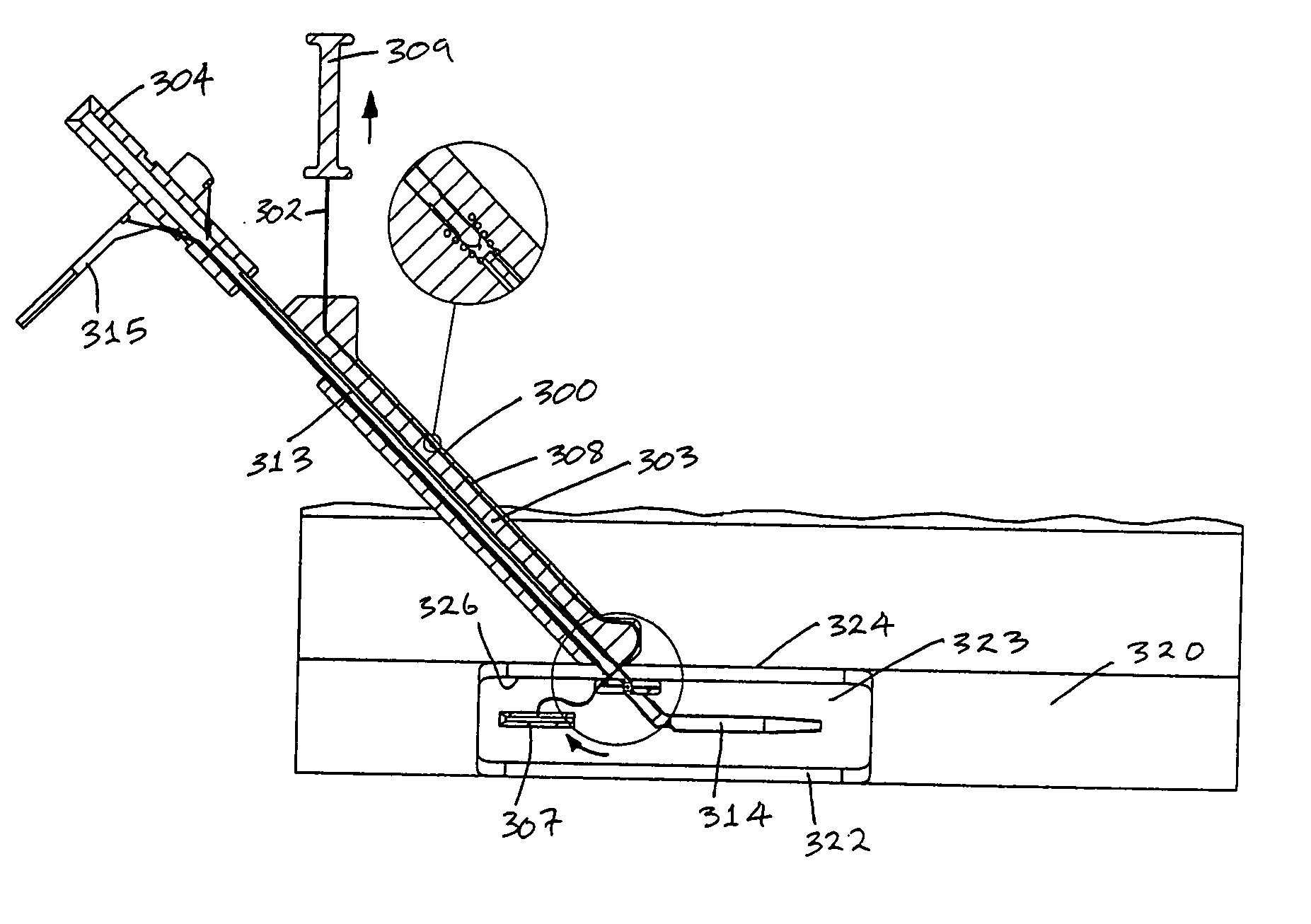
[0029] moving the closure element through a first part of the tissue wall on a first side of the opening, across the opening, and through a second part of the tissue wall on a second side of the opening to assist in closure of the opening.
[0132] During the vessel entry procedure, a trapping element may be deployed in the vessel to secure both leaves of the incision. This effectively, at the beginning of the procedure, provides the mechanism for subsequently closing the vessel wall opening at the end of the procedure, without having to use another closure device. This contrasts with conventional devices which address the problem of the puncture wound, created by the blunt introducer / dilator combination, at the end of the procedure.

Problems solved by technology

The time required for the manual compression may be long and this may lead to considerable patient discomfort and extend the time to ambulation and the duration of stay in the hospital.
This may increase hospital staff time and the cost of health care for the patient.
Complications may arise with compressive techniques, as too much pressure may restrict or occlude the blood vessel, potentially leading to ischemia and thrombus formation in the vessel.
These techniques can have unpredictable post procedural haemorrhaging requiring additional interventions by the doctors and nurses.
However these vascular sealing devices suffer from a number of disadvantages, such as:
These devices may lead to puckering of the vessel wall after deployment, leading to the risk of thrombosis and vessel lumen narrowing;
Considerable patient discomfort may be caused during the deployment of the devices;
It may be difficult to effect good closure if the device is poorly placed and it restricts access options for repairs;
There is a risk of embolisation of the gel or foam material in the blood stream due to inaccurate deployment or migration post delivery to the tissue tract;
Ineffective closure of the puncture wound in the blood vessel may occur as the material is deployed in the tissue tract with an increased risk of haemotoma;
These devices are often designed to leave materials on the inside of the blood vessel, thereby increasing the risk of blood flow restriction or occlusion;
A disk may detach from an anchor element and travel distal to the wound site and cause blood flow restrictions or occlusions, potentially requiring surgical intervention;
There is a risk of haematoma forming in the tissue tract;
There is a risk of haemorrhage due to continued patency of the puncture in the vessel wall;
These devices may require significant manual compression before and during use.

Method used


Image

  • Interventional medical closure device
  • Interventional medical closure device
  • Interventional medical closure device

Examples


Embodiment Construction

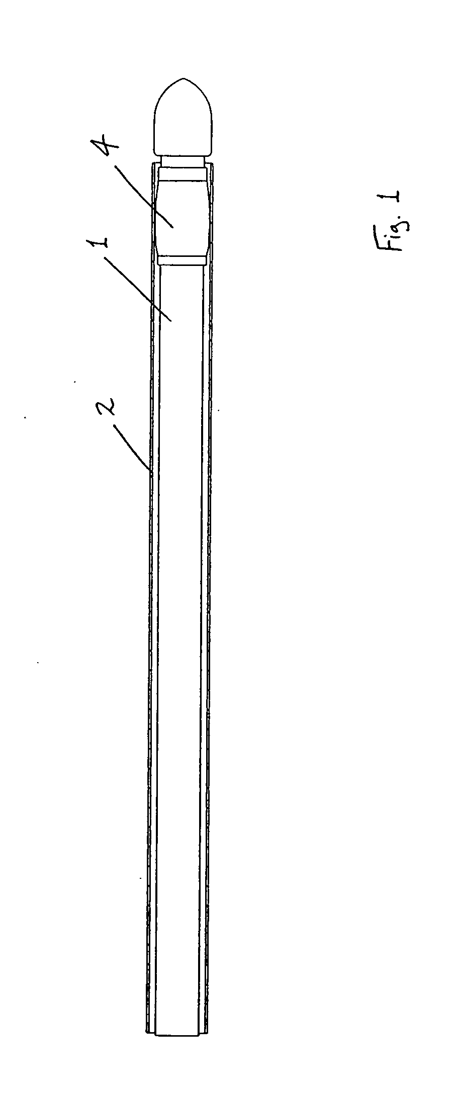
[0183] Referring to the drawings and initially to FIGS. 1 to 35(a) thereof, there is illustrated a medical device according to the invention, which is suitable for use in accessing an interior of a body part. The device is particularly suitable for accessing the interior of a blood vessel 6. The medical device comprises an elongate engagement element 1, a protective sheath 2, as illustrated in FIGS. 1 to 5, and an incising element 3, as illustrated in FIGS. 6 to 9.

[0184] The engagement element 1 comprises a retaining element, which is provided in this case as a balloon part 4, which is expandable, in this case inflatable, from a low-profile delivery configuration (FIGS. 1 and 2) to a deployed configuration (FIGS. 3 and 4), and is returnable, in this case deflatable, from the deployed configuration to the delivery configuration (FIG. 5). In the delivery configuration, the engagement element 1 is piercable through a flap 7 of tissue wall and through a wall 8 of the blood vessel 6. In...


PUM


Abstract

An interventional medical closure device is suitable for assisting closure of an opening through a blood vessel wall after completion of an interventional procedure within the internal lumen of the blood vessel. The device comprises a closure element, a grasping element for grasping the closure element, and a delivery element for delivering the closure element into the internal lumen of the blood vessel. The closure element comprises a suture, and an engagement foot at a distal end of the suture. The grasping element is inter-engageable with the closure element to grasp the closure element. The delivery element comprises a main body portion, and a distal nose. The distal nose defines a reception space for carrying the engagment foot of the closure element. The delivery element also comprises an engagement foot which is movable relative to the main body portion between a low-profile delivery configuration and a protruding engagement configuration.

Description

RELATED APPLICATION [0001] This application claims the benefit of U.S. Provisional Patent Application Ser. No. 60 / 676,279, filed Apr. 29, 2005, the entire content of which are incorporated herein by reference.FIELD OF INVENTION [0002] This invention relates to an interventional medical closure device, and to a method of performing an interventional procedure. This invention also relates to a medical device suitable for use in accessing an interior of a body part, and to a method of accessing an interior of a body part. In one embodiment this invention relates to a vessel access and closure device for making and closing access sites in human and animal tissue, and to a related method. A particular example of the invention is a vascular lumen access and closure. In further detail, this invention generally relates, in one example, to the field of minimally invasive vascular catheterisation, and more specifically to vascular vessel access and closure methods and devices which are used t...

Claims


Application Information

Patent Timeline
IPC IPC(8): A61B17/08
CPCA61B17/0057A61B2017/22048A61B17/0467A61B17/32053A61B2017/00004A61B2017/00336A61B2017/00349A61B2017/00637A61B2017/00654A61B2017/00659A61B2017/0406A61B2017/0458A61B2017/0464A61B2017/22038A61B17/0401
Inventor BRETT, GERARDRONAN, DAVIDMULLOY, LIAMHORAN, STEVEN
Owner VIVASURE MEDICAL LTD
Learn More