
Method for pre treatment verification in radiation therapy

Active Publication Date: 2006-09-14
NILSSON GORGEN
View PDF3 Cites 39 Cited by
  • Summary
  • Abstract
  • Description
  • Claims
  • Application Information

AI Technical Summary

Benefits of technology

[0024] Thus, an object of the present invention is to provide an efficient pre-treatment measurement method that sufficiently and accurately verifies the dose distribution from a complete treatment fraction (all beam projections) to be delivered to a patient.
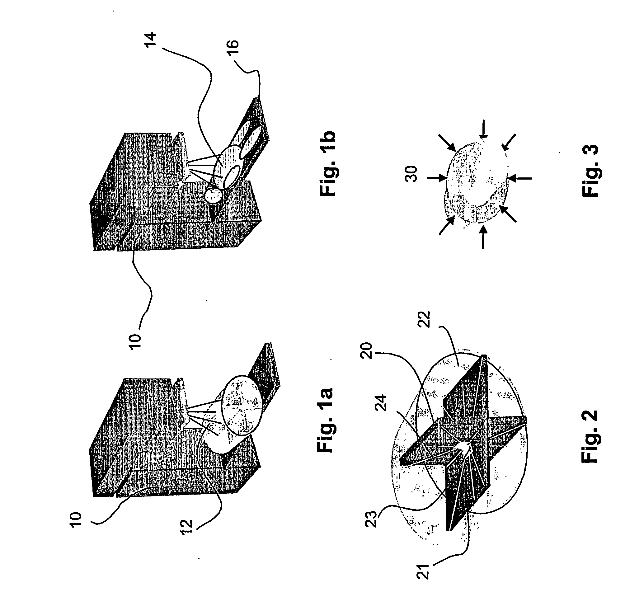
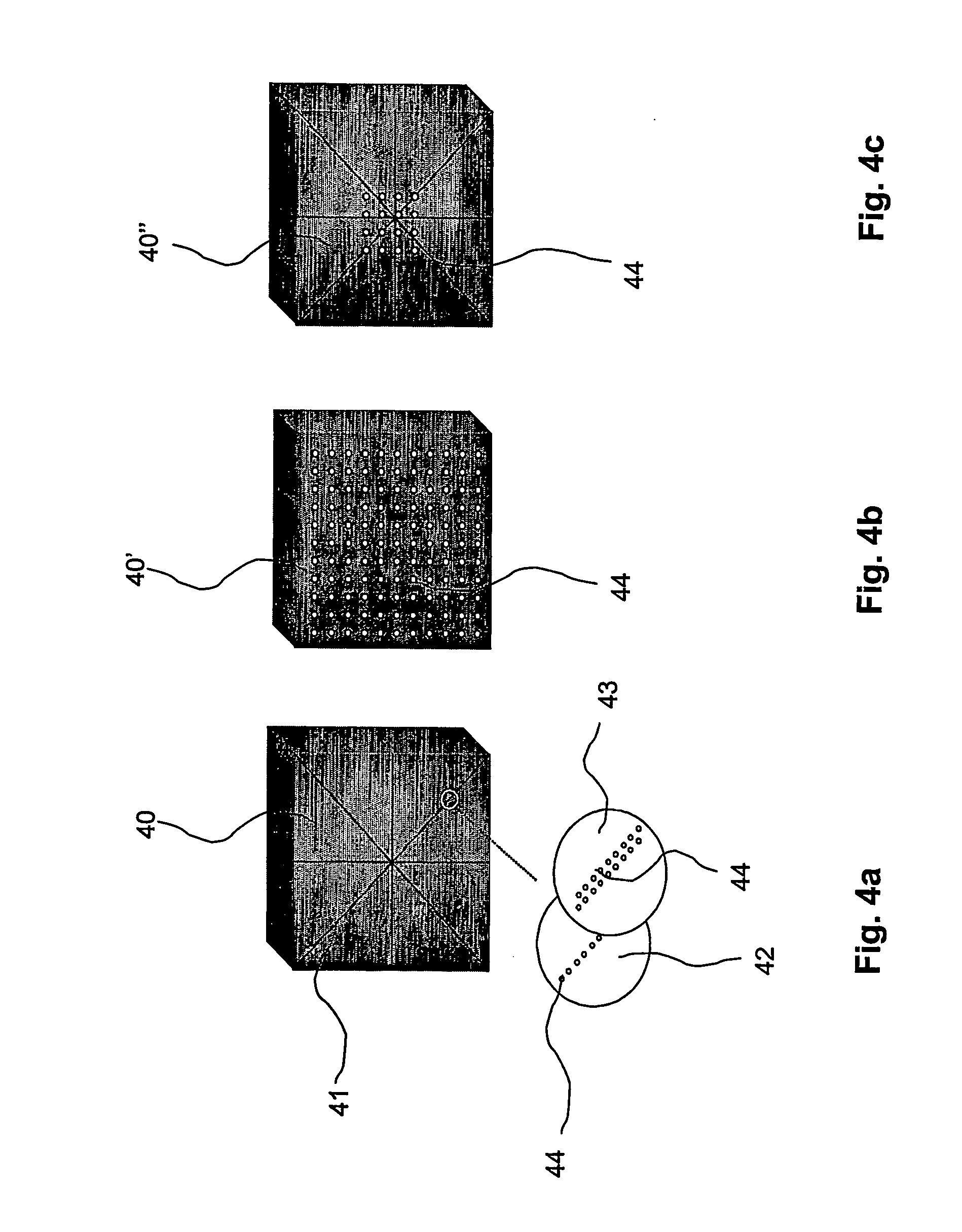
[0030] Thus, the invention is based on the idea of a configuration of diode detectors in two or more non-parallel planes in a phantom e.g. body-phantom (without connection to the gantry rotation, i.e. the rotation of the device applying the irradation), wherein the measurement is divided in time-intervals. The special configuration of the detectors makes it possible to verify the intensity modulation across the beam in any beam projection and at the same time totalise the dose from all projections in the fixed measurement-points in the phantom. In addition, the overall measurement accuracy is significantly improved by dividing the measurement in time-intervals. Obviously, this is clear advantages of the present invention compared to existing solutions. For example, a placement of the detectors in a 3D matrix would require an extensively increased number of detectors, which, in turn, would entail very high costs and which also would require significant processing times in order to process the information or data obtained from the detectors during the measurements. Furthermore, the division into time-interval enables use of individual correction factors for each time interval. In addition, this facilitates an evaluation of discrepancies in dynamic fields and / or ARC-therapy and to reduce the directional and / or energy dependency of the detectors.
[0034] According to another embodiment of the present invention, the measurements are synchronized with the respiratory cycle of the patient for which the patent specific treatment is intended. External signals obtained by means of, for example, an X-ray unit, which indicates the phase of the respiratory cycle, e.g. used to turn the irradiation beam on and off, is stored with each measurement. Thereby, the dose delivered in the various phases of the respiratory cycle can be determined. The measured dose per pulse can be grouped in various time-intervals due to the requirements at the specific set-up. Furthermore, the IMRT treatment technique increases the requirement on reproducible patient positioning. The synchronisation with the respiratory cycle, so called respiratory gating, decreases the irradiation of healthy tissue. That is, the margins that are added in order to ensure that the tumour without doubt is within the applied field can be reduced by using respiratory gating and thereby the proportion of irradiated healthy tissue can be decreased.
[0041] According to embodiments of the present invention, each detector plane may be provided with detectors having a thickness in a range less than the range of the electrons of the maximum energy in the range where the dependency is significant. (<200 keV corresponding to approximately 200 μm in silicon). Thereby, the energy and / or directional dependency of the detectors are reduced significantly, which mainly is caused by differences in the photons mass-attenuation in the detectors compared with the media they are arranged in.

Problems solved by technology

The major reasons for this have been the inability to control the primary tumour or the occurrence of metastases.
The treatment technique to deliver this optimised treatment is more complicated than conventional treatments because each field must be modulated laterally in intensity and thereby compensate for the patients contour and anatomic heterogeneity, the technique is called IMRT—Intensity Modulated Radiation Therapy.
However when using several projections (4-8), each projection requiring individual compensators, this technique is time-consuming and requires a lot of effort.
Additionally the attenuation of the beam in the filter causes unwanted change of the beams spectral distribution, thereby complicating the whole process.
Both methods have implementations using traditional measurement techniques and both of them have important limitations both in methodology and in measurement accuracy.
The shortcoming of the first method is: Complicated and time-consuming to verify each projection individually rather than the total contribution from all projections in one comparison.
Unnecessary efforts invested in correcting minor errors per projection that would have shown to be neglectable if all projections could be totalised.
It is not useful in ARC-therapy (described above).
Lack of time resolution in the measurements disables the possibility to analyse the course of a measured deviation, e.g. in sub fields or segments of a field without updated measurement.
Additionally there is no possibility to distinguish whether a dose is delivered when expected during the respiratory cycle.
However the requirement to measure with high accuracy outside the primary field, described above, raises several demands on the detectors and one of the hardest to fulfil for semiconductors is energy independency.
Error in delivery e.g. in MLC position, gantry angle, collimator rotation etc. will cause similar dose discrepancy in the phantom as in the patient.
However, across the field, where the beam is modulated, the method is limited to measure along the film (1D).
Additionally, film is an integrating detector and thereby the film-data has no time resolution and thereby analyses of the cause of a deviation between measurement and the treatment plan often become more or less impossible.
Ideal point detectors would measure the point-dose accurately, however a few point detectors will not enable verification of the intensity modulated beam in the various projections.
Ideal detectors do not exist and currently used measuring methods that have no time resolution and / or synchronisation or documentation to the treatment phase makes it impossible to apply relevant corrections to the measurement and thereby improve the result.
However, the spatial resolution of the ionisation chamber is rather limited, normally about 3-4 mm, which is a major limitation in the applications discussed herein.
Nearly 10 years ago scintillation detectors was proposed for radiation therapy, but is has however not been possible to make this technology work in practice.
Proposals using dummy fibre optics has been presented, but the underlying technical problems has not been possible to solve.
The major drawback with the disclosed MOSFET detector system is however the limited lifespan of the detectors, which is mainly caused by radiation damage.
Moreover, the absorbed dose in a MOSFET can be read directly or after use, but not in real time applications.
Both MOSFET detectors and ionisation chambers require a bias, which complicates a system with an extensive number of detectors.
The main limitation is the energy dependence and the long term stability even though the latter has been improved during the recent years.

Method used


Image

  • Method for pre treatment verification in radiation therapy
  • Method for pre treatment verification in radiation therapy
  • Method for pre treatment verification in radiation therapy

Examples


Embodiment Construction

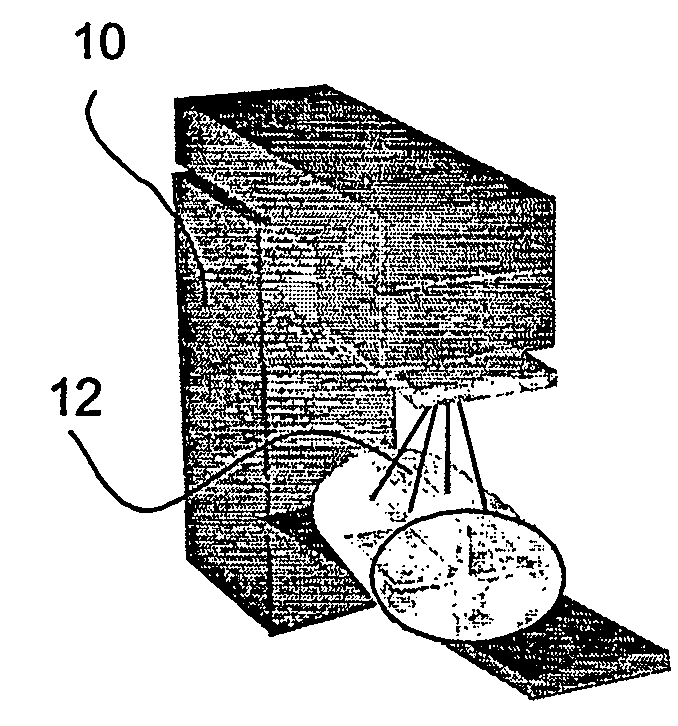
[0052] A radiotherapy device (gantry) utilised for treating tumours with radiation are shown schematically in FIGS. 1a and 1b and is generally denoted with reference numeral 10. The device comprises a radiotherapy system capable of emitting a beam 12 of electrons or photons from a treatment head. The radiotherapy system is provided with conventional field-shaping device (not shown), for example an MLC, for allowing the lateral shape of the beam to be altered so as to shield off non-affected areas of the body, intensity modulate the beam and concentrate the beam to the tumour. The radiotherapy system comprises control and measurement means (not shown) including processor and storage means, for example, a central processing unit CPU for performing the steps of the method according to the invention.

[0053] A treatment couch 16 is arranged for a patient 14 to lie on, see FIG. 1b. The couch can rotate around a vertical axis, and move horizontally, vertically and longitudinally in order t...


PUM


Abstract

The present invention relates to a method to measure dose distribution in a patient-shaped phantom with high accuracy. The invention consists of a method of measuring dose distribution in a phantom for radiation therapy treatment verification, a detector configuration in such a phantom, detector improvement and measurement methodology to enable application of correction factors in an accurate way.

Description

TECHNICAL FIELD [0001] The present invention relates generally to radiation therapy treatment verification. In particular the invention pertains to methods, a detector configurations, a detector and a computer readable medium for verifying that a patient specific cancer treatment using radiation therapy, and in particular intensity modulated radiation therapy, is delivered as planned. BACKGROUND OF THE INVENTION [0002] Radiotherapy has been used to treat cancer in the human body since early 1900. Even though radiation of cancer tumours is known to be efficient, mortality rate for many cancers remained virtually unchanged for a long time. The major reasons for this have been the inability to control the primary tumour or the occurrence of metastases. Only by improving the local control may the treatment be more effective. In the last years Treatment Planning Systems, TPS, in Radiation Therapy have developed extensively and is now able to take into account the anatomy of the specific ...

Claims


Application Information

Patent Timeline
IPC IPC(8): G01D18/00A61BA61B6/00A61NA61N5/10G01T1/16
CPCA61B6/542A61B6/583A61N5/1048A61N2005/1076
Inventor NILSSON, GORGEN
Owner NILSSON GORGEN
Learn More