
Perfusion and/or preservation solution for organs

a technology for organs and organs, applied in the field of organ preservation, perfusion and/or reperfusion, can solve the problems of tissue and cellular damage to other organs, affecting the survival of organs, and limiting the success of organ transplantation,

Inactive Publication Date: 2006-07-20
PHILADELPHIA COLLEGE OF OSTEOPATHIC MEDICINE
View PDF5 Cites 14 Cited by
  • Summary
  • Abstract
  • Description
  • Claims
  • Application Information

AI Technical Summary

Benefits of technology

[0008] The present invention provides a solution for preservation, perfusion, and / or reperfusion of an organ, especially the heart, containing peptide inhibitor(s) of protein kinase C βII (PKC βII) and / or of protein kinase C ζ (PKC ζ) and / or peptide activator(s) of protein kinase C δ (PKC δ). The solution protects organ tissues and cells from damage while the organ is isolated from the circulatory system or is experiencing decreased blood flow (ischemia). The present inventor has discovered that the peptide inhibitors of PKC βII and / or PKC ζ, and / or peptide activator(s) of PKC δ enhance NO release or inhibit endothelial / PMN superoxide release, which can exert protective effect in organs undergoing ischemia / reperfusion.

Problems solved by technology

Successful organ transplantation is often limited due to ischemic / reperfusion injury.
Isolated human hearts deprived of oxygen for more than four hours progressively loose vigor and often do not survive in recipient hosts.
Other organs such as the kidney, liver, pancreas and lung are also subject to tissue and cellular damage when removed from their hosts prior to transplantation.
This damage is due to hypoxic conditions and a lack of circulation, which normally delivers physiological concentrations of oxygen and nutrients, and removes toxic compounds produced by an organ's cells.
Nevertheless, organ rejections still remains due to deterioration in the condition of the transplanted organ between the time of removal and the restoration of blood flow in the recipient.
However, reperfusion of blood flow induces endothelium and myocyte injury, resulting in organ dysfunction (Buerke et al., Am J Physiol 266: H128-136, 1994; Lucchesi and Mullane, Ann Rev Pharmacol Toxicol 26: 2011-2024, 1986; and Lucchesi et al., J Mol Cell Cardiol 21: 1241-1251, 1989).

Method used


Image

  • Perfusion and/or preservation solution for organs
  • Perfusion and/or preservation solution for organs
  • Perfusion and/or preservation solution for organs

Examples


example 1

Effects of Peptide Inhibitor of PKC ζ

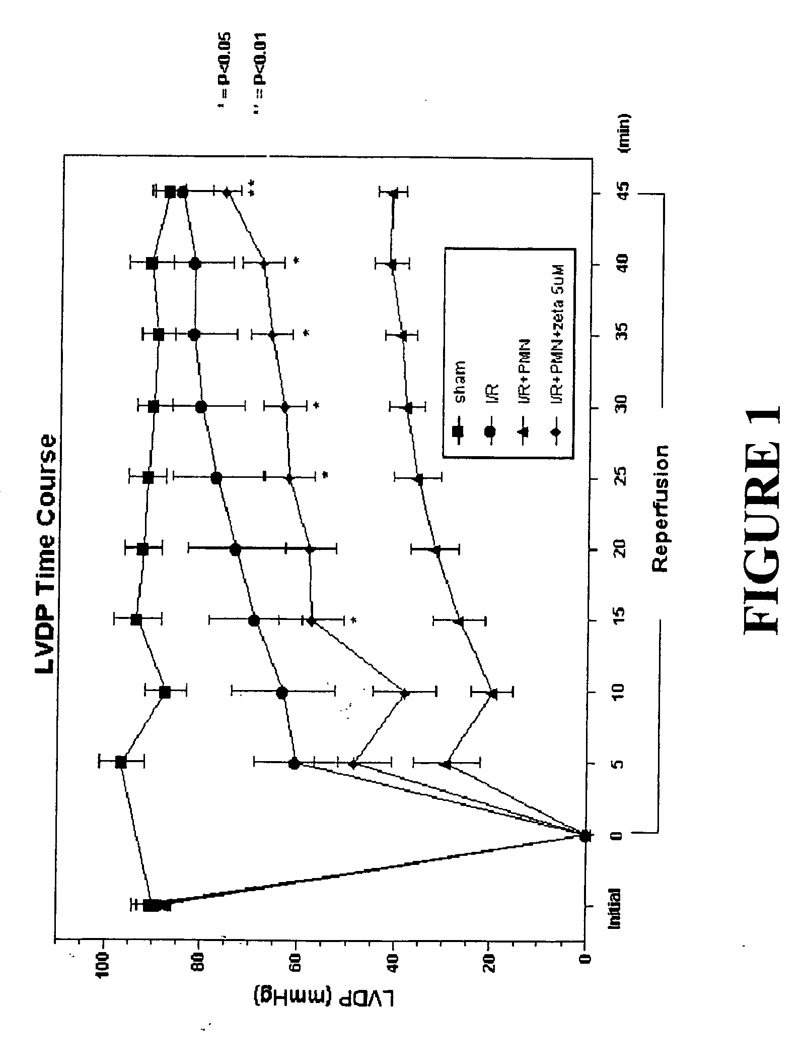
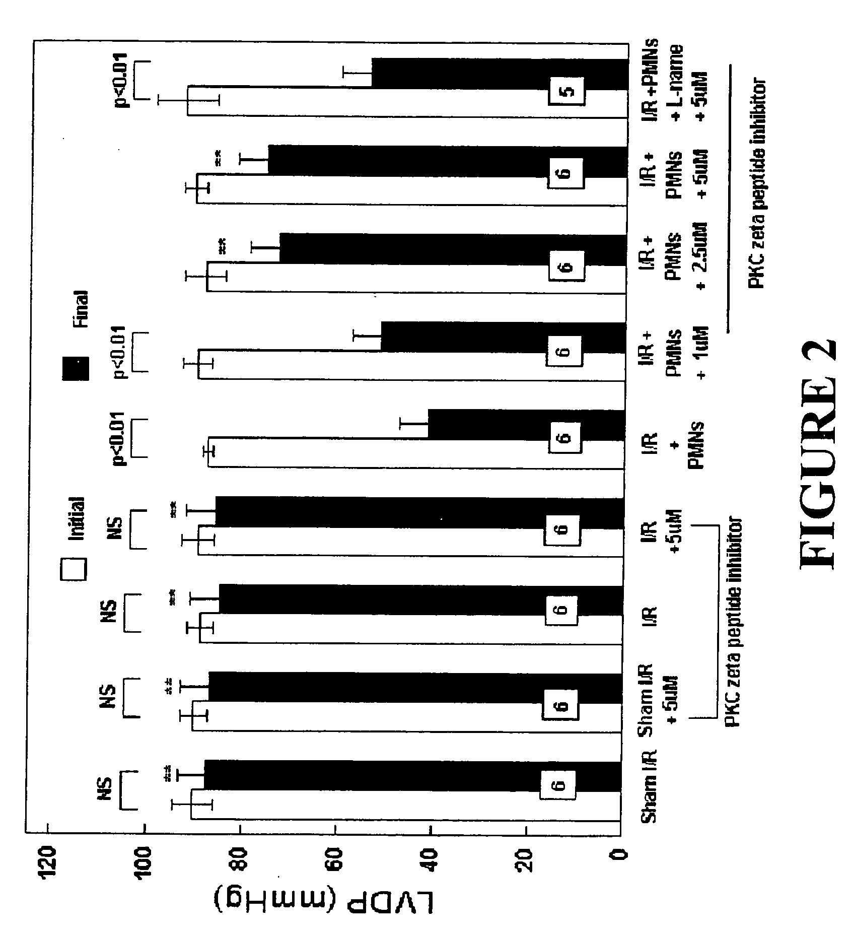
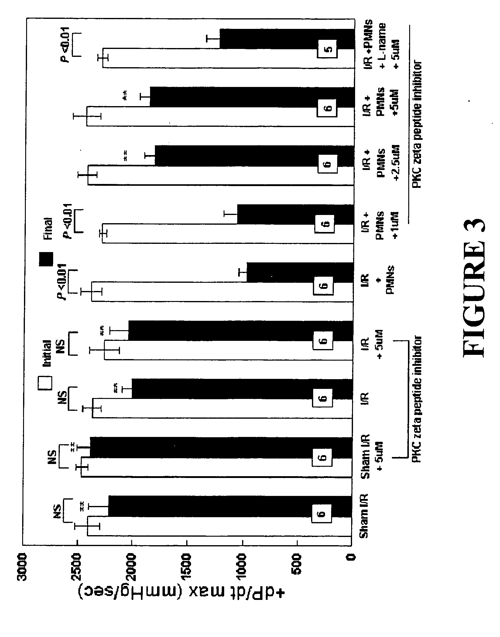
[0050] Male Sprague Dawley rats (275-325 g, Ace Animals, Boyertown, Pa.) were anesthetized with 60 mg / kg pentobarbital sodium intraperitoneally (i.p.). Sodium heparin (1,000 U) was also administered i.p. The hearts were rapidly excised, the ascending aortas were cannulated, and retrograde perfusion of the heart was initiated with a modified Krebs buffer maintained at 37° C. at a constant pressure of 80 mmHg. The Krebs buffer had the following composition (in mmol / l): 17 dextrose, 120 NaCl, 25 NaHCO3, 2.5 CaCl2, 0.5 EDTA, 5.9 KCl, and 1.2 MgCl2. The perfusate was aerated with 95% O2 and 5% CO2 and equilibrated at a pH of 7.3-7.4. The two side arms in the perfusion line proximal to the heart inflow cannula allowed PMNs, plasma without PKC ζ peptide inhibitor (control hearts) or plasma containing different concentrations of PKC ζ peptide inhibitor (1, 2.5 or 5 μM) to be directly infused into the coronary inflow line. Coronary flow was monitored by a...

example 2

Effects of Peptide Inhibitor of PKC βII

[0069] Experiments with PKCβ II peptide inhibitors were performed substantially as described in Example 1 for PKCζ peptide inhibitors.

[0070] The following groups of isolated perfused rat hearts were used:

[0071] Group 1: Sham I / R hearts were not subjected to ischemia and were not perfused with PMNs, but were perfused with 5 mL of plasma (1 mL / min) at 35 minutes into perfusion (the same time point that I / R hearts would be given 5 mL of plasma, 15 minutes of baseline recordings plus 20 minutes ischemia). These hearts represented a control group to determine if the isolated rat heart can maintain LVDP and +dP / dtmax throughout the 80-minute protocol (n=6).

[0072] Group 2: Sham I / R+PKC βII peptide inhibitor (10 μM) hearts were not subjected to ischemia and not perfused with PMNs. These hearts were administered the PKC βII peptide inhibitor (10 μM, dissolved in plasma from a 5 mM stock in H2O) 35 minutes into perfusion. This group was employed to d...

example 3

Effects of Peptide Inhibitors of PKC βII and PKC ζ in Combination

[0089] Experiments with PKC βII and PKC ζ peptide inhibitors were performed substantially as described in Examples 1 and 2.

[0090] The following groups of isolated perfused rat hearts were used:

[0091] Group 1: Sham I / R hearts were not subjected to ischemia and were not perfused with PMNs, but were perfused with 5 mL of plasma (1 mL / min) at 35 minutes into perfusion (the same time point that I / R hearts would be given 5 mL of plasma, 15 minutes of baseline recordings plus 20 minutes ischemia). These hearts represented a control group to determine if the isolated rat heart can maintain LVDP and +dP / dtmax throughout the 80-minute protocol (n=6).

[0092] Group 2: Sham I / R+PKC βII (10 μM)+PKC ζ (5 μM) peptide inhibitors hearts were not subjected to ischemia and not perfused with PMNs. These hearts were administered the PKC βII and PKC ζ peptide inhibitor (10 μM and 5 μM, respectively, dissolved in plasma from a 5 mM stock i...


PUM


Abstract

The present invention relates to a solution for preservation, perfusion, and / or reperfusion of an organ, especially the heart, for transplantation. The solution contains peptide inhibitor(s) of protein kinase C βII (PKC βII) and / or of protein kinase C ζ (PKC ζ) and / or peptide activator(s) of protein kinase C δ (PKCδ). Methods for using the inventive solution are also disclosed, including methods for preserving an organ for transplantation, for protecting an ischemic organ from damage, for attenuating organ dysfunction after ischemia, for maintaining nitric oxide release and / or inhibiting superoxide release in an ischemic organ, and for protecting an organ from damage when isolated from the circulatory system.

Description

FIELD OF THE INVENTION [0001] The present invention relates to a solution for preservation, perfusion, and / or reperfusion of an organ, especially the heart, for transplantation. The solution contains peptide inhibitor(s) of protein kinase C beta II (PKC βII) and / or of protein kinase C zeta (PKC ζ) and / or peptide activator(s) of protein kinase C delta (PKC δ). BACKGROUND OF THE INVENTION [0002] Successful organ transplantation is often limited due to ischemic / reperfusion injury. Isolated human hearts deprived of oxygen for more than four hours progressively loose vigor and often do not survive in recipient hosts. Other organs such as the kidney, liver, pancreas and lung are also subject to tissue and cellular damage when removed from their hosts prior to transplantation. This damage is due to hypoxic conditions and a lack of circulation, which normally delivers physiological concentrations of oxygen and nutrients, and removes toxic compounds produced by an organ's cells. Organ transp...

Claims


Application Information

Patent Timeline
IPC IPC(8): A01N1/02C12N5/07C12N5/071
CPCA01N1/0226A01N1/02A01N1/00A61P7/08
Inventor YOUNG, LINDON
Owner PHILADELPHIA COLLEGE OF OSTEOPATHIC MEDICINE
Learn More