Eureka AIR delivers breakthrough ideas for toughest innovation challenges, trusted by R&D personnel around the world.

Membrane scaffold proteins

a membrane scaffold and protein technology, applied in the field of molecular biology and membrane technology, can solve the problems of random orientation of the protein with respect, no definitive means of distinguishing between them, no compelling direct evidence as to which model is correct, etc., to achieve the effect of increasing the stability and monodispersity of self-assembled nanoparticles, reducing the size of the msp, and high throughpu

Inactive Publication Date: 2006-03-16
THE BOARD OF TRUSTEES OF THE UNIV OF ILLINOIS
View PDF6 Cites 4 Cited by
  • Summary
  • Abstract
  • Description
  • Claims
  • Application Information

AI Technical Summary

Benefits of technology

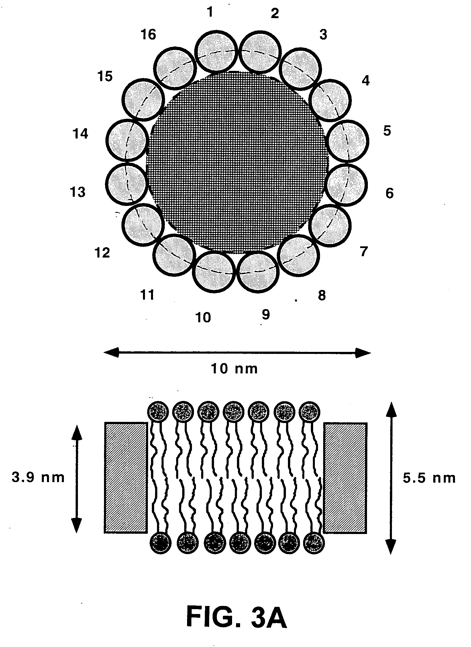
[0015] The present invention further provides the use of the nanometer scale phospholipid bilayer structures or nanodiscs formed using the MSPs of the present invention are useful for the incorporation of additional hydrophobic or partially hydrophobic protein molecules. Those additional proteins can be solubilized, for example, with the use of detergent, and the solubilized proteins can be added to a solution of MSP, with or without phospholipid(s), and the nanoscale particles self assemble so that the MSPs and the additional proteins are incorporated into a stable and soluble particle. Subsequently, any detergent can be removed by dialysis. Those proteins, found in nature in the various membrane structures of a living organism, are solubilized in the MSP supported nanobilayer or nanodisc, and the native structure and activity of the target protein are preserved in these MSP-supported structures. Besides hydrophobic or partially hydrophobic proteins, membrane fragments or disrupted membranes can be assembled with the MSPs of the present invention.
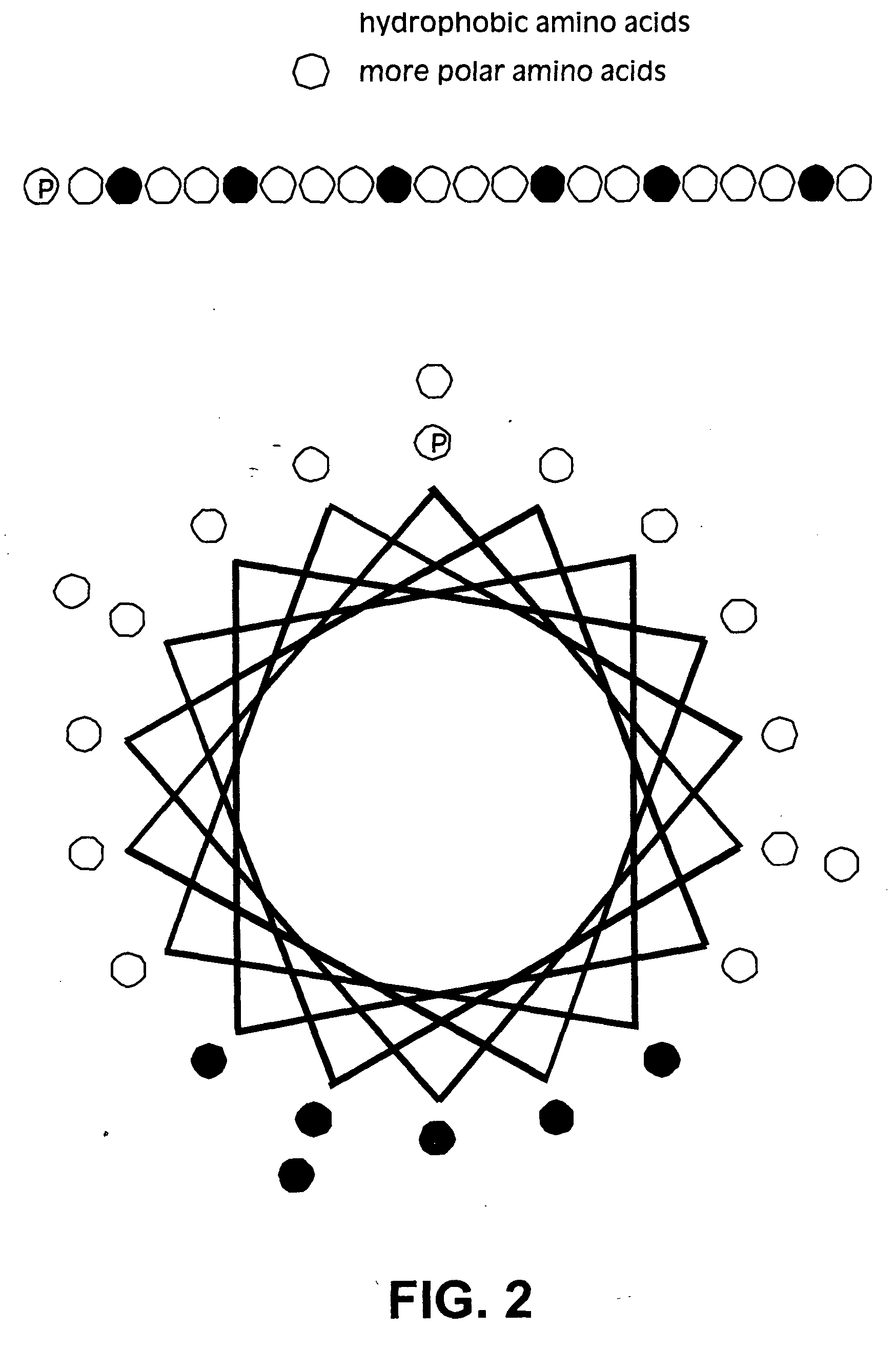
[0018] We developed nanodiscs for use in structural, biochemical and pharmaceutical techniques by engineering the scaffold protein (MSP) for greater stability, size homogeneity and useful functionalities in the resultant nanoscale lipoprotein particle. These particles can form tags for purification and physical manipulation of disks such as in hydrogels or on a gold biosensor surface, and they can serve as entities for rapid and reproducible assay and crystallization. The nanoparticles and membrane protein scaffolds are useful in biotechnology, the pharmaceutical industry as well as in basic research. In addition, the structural and functional principles uncovered through our discovery and the related techniques facilitate understanding the interactions of proteins with lipid bilayers at the molecular level.
[0020] The present invention further provides materials and methods using genetically engineered MSPs to minimize the size of the MSP with respect to membrane particle formation and to increase the stability and monodispersity of the self-assembled nanoparticle by altering the sequence of the parent molecule. In particular, we have developed MSPs for the study of G-protein coupled receptors. G-protein coupled receptors (GPCRs) are an important and diverse class of pharmaceutical targets in mammalian cellular membranes where they function as signal transducing elements, bind several classes of bioactive ligands and transmit information to the intracellular machinery. The artificial MSP of the present invention stabilizes and solubilizes the membrane-associated form of GPCR to allow purification and manipulation in solution or on a solid support for use in high throughput screening applications and on surfaces for surface-plasmon biosensor and scanning-probe techniques. The artificial MSP of the present invention can be used to facilitate expression and purification of recombinant membrane proteins in soluble form.

Problems solved by technology

Both models are based primarily on indirect experimental evidence, and no definitive means of distinguishing between them has emerged.
So far, there is no compelling direct evidence as to which model is correct, even though a low resolution x-ray crystal structure for apo A-I crystallized without lipid (Borhani et al., 1997) has been obtained.
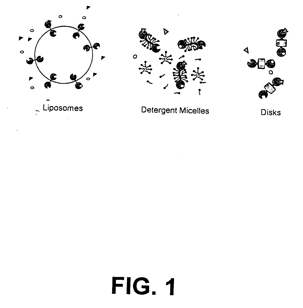
Reconstitution into liposomes by detergent dialysis or other methods typically results in random orientation of the protein with respect to outer and lumenal spaces.
Since both sides of the liposomal bilayer are not accessible to bulk solvent, coupling effects between domains on opposite sides of the bilayer cannot be studied.
Liposomes are also prone to aggregation and fusion and are usually unstable for periods of more than about a week or under certain physical manipulations, such as stopped flow or vigorous mixing.
Liposomes also may present difficulties due to light scattering, and aggregation of membrane proteins present in the bilayer.
Detergent micelles are dynamic and undergo structural fluctuations that promoter subunit dissociation and often present difficulty in the ability to handle proteins in dilute solutions.
Detergents can also be mildly denaturing and often do not maintain the properties found in a phospholipid bilayer system.

Method used


Image

  • Membrane scaffold proteins
  • Membrane scaffold proteins
  • Membrane scaffold proteins

Examples


example 1

Construction of Recombinant DNA Molecules for Expression of MSPs

[0088] The human proapoAI coding sequence as given below was inserted between Nco I and Hind III sites (underlined) in pET-28 (Novagen, Madison, Wis.). Start and stop codons are in bold type. The restriction endonuclease recognition sites used in cloning are underlined.

TABLE 1ProApoAI coding sequence (SEQ ID NO:1) Restrictionsites used in cloning are underlined, and thetranslation start and stop signals are shownin bold.CCATGGCCCATTTCTGGCAGCAAGATGAACCCCCCCAGAGCCCCTGGGATCGAGTGAAGGACCTGGCCACTGTGTACGTGGATGTGCTCAAAGACAGCGGCAGAGACTATGTGTCCCAGTTTGAAGGCTCCGCCTTGGGAAAACAGCTAAACCTAAAGCTCCTTGACAACTGGGACAGCGTGACCTCCACCTTCAGCAAGCTGCGCGAACAGCTCGGCCCTGTGACCCAGGAGTTCTGGGATAACCTGGAAAAGGAGACAGAGGGCCTGAGGCAAGAGATGAGCAAGGATCTGGAGGAGGTGAAGGCCAAGGTGCAGCCCTACCTGGACGACTTCCAGAAGAAGTGGCAGGAGGAGATGGAGCTCTACCGCCAGAAGGTGGAGCCGCTGCGCGCAGAGCTCCAAGAGGGCGCGCGCCAGAAGCTGCACGAGCTGCAAGAGAAGCTGAGCCCACTGGGCGAGGAGATGCGCGACCGCGCGCGCGCCCATGTGGACGCGCTGCGCACG...

example 2

Construction of Synthetic MSP Gene

[0105] A synthetic gene for MSP1 is made using the following overlapping synthetic oligonucleotides which are filled in using PCR. The codon usage has been optimized for expression in E. coli, and restriction sites have been introduced for further genetic manipulations of the gene.

Synthetic nucleotide taps1aTACCATGGGTCATCATCATCATCATCACATTGAGGG(SEQ ID NO:30)ACGTCTGAAGCTGTTGGACAATTGGGACTCTGTTACGTCTASynthetic nucleotide taps2aAGGAATTCTGGGACAACCTGGAAAAAGAAACCGAGG(SEQ ID NO:31)GACTGCGTCAGGAAATGTCCAAAGATSynthetic nucleotide taps3aTATCTAGATGACTTTCAGAAAAAATGGCAGGAAGAG(SEQ ID NO:32)ATGGAATTATATCGTCAASynthetic nucleotide taps4aATGAGCTCCAAGAGAAGCTCAGCCCATTAGGCGAAG(SEQ ID NO:33)AAATGCGCGATCGCGCCCGTGCACATGTTGATGCACTSynthetic nucleotide taps5aGTCTCGAGGCGCTGAAAGAAAACGGGGGTGCCCGCT(SEQ ID NO:34)TGGCTGAGTACCACGCGAAAGCGACAGAASynthetic nucleotide taps6aGAAGATCTACGCCAGGGCTTATTGCCTGTTCTTGAG(SEQ ID NO:35)AGCTTTAAAGTCAGTTTTCTSynthetic nucleotide taps1bCAGAATTCCTGCGTCACG...

example 3

Expression of Recombinant MSPs

[0111] To express MSP proteins, the nucleic acid constructs were inserted between the NcoI and HindIII sites in the pET28 expression vector and transformed into E. coli BL21(DE3). Transformants were grown on LB plates using kanamycin for selection. Colonies were used to inoculate 5 ml starter cultures grown in LB broth containing 30 μg / ml kanamycin. For overexpression, cultures were inoculated by adding 1 volume overnight culture to 100 volumes LB broth containing 30 μg / ml kanamycin and grown in shaker flasks at 37° C. When the optical density at 600 nm reached 0.6-0.8, isopropyl b-D-thiogalactopyranoside (IPTG) was added to a concentration of 1 mM to induce expression and cells were grown 3-4 hours longer before harvesting by centrifugation. Cell pellets were flash frozen and stored at −80° C.


PUM

PropertyMeasurementUnit
diameteraaaaaaaaaa
diameteraaaaaaaaaa
heightaaaaaaaaaa

Abstract

Membrane proteins are difficult to express in recombinant form, purify, and characterize, at least in part due to their hydrophobic or partially hydrophobic properties. Membrane scaffold proteins (MSP) assemble with target membrane or other hydrophobic or partially hydrophobic proteins or membrane fragments to form soluble nanoscale particles which preserve their native structure and function; they are improved over liposomes and detergent micelles. In the presence of phospholipid, MSPs form nanoscopic phospholipid bilayer disks, with the MSP stabilizing the particle at the perimeter of the bilayer domain. The particle bilayer structure allows manipulation of incorporated proteins in solution or on solid supports, including for use with such surface-sensitive techniques as scanning probe microscopy or surface plasmon resonance. The nanoscale particles facilitate pharmaceutical and biological research, structure / function correlation, structure determination, bioseparation, and drug discovery.

Description

CROSS REFERENCE TO RELATED APPLICATIONS [0001] This application is a Continuation-in-Part of U.S. Provisional Application No. 60 / 252,233, filed Nov. 20, 2000.ACKNOWLEDGMENT OF FEDERAL RESEARCH SUPPORT [0002] This invention was made, at least in part, with funding from the National Institutes of Health (Grant Nos. (R01GM31756, R01GM33775, GM63574 and 5F32GM19024). Accordingly, the United States Government has certain rights in this invention.BACKGROUND OF THE INVENTION [0003] The field of present invention encompasses molecular biology and membrane technology. Specifically, the present invention relates to artificial membrane scaffold proteins (MSPs), sequences encoding them, vectors and recombinant host cells, methods for recombinant production of them, and methods of using the membrane scaffold proteins to stabilize, disperse and solubilize fully or partially hydrophobic proteins such as tethered, embedded or integral membrane proteins while maintaining the biological activities of...

Claims


Application Information

Patent Timeline
Patent Type & Authority Applications(United States)
IPC IPC(8): C07K14/72C12P21/06C07K14/00G01N33/50C07K14/47C07K14/705C07K19/00C12N1/19C12N1/21C12N5/10C12N15/09G01N33/15
CPCC07K14/47
Inventor SLIGAR, STEPHENBAYBURT, TIMOTHY
Owner THE BOARD OF TRUSTEES OF THE UNIV OF ILLINOIS
Why Eureka
Eureka Blog
Learn More