Mouse model for psoriasis and psoriatic arthritis
a mouse model and psoriatic arthritis technology, applied in the field of psoriasis, can solve the problems of severely hampered research into the pathogenesis of this common skin disorder, and the efforts to transgeneically deliver inflammatory mediators or keratinocyte growth factors to the skin have not completely reproduced the psoriatic phenotyp
- Summary
- Abstract
- Description
- Claims
- Application Information
AI Technical Summary
Problems solved by technology
Method used
Image
Examples
example 1
Generation of c-junΔep*; junBΔep* Mice
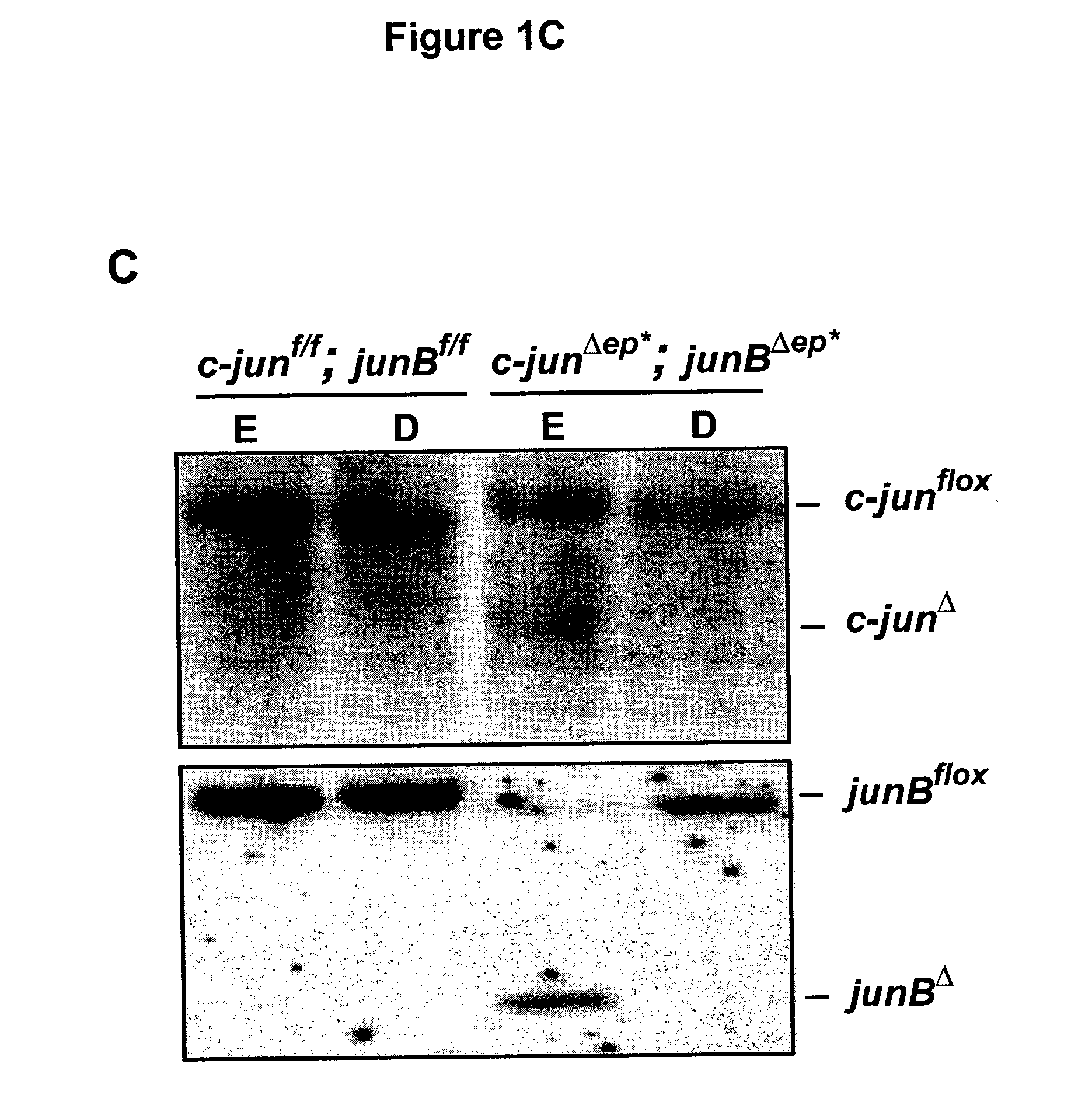
[0072] c-junf / f; junBf / f mice were crossed to K5-Cre-ER transgenic mice and heterozygous progenies were intercrossed to get c-junf / f; junBf / f; K5-Cre-ER and c-junf / f; junBf / f mice. The approach to delete c-jun and junB in the epidermis is outlined in FIGS. 1A and 1B. In brief, 8 weeks old experimental mice (c-junf / f; junBf / f; K5-Cre-ER) and control mice (c-junf / f; junBf / f) were injected intraperitoneally 5 times with 1 mg tamoxifen. 2 weeks after the last injection the mice were analyzed. The deletion of c-jun and junB was analyzed by Southern blot and RPA (RNAse protection assay). Southern blot analyses for c-jun and JunB deletion (FIG. 1C) showed deletion in both cases. The remaining signal for floxed c-jun and floxed junB is explained by incomplete deletion and the inflammatory infiltrate seen in the epidermis of c-junΔep*; junBΔep* mice (FIG. B). Quantification of AP-1 mRNA in the epidermis of c-junΔep*; junBΔep* mice showed significant dow...
example 2
Characterization of c-junΔep*; junBΔep* Mice
[0073] Mice obtained according to the treatment described in Example 1 develop a phenotype resembling psoriasis within 2 weeks. Hairless skin like ear, tail and feet were dramatically affected (FIG. 2A). Histological examination (FIG. 2B) revealed abnormally thickened epidermis, parakeratosis (nucleated keratinocytes in the cornified layer), thickened keratinized upper layer (hyperkeratosis), and fingerlike epidermal projections into the dermis (rete ridges). Furthermore, epidermal microabscesses and the typical inflammatory cell infiltrate are seen: intraepidermal T-cells (CD3 staining), increased numbers of neutrophils in the epidermis (esterase staining), and macrophages in the dermis (esterase- and F4 / 80-staining). H&E staining from affected mouse finger demonstrating granulocytic infiltrates into the joint region resembling psoriatic arthritis (FIG. 2C).
PUM
 Abstract
Description
Claims
Application Information
 - R&D
- Intellectual Property
- Life Sciences
- Materials
- Tech Scout
- Unparalleled Data Quality
- Higher Quality Content
- 60% Fewer Hallucinations
Browse by: Latest US Patents, China's latest patents, Technical Efficacy Thesaurus, Application Domain, Technology Topic, Popular Technical Reports.
© 2025 PatSnap. All rights reserved.Legal|Privacy policy|Modern Slavery Act Transparency Statement|Sitemap|About US| Contact US: help@patsnap.com


