
Quantitative alkaline-phosphatase precipitation reagent and methods for visualization of protein microarrays

a technology of alkaline phosphatase and precipitation reagent, which is applied in the field of quantitative alkaline phosphatase precipitation reagent and methods for visualization of protein microarrays, can solve the problems of ccds consuming relatively large amounts of power, single microscope objective typically has multiple lenses that are generally very expensive, and the reaction rate of this enzyme remains linear, so as to improve sensitivity and facilitate visualization. , the effect of rapid and economical method

Inactive Publication Date: 2005-06-09
MIRAGENE
View PDF40 Cites 11 Cited by
  • Summary
  • Abstract
  • Description
  • Claims
  • Application Information

AI Technical Summary

Benefits of technology

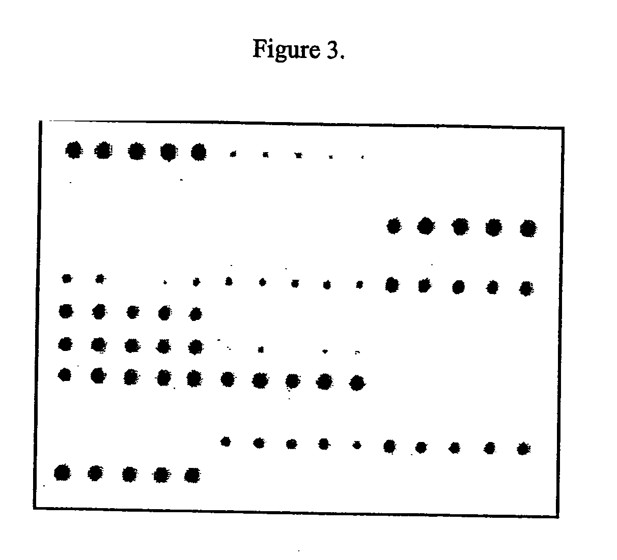
[0014] In one embodiment, the present invention is related to a rapid and economical method for visualization of microarrays. In a preferred mode, the method is adapted for analysis of protein arrays. Briefly, proteins are spotted on a suitable surface in an addressed format with an opaque background, preferably a solid white background. The protein, DNA, or antibody array is incubated with molecules of interest (antibodies, serum, proteins, drugs or other molecules) washed and then incubated with a detector (secondary antibody labeled with alkaline phosphatase or biotin) or other suitable detection system that can produce a color change at reactive sites. The detector is then visualized using an alkaline phosphatase (an enzyme isolated from calf intestines) catalyzed biotin / streptavidin precipitation reaction. The precipitation reaction results in a sharp color that appears only where AP has been immobilized. The reaction rates for this enzyme remain linear over time, and sensitivity can therefore be improved by allowing the reaction to proceed for longer periods of time.
[0015] In another embodiment, visualization of the aforementioned method is enhanced by colorimetry (or, a type of method used to measure color and to define the results of the measurements). The color is digitally captured using a scanning apparatus in conjunction with novel software. This allows for a lumens analysis of the color density, which directly correlates to interactions between immobilized biological samples and various test substances. This data can then be quantified and corrected-using a standard curve and calibration markers, so as to convert the color data to molecular data.

Problems solved by technology

These conventional microscopes are sophisticated and expensive instruments that require training and maintenance.
A single microscope objective typically has multiple lenses that are generally very expensive.
One of the challenges in microscopy is making the microscope as efficient as possible in capturing all of the light that leaves the sample surface so that an optimal image can be captured.
Unfortunately, CCDs consume relatively large amounts of power, usually work over a smaller area, and are limited in their multi-color capabilities.
The costly instrumentation conventionally used to image biological samples, e.g., protein and DNA chips, impedes the broad usage of such technologies.

Method used


Image

  • Quantitative alkaline-phosphatase precipitation reagent and methods for visualization of protein microarrays
  • Quantitative alkaline-phosphatase precipitation reagent and methods for visualization of protein microarrays
  • Quantitative alkaline-phosphatase precipitation reagent and methods for visualization of protein microarrays

Examples


Embodiment Construction

[0043] In a preferred embodiment, the present invention provides inexpensive methods for resolving colorimetric density representative of interactions between immobilized biological samples (e.g., protein or nucleic acid spots on a microarray) and various test substances. As used herein, biological samples refers to biological material (proteins, nucleic acids, tissues, etc.) associated with a biological material holding structure (e.g., a microarray substrate such as a protein or DNA chip substrate, a gel, etc.) in a manner that allows for detection of the biological material, or portions thereof (e.g., with the use of markers such as dyes, tags, labels, or stains), such as through the use of imaging (e.g., direct mapping).

[0044] One or more embodiments of the present invention are operable for use in multiple imaging applications, e.g., imaging of two-dimensional and three-dimensional objects, such as fluorescence imaging, reflective imaging, bar code imaging, densitometry, gel d...


PUM


Abstract

A system and method are disclosed for the rapid, reproducible and inexpensive visualization, imaging and digital analysis of molecular interactions between ligands and proteins immobilized on an addressable two-dimensional microarray.

Description

RELATED APPLICATIONS [0001] This application claims priority under 35 U.S.C. § 119(e) to U.S. Provisional Application No. 60 / 527,242, filed on Dec. 5, 2003 which claims priority under 35 U.S.C. §120 to U.S. patent application Ser. No. 10 / 431,686 filed on May 8, 2003, which claims priority under 35 U.S.C. §119(e) to U.S. Provisional Application No. 60 / 379,326 filed on May 9, 2002.BACKGROUND OF THE INVENTION [0002] 1. Field of the Invention [0003] In one aspect of the present invention, a method is disclosed for the rapid, reproducible and inexpensive visualization, imaging and digital analysis of molecular interactions between ligands and proteins immobilized on an addressable two-dimensional microarray. [0004] 2. Description of the Related Art [0005] Various conventional approaches have been used to visualize the surface of biological samples, e.g., DNA spots of a microarray such as a DNA chip, protein bands in a one dimensional (1-D) or two dimensional (2-D) gel, etc. For example, ...

Claims


Application Information

Patent Timeline
IPC IPC(8): G01N33/68
CPCG01N33/6839
Inventor LEBRUN, STEWART
Owner MIRAGENE
Learn More