
Semi-automated measurement of anatomical structures using statistical and morphological priors

a morphological prior and anatomical structure technology, applied in the field of medical image system and method, can solve the problems of manual and subjective acquisition of data points, manual tracing, and high error rate, and achieve the effect of minimizing both intra-operator and inter-operator variation and rapid and accurate operation

Active Publication Date: 2005-03-31
VIRTUALSCOPICS
View PDF5 Cites 22 Cited by
  • Summary
  • Abstract
  • Description
  • Claims
  • Application Information

AI Technical Summary

Benefits of technology

"The present invention is a system and method for accurately identifying and measuring various anatomical structures in medical images. It uses statistical information and a shape model to quickly and accurately identify and measure these structures. The system minimizes both intra- and inter-operator variation and can be adapted to identifying a wide variety of normal and abnormal biological structures in medical images."

Problems solved by technology

Current standard methods for obtaining these data points are largely manual and subjective, and are therefore both error-prone and subject to inter- and intra-operator variability.
In addition, manual tracing of structures such as the vascular system, which may appear on up to 800 images in a single study, requires both considerable expertise and a great deal of time.
Significant research effort has been devoted to the subject of identification of curvilinear and poorly defined structures in medical images, but there is at this time no generally accepted solution. de Bruijne et al.
However, they have difficulty when these assumptions break down, as at bifurcations.
In addition, these methods are not able to identify associated structures such as thrombus or calcifications, and have not been demonstrated to be effective in cases where significant artifacts are present, as in post-aortic endograft CT images.

Method used


Image

  • Semi-automated measurement of anatomical structures using statistical and morphological priors
  • Semi-automated measurement of anatomical structures using statistical and morphological priors
  • Semi-automated measurement of anatomical structures using statistical and morphological priors

Examples


Embodiment Construction

A preferred embodiment of the present invention, and experimental results therefrom, will now be set forth in detail with reference to the drawings.

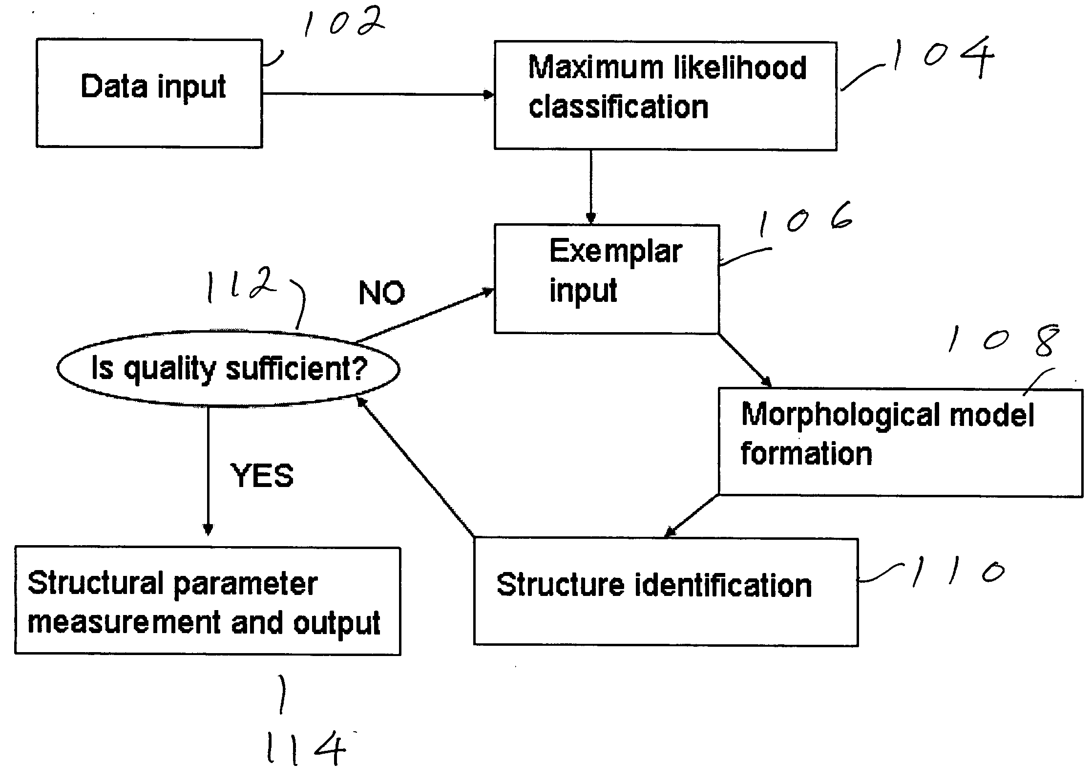
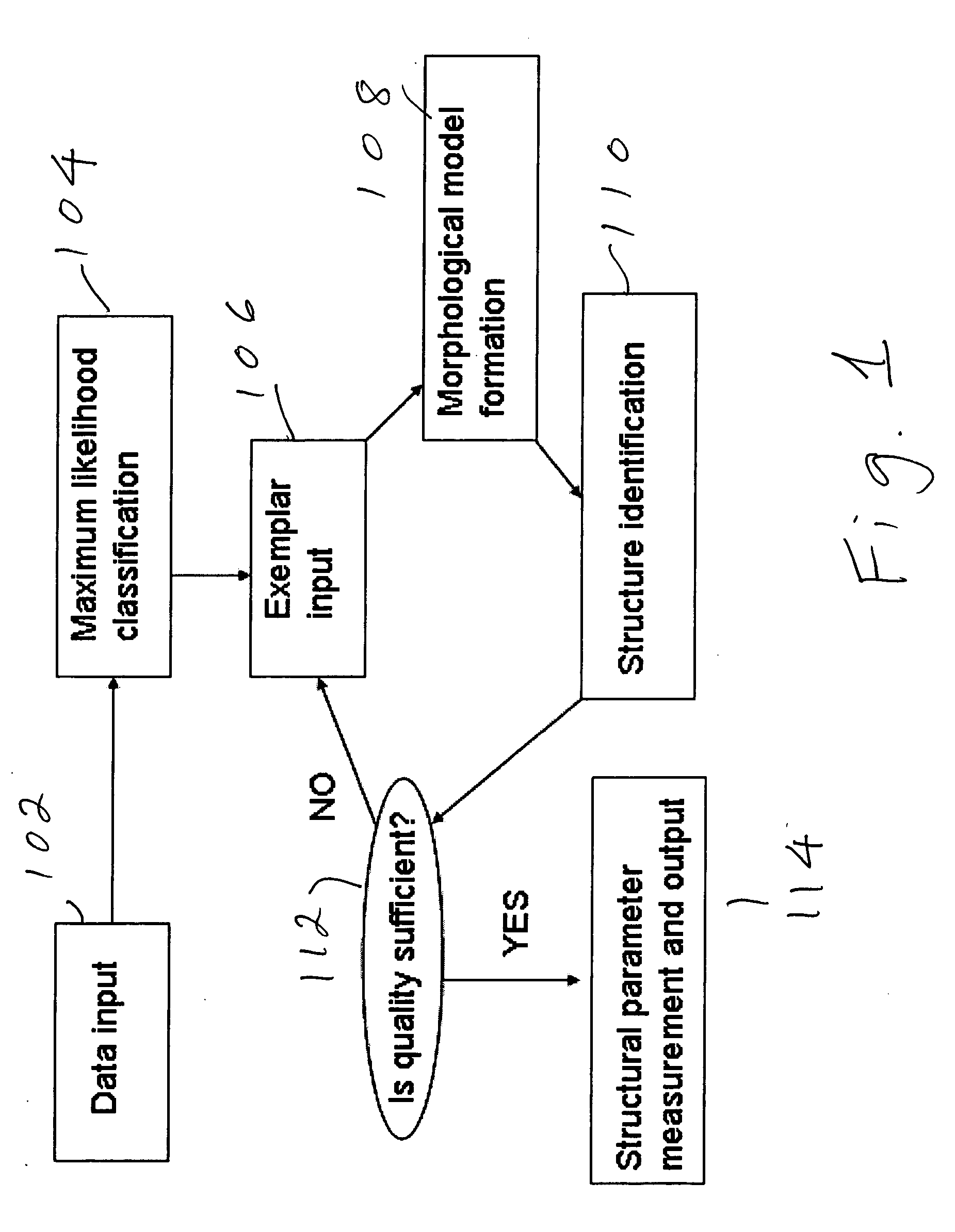
FIG. 1 shows a flow chart of the operational steps of the preferred embodiment. In step 102, data of an image or a sequence of images are input from a suitable source, e.g., a storage medium on which MRI data have been stored.

The maximum likelihood classification (MLC) of step 104 refers to the process of optimally separating an image into areas of similar statistical behavior. It is assumed that regions of similar statistical behavior will correspond to different tissue types. The goal of the MLC algorithm used in this invention is to globally maximize one of the following discriminant functions: gi⁡(x)=ln⁢pi-12⁢ln⁢Ri-12⁢(x-mi)t⁢Ri-1⁡(x-mi)(1)

where Ri is the covariance matrix for class i, mi is the mean vector for class i, pi is the a priori probability of class i appearing at the voxel under consideration, and x is the value vector...


PUM


Abstract

Structures are delineated in medical or other images. First, various tissue types present in the image are statistically described using a maximum likelihood classifier. Second, the tissue of interest is described using an exemplar, which is derived either from an anatomical atlas or from user input. Third, the structure of interest is morphologically described. The process can be iterated until a desired level of accuracy is achieved.

Description

FIELD OF THE INVENTION The present invention is directed to a system and method for measuring anatomical structures in medical images, such as those generated by magnetic resonance imaging (MRI), computed tomography (CT), positron emission tomography (PET), etc, and more particularly to a system and method in which the measurement is semi-automated and makes use of statistical and morphological priors in the form of user-defined exemplars, seed regions, shape models, or other guiding information. DESCRIPTION OF RELATED ART Accurate identification and measurement of various anatomical structures is a vital tool both for surgical planning and for evaluation of disease progression and patient response to therapy for numerous diseases. Measurement of hippocampal volume is an important endpoint for diagnosing and monitoring both intractable temporal lobe epilepsy and Alzheimer's disease. Identification of the aorta and associated vessels and measurement of various related parameters ar...

Claims


Application Information

Patent Timeline
Patent Type & Authority Applications(United States)
IPC IPC(8): A61BG06K9/00G06K9/62G06T7/00
CPCG06K9/6278G06T2207/30004G06T7/0012G06F18/24155
Inventor ASHTON, EDWARD
Owner VIRTUALSCOPICS
Learn More