Kits and methods for preparing gell samples optmimized for dual staining
a technology of gell samples and kits, applied in the field of kits and methods for preparing cell samples optimized for dual staining, can solve the problems of difficult to locate cells on the slide, cell smearing is fast and easy to perform, and the cell is often not evenly distributed on the slid
- Summary
- Abstract
- Description
- Claims
- Application Information
AI Technical Summary
Benefits of technology
Problems solved by technology
Method used
Image
Examples
example 1
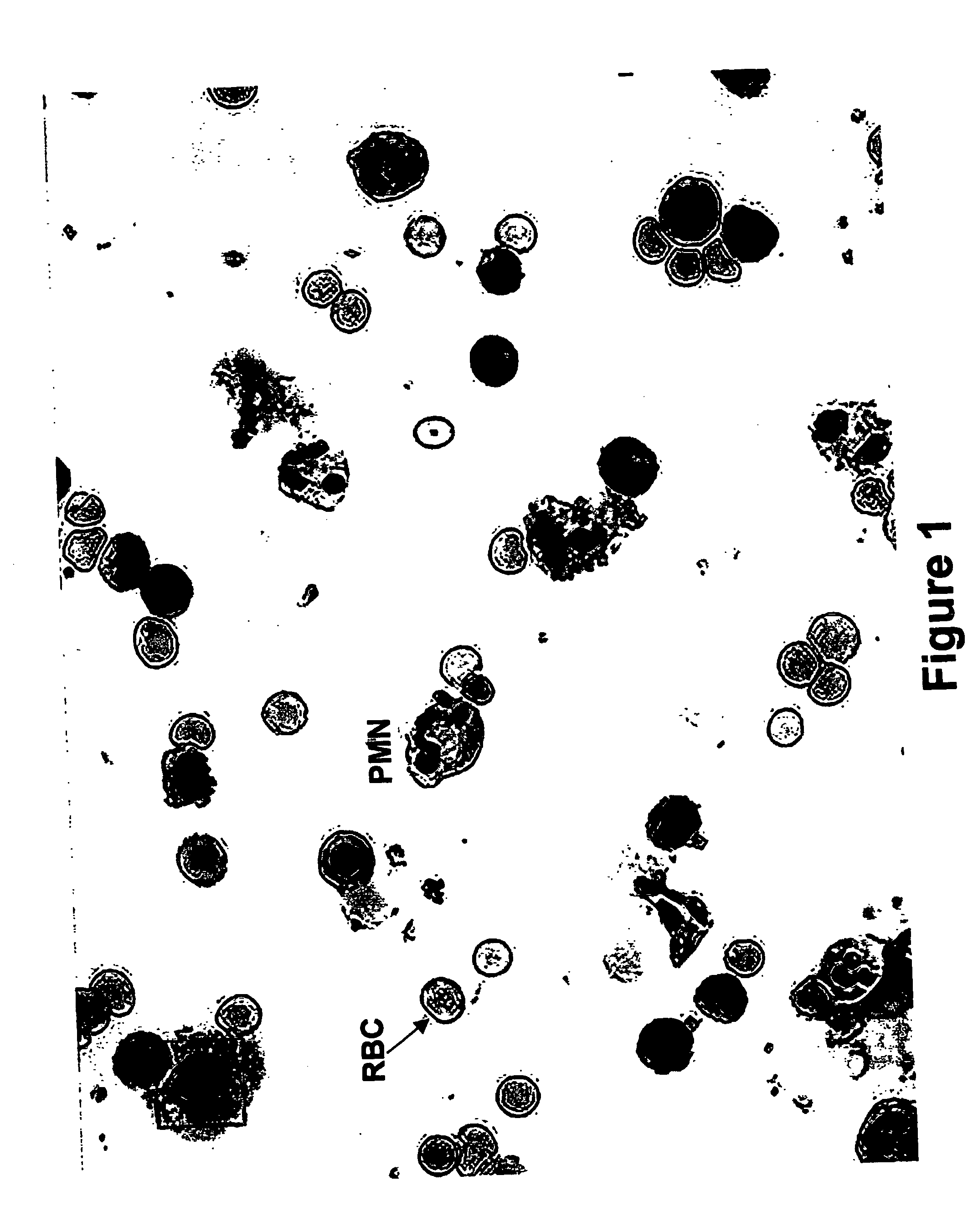
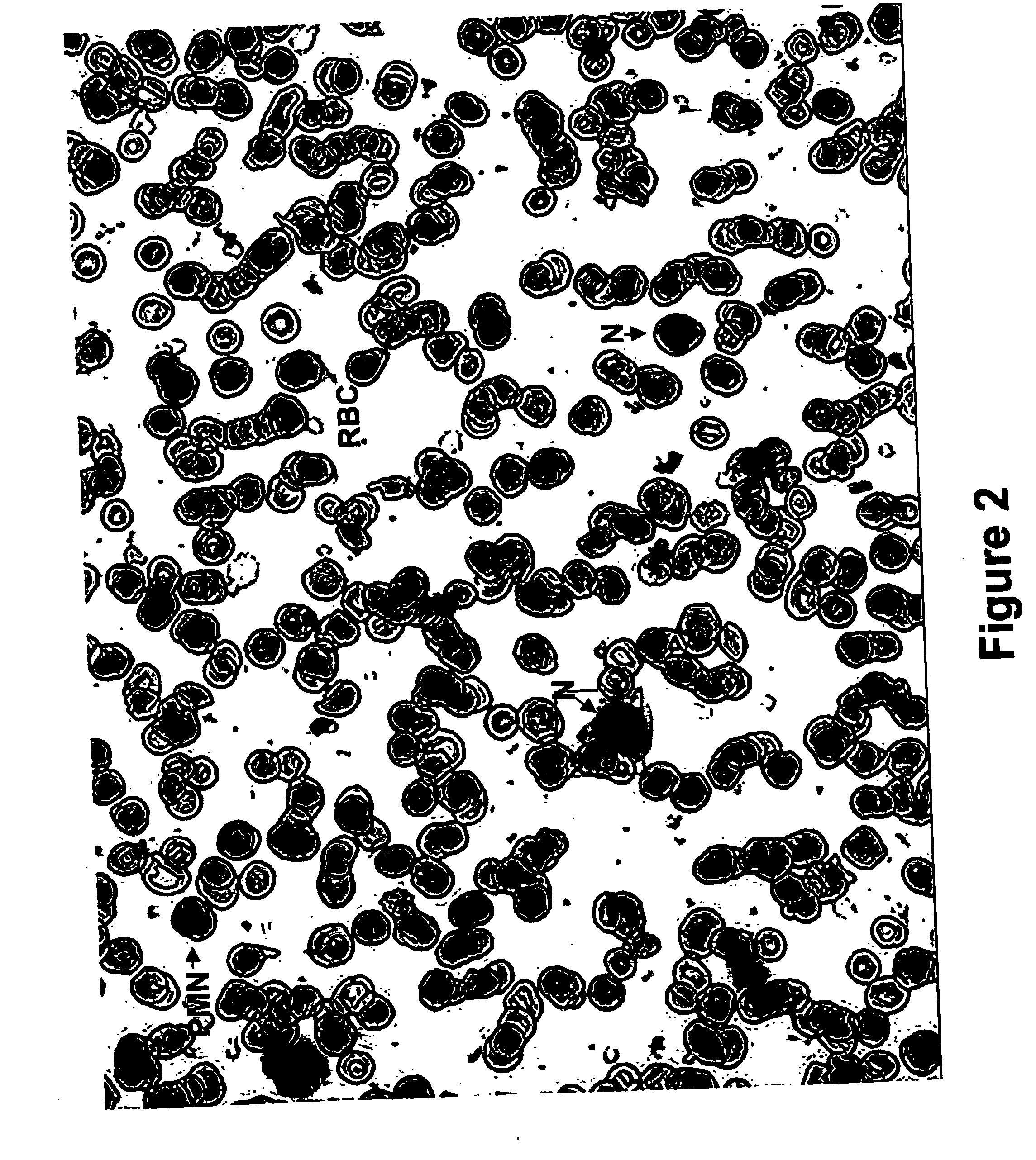
[0126] Preparation of Cytospin Slides of Peripheral Blood and Bone Marrow Cells
[0127] Sample dilution: Peripheral blood (PB) or bone marrow (BM) cells are diluted with equal volume (up to 3 ml) of wash solution (Bio View Cat. # BV-000-05).
[0128] White blood cells (WBC) separation: Six ml of a Ficoll-based density gradient WBC separation reagent (Bio View Cat. # BV-000-09) are poured into a 15 ml culture tube (Corning, N.Y., USA). The diluted PB or BM sample is then carefully layered over the WBC separation reagent and the tubes are centrifuged for 30 minutes at 400×g at room temperature (20-25° C.). Following centrifugation, the upper layer is carefully removed with a Pasteur pipette up to a distance of 0.5 cm from the opaque interface containing the white blood cells. The opaque interface is transferred with a Pasteur pipette into a clean 15 ml conical test tube (Falcon, N.J., USA) and is gently mixed with 5 ml of the wash buffer. The cells are then centrifuged for 10 minutes at ...
example 2
A Kit for Cell Preparation of Cyto-Spin Slides of Blood or Bone Marrow Samples
[0144] The following kit (Table 1) is designed for the preparation of cyto-spin slides for the evaluation of the cells' morphology and for further use of the same slides for ICC and FISH assays. Slides can be scanned with the automated scanning system described in PCT / IL00 / 00101.
TABLE 1Kit for preparing cyto-spin slides of blood or bone marrow samplesReagents required forpreparation of kit'sInstructions forKit'sBioViewcomponent (includingpreparation of kit'sSpecialcomponentsLtd Cat. #supplier's Cat. #)componentNotificationsWashBV-000-0520 X PBS (Cat. # I 291,Diluted 1:20 inWork in a sterileSolutionSavyon, Israel)double distilledenvironmentwaterWBCBV-000-09Histopaque 1.077 (Cat. #250 ml ofWork in a sterileSeparation1077-1, Sigma, USA);Histopaque 1.077tent.ReagentHistopaque 1.119 (Cat. #are mixed with 750 ml1119-1, Sigma, USA)of Histopaque1.119.RBC lysisBV-000-12Water, tissue culture gradeSodium AzideWork...
example 3
A Kit for an Immunohistochemistry Assay on Slides
[0145] The following kit (Table 2) is designed for an immunocytochemistry (ICC) assay. Following ICC slides can be scanned with the automated scanning system described herein above.
TABLE 2A kit for Immunohistochemistry assayReagents required forpreparation of kit'sInstructions forBioView Ltd.component (includingpreparation ofit's componentsCat. #supplier's Cat. #)kit's componentpecial notificationsash solutionBV-020-05TBS (Cat. # T6664,TBS powder is—Sigma, USA)mixed in 1 liter ofdistilled water.locking reagentBV-020-08TBS (Cat. # T6664,TBS is preparedork in a sterileSigma, USA)as mentionednvironment and use aNormal goat serum (Cat.herein above.embrane for# 005-000-121, JacksonGoat serumiltrationUSA)(freeze-dried) isreconstituted with10 ml of water atroom temperaturefor 2 hours.Reconstitute goatserum (10 ml) ismixed with TBS(90 ml) andSodium Azide(0.1 gr) is added.ntibody diluentBV-020-04TBS (Cat. # T6664,TBS and goatork in a steril...
PUM
 Abstract
Description
Claims
Application Information
 - R&D
- Intellectual Property
- Life Sciences
- Materials
- Tech Scout
- Unparalleled Data Quality
- Higher Quality Content
- 60% Fewer Hallucinations
Browse by: Latest US Patents, China's latest patents, Technical Efficacy Thesaurus, Application Domain, Technology Topic, Popular Technical Reports.
© 2025 PatSnap. All rights reserved.Legal|Privacy policy|Modern Slavery Act Transparency Statement|Sitemap|About US| Contact US: help@patsnap.com


