
Urethral muscle controlled micro-invasive sphincteric closure device

a urethral muscle and micro-invasive technology, applied in the field of urethral muscle controlled micro-invasive sphincteric closure devices, can solve the problems of inconvenient use, high indirect cost, and high indirect cost, and achieve the effect of reducing device migration

Inactive Publication Date: 2004-02-12
YEUNG JEFFREY E +1
View PDF31 Cites 119 Cited by
  • Summary
  • Abstract
  • Description
  • Claims
  • Application Information

AI Technical Summary

Benefits of technology

[0042] The arch, needle and plunger assemblies are parts of a delivery device with a balloon behind an indented pocket or recessed region. After insertion of the device into the urethra, the balloon is inflated from behind the indented recess, pushing and embedding the recess into the mucosal tissue. The indented recess holds or shelters a portion of mucosa and other soft urethral tissue from being flattened by the compression. The needle and the arch are then advanced through a hole in the proximal recessed wall to longitudinally penetrate beneath the surface of the loosely packed mucosa within the recessed pocket. To deploy the resiliently straightened arch, the needle is withdrawn while the plunger is held stationary behind the arch. As a result, the arch resumes the pre-disposed curvature toward the lumen, beneath the surface of the mucosa. After the balloon is deflated, the delivery device is withdrawn. The curvature or bend of the arch protrudes from within the urethral wall, elevating or lifting the mucosa to narrow or close the lumen, preventing or minimizing urine leakage. For voiding, the urethral muscles stiffen the urethra and significantly widen the lumen beyond the closing range of the arch. As a result, urine passes.Arch Tube Closure Device
[0132] FIG. 75 depicts anchoring devices 149 and tissue ingrowth openings 150 on the magnet 132 to minimize device migration.

Problems solved by technology

Urinary incontinence is costly to patients and health care systems.
The indirect cost is likely to be much higher, in fact incalculable.
The careers of the sufferers are often prematurely terminated or adversely affected by the offensive odor.
However, unlike the valves of a heart, the urethral sphincter cannot be identified with the naked eye or even under a microscope.
It is widely believed that a leading cause of urinary incontinence is the loss of structural support for the urethra, especially behind the posterior urethral wall, which is indicated by hypermobility of the urethra.
Gravity and / or pregnancy may adversely affect the structural support.
Urethral hypermobility is often caused by a weakness of pelvic floor support.
If the leakage stops as soon as the stress is over, it is stress incontinence.
If voiding continues after the stress ceases, it is likely detrusor hyperactivity, or hyper-reflexia.
Drug therapy has very limited success and significant side effects.
Urethral removable plugs (U.S. Pat. No. 5,562,599 to Beyschlag, U.S. Pat. No. 4,457,299 to Cornwell, U.S. Pat. No. 5,131,906 to Chen, U.S. Pat. No. 5,906,575 to Conway et al., U.S. Pat. No. 5,885,204 to Vergano) are uncomfortable and troublesome to use, and their use increases the possibility of urinary tract infections.
Penile clamping devices (U.S. Pat. No. 4,942,886 to Timmons) are also highly uncomfortable and unnatural and may even cut off blood supply.
To be effective, the compression has to be strong and uncomfortable.
Similar to the urethral plugs, pessary devices increase the possibility of infections and are troublesome to use, messy during menstrual periods.
Without a direct view of the surgical site, one of the major potential problems with the devices is the uncertainty of suture tension, let alone obtaining the optimal suture tension.
If the suture is too tight, the urethra is too restricted, and urinary obstruction occurs.
If the tension is too loose, incontinence continues.
For intrinsic sphincter dysfunction, merely anatomic correction supported by a wall of soft vaginal tissue is inadequate.
Most of these complications are once again due to improper tension of the suture or sling.
If the sling is too tight, the urethra is obstructed; if it is too loose, incontinence continues.
Unfortunately, no standard parameters exist to identify the appropriate sling tension.
In many failed sling procedures in the past, sutures attaching the urethra to the abdominal ligaments were too close to the urethra.
Due to the close proximity of the suture and the pliable nature of the urethra, the tension of the suture created kinks in the urethra, resulting in urinary obstruction.
Furthermore, the rubbing of the abdominally anchored suture onto the urethra is presumably the cause of fibrotic tissue formation around the urethra and sometimes urethral erosion to the point of severance.
Furthermore, all these bulking agents migrate or metabolize away, some in less than a few months.
Tissue atrophy, a natural result of cuff compression over time, is often followed by cuff erosion with symptoms of pain, swelling, infection and / or bloody discharge.
However, alpha-blockers have the side effect of hypotension, characterized by dizziness.
However, given time, epithelial tissues grow into the lumen of the stents, requiring traumatic surgical removal.

Method used


Image

  • Urethral muscle controlled micro-invasive sphincteric closure device
  • Urethral muscle controlled micro-invasive sphincteric closure device
  • Urethral muscle controlled micro-invasive sphincteric closure device

Examples


Embodiment Construction

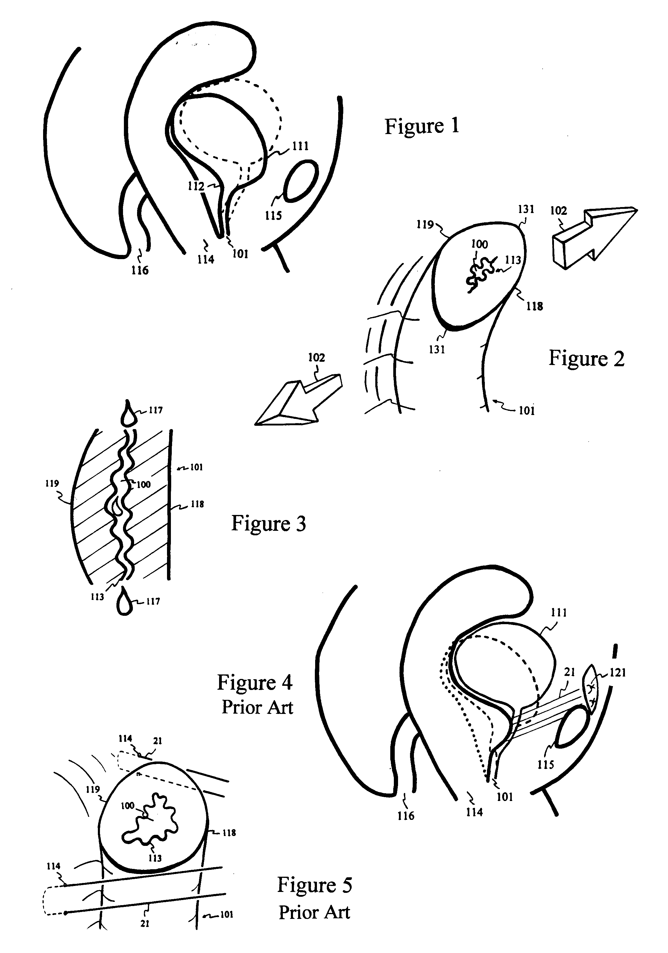
[0133] It is widely believed that most of the urinary incontinence in women is related to the descended position of the bladder 111, the funnneling of the bladder neck 112 and / or diminishing posterior 119 urethral support. The dashed line of FIG. 1 indicates the normal position and the solid line depicts the descended position of the bladder 111 with its funnel-shaped bladder neck 112. FIG. 2 shows a failed lumen 100 closure and hypermobility under stress with the urethropelvic ligament 102 pulling the lateral walls 131 of the poorly supported urethra 101. The mid-longitudinal view of FIG. 2 during stress is shown in FIG. 3, with urethropelvic ligaments pulling perpendicularly above and below the plane of the page. A section of poorly-supported posterior wall 119 withdraws from mucosal 113 coaptation, leading to urine 117 leakage.

[0134] Numerous prior art surgical procedures are designed to treat urinary incontinence. The traditional surgical treatment for urinary incontinence is to...


PUM


Abstract

A rod-like or tube-like sphincteric closure device is micro-invasively implanted beneath the surface of the posterior mucosal wall to elevate the mucosa and close the lumen, by using a delivery device operated through the urethra. To initiate voiding, the muscle-rich anterior urethral wall contracts and widens the lumen beyond the closing or elevating range of the sphincteric closure device. For urethral obstruction, repelling magnets are micro-invasively implanted beneath the surface of mucosa with the delivery device through the urethra, to widen the lumen with magnetic force.

Description

[0001] This invention relates to rod-like or tube-like sphincteric closure devices implanted beneath the surface of the mucosa through the urethra with a micro-invasive delivery device. The sphincteric closure devices narrow or close the lumen opening to prevent urine leakage without interfering with the process of voluntary voiding.BACKGROUND, TRADITIONAL TREATMENTS AND PRIOR INVENTIONSPrevalence and Cost of Urinary Incontinence[0002] Urinary incontinence is one of the most common urinary dysfunctions. The number of people living with urinary incontinence is far higher than estimated, even by most primary care physicians. A report published by the Agency for Health Care Policy and Research of the U.S. Public Health Service estimates that at least 10 million, more likely 20 million, adult Americans are affected by urinary incontinence. Many patients, especially women, do not mention their incontinence problems to their physicians. One of the reasons is that women are accustomed to u...

Claims


Application Information

Patent Timeline
Patent Type & Authority Applications(United States)
IPC IPC(8): A61F2/00
CPCA61B17/06066A61B17/06109A61B2017/00805A61F2/0036A61B2017/2925A61F2/0018A61B2017/00876
Inventor YEUNG, JEFFREY EYEUNG, TERESA T
Owner YEUNG JEFFREY E
Learn More