
Methods and systems for X-ray tube conditioning

a technology of computed tomography and x-ray tube, which is applied in the field of diagnostic medical imaging, can solve problems such as stress fractures and target degradation, and achieve the effects of improving the accuracy of diagnosis and treatment results

Active Publication Date: 2021-10-19
GE PRECISION HEALTHCARE LLC
View PDF15 Cites 1 Cited by
  • Summary
  • Abstract
  • Description
  • Claims
  • Application Information

AI Technical Summary

Benefits of technology

The patent describes a method for an imaging system where the system generates x-rays to condition the x-ray tube for a diagnostic scan. This helps to adjust the temperature of the x-ray tube for the scan based on its initial temperature and the desired temperature for the scan. The technical effect of this method is to improve the accuracy and quality of the diagnostic scan by ensuring the optimal conditioning of the x-ray tube.

Problems solved by technology

Below the desired temperature range, the target material may be brittle whereby a high-energy electron beam impinging on the target may result in stress fractures such that a target degradation might ensue.

Method used


Image

  • Methods and systems for X-ray tube conditioning
  • Methods and systems for X-ray tube conditioning
  • Methods and systems for X-ray tube conditioning

Examples


Embodiment Construction

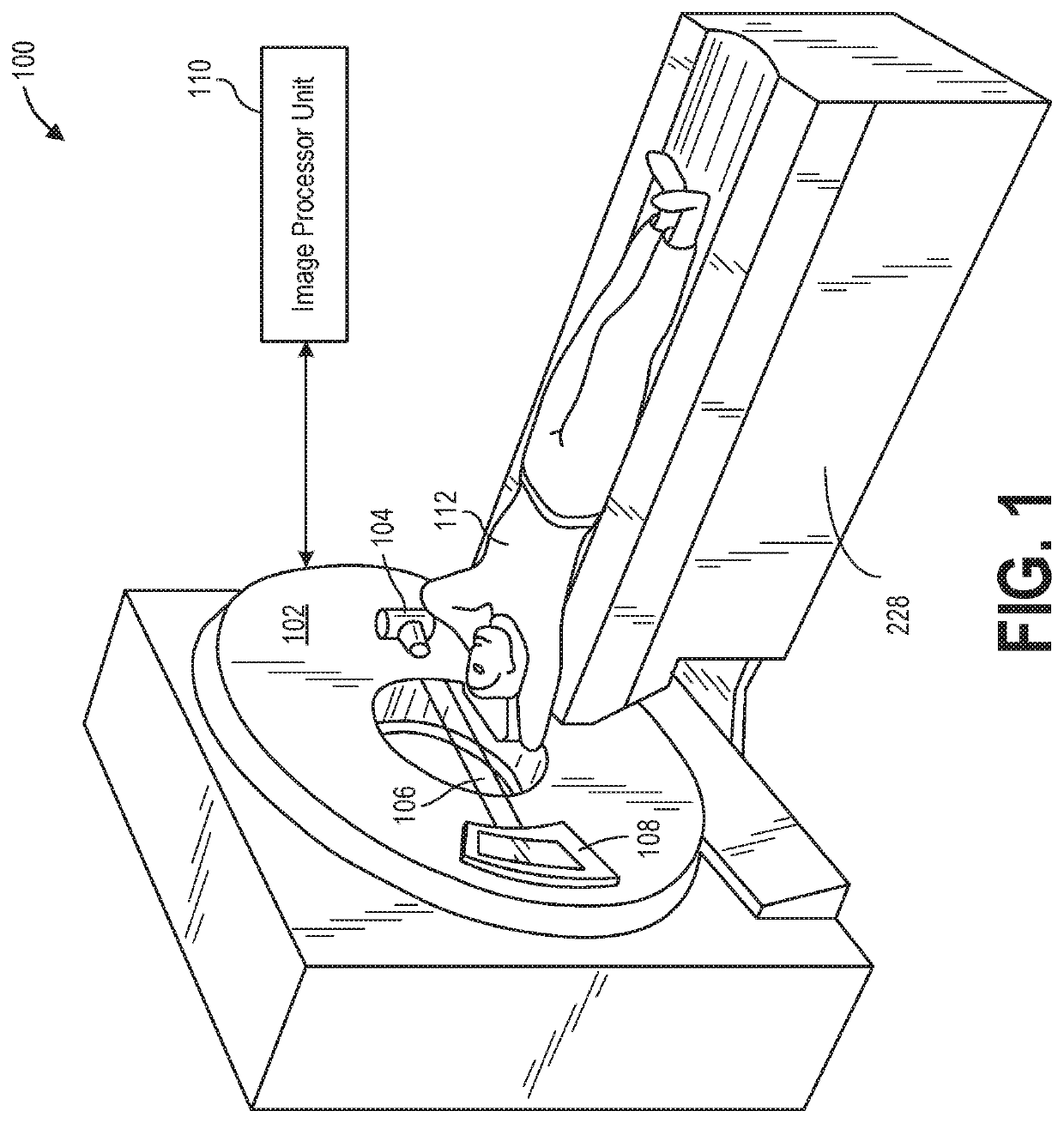
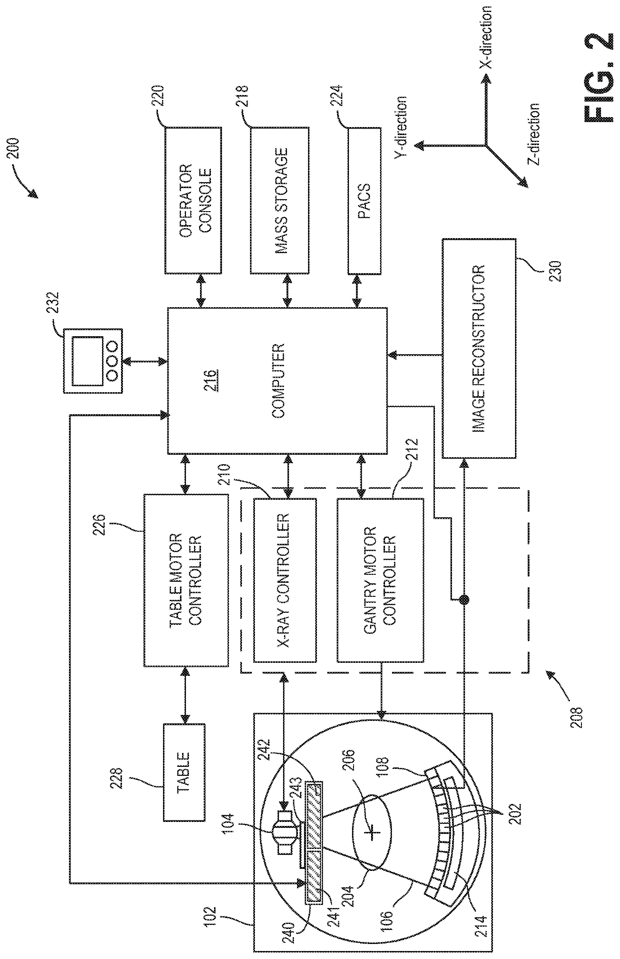
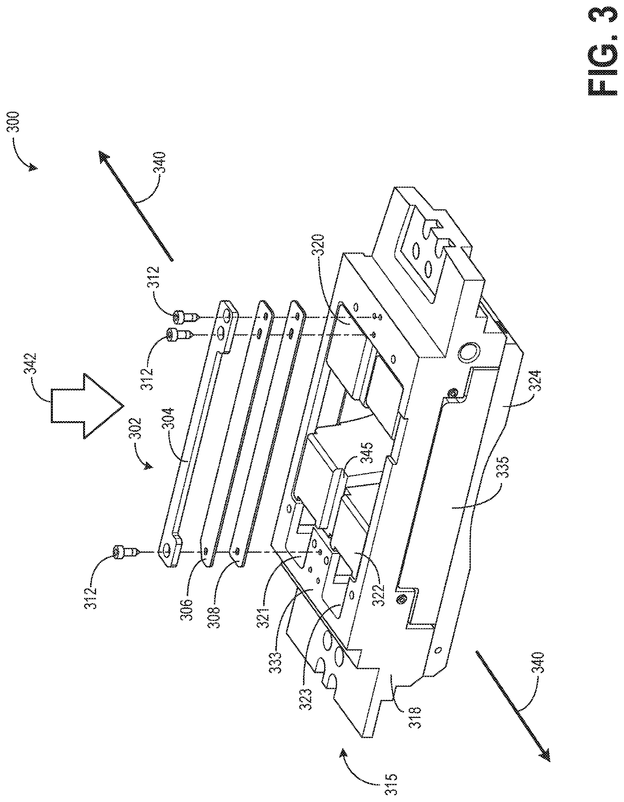
[0029]The following description relates to various embodiments of x-ray imaging of a subject. In particular, systems and methods are provided for CT imaging using one or more of a hardening filter and bowtie filters. FIGS. 1-2 show an example embodiment of an imaging system, wherein the one or more filters are positioned between the radiation source and the imaging subject. Different filters may be selected based on the anatomy of the imaging subject being imaged. FIG. 3 shows an example of an integrated filter assembly including a carriage, a hardening filter, and a plurality of bowtie filters which may be positioned to adjust a spatial distribution and condition the beam reaching the subject. As an example, in a single carriage, two bowtie filters may be positioned next to each other with a hardening filter also coupled to the same carriage between the two bowtie filters. A single bowtie filter or a combination of a hardening filter and a bowtie filter may be positioned in a path ...


PUM


Abstract

Various methods and systems are provided for x-ray tube conditioning for a computed tomography imaging method. In one embodiment, x-ray may be generated in an x-ray tube of a radiation source prior to a diagnostic scan to warmup the x-ray tube to a desired temperature for the diagnostic scan. The power delivered to the x-ray tube during warmup may be adjusted in a closed loop system based on an initial temperature of the x-ray tube and the desired temperature for the diagnostic scan. During tube warmup, by placing a blocking plate coupled to a collimator blade in a path of the x-ray beam, the x-ray beam may be blocked from exiting a collimator.

Description

FIELD[0001]Embodiments of the subject matter disclosed herein relate to diagnostic medical imaging, and more particularly, to x-ray tube conditioning for computed tomography imaging method.BACKGROUND[0002]Noninvasive imaging modalities may transmit energy in the form of radiation into an imaging subject. Based on the transmitted energy, images may be subsequently generated indicative of the structural or functional information internal to the imaging subject. In computed tomography (CT) imaging, radiation transmits from a radiation source to a detector through the imaging subject. The X-Ray tubes that power CT systems generate x-rays by accelerating and focusing a high-energy beam of electrons onto a rotating target. As individual electrons strike the target, the energy released by interacting with the atoms of the target produces x-ray photons isotropically under a polychromatic spectrum, a maximum energy of the x-ray photons matching that of the incident electrons. The x-ray photo...

Claims


Application Information

Patent Timeline
Patent Type & Authority Patents(United States)
IPC IPC(8): A61B6/00A61B6/06A61B6/03
CPCA61B6/54A61B6/06A61B6/032A61B6/4035A61B6/488A61B6/40A61B6/52A61B6/035H05G1/36A61B6/4488H05G1/56G21K1/10G21K1/04H01J35/26H01J2235/085
Inventor THIBAULT, JEAN-BAPTISTEHSIEH, JIANGSTRONG, GARY
Owner GE PRECISION HEALTHCARE LLC
Learn More