
Use of cells derived from first trimester umbilical cord tissue

a technology of umbilical cord tissue and stem cells, which is applied in the field of human stem cells, can solve the problems of insufficient perfusion of the heart, permanent damage to the heart tissue, and morbidity and mortality

Active Publication Date: 2021-02-23
REPROBIOGEN INC
View PDF7 Cites 0 Cited by
  • Summary
  • Abstract
  • Description
  • Claims
  • Application Information

AI Technical Summary

Benefits of technology

This method provides a large quantity of pluripotent cells with the ability to differentiate into various cell types, offering a viable alternative for treating cellular damage and diseases by using readily available first trimester umbilical cord tissue, enhancing cell viability and therapeutic potential.

Problems solved by technology

Cardiovascular disease is the leading cause of mortality and morbidity globally.
During an episode of acute myocardial infarction (MI), impaired blood flow due to an obstructed coronary artery triggers an ischemic cascade leading to insufficient perfusion of the heart.
The hypoxic injury results in permanent damage to heart tissue and, without successful intervention, frequently leads to CHF.
In addition to drug-based therapies, major surgical procedures such as coronary artery bypass and medical devices providing mechanical augmentation for the ventricular myocardium are frequently administered, with significant risk of complications.
Heart transplantation for post-MI patients with congestive heart failure is the most invasive procedure, considered extremely high-risk, and is limited by both organ availability and cost, making it unsustainable as a large scale solution.
Age-related limitations in the regenerative capacity of the implanted and endogenous progenitor cells are considered to be a major factor in the limited efficacy.
However, the underlying cellular mechanisms required to achieve this are multifaceted and complex in nature.
After cardiac injury, the disruption of functional intercellular connections due to cell loss or separation by pathological ECM often leads to akinetic or arrhythmic tissue.

Method used


Image

  • Use of cells derived from first trimester umbilical cord tissue
  • Use of cells derived from first trimester umbilical cord tissue
  • Use of cells derived from first trimester umbilical cord tissue

Examples


example 1

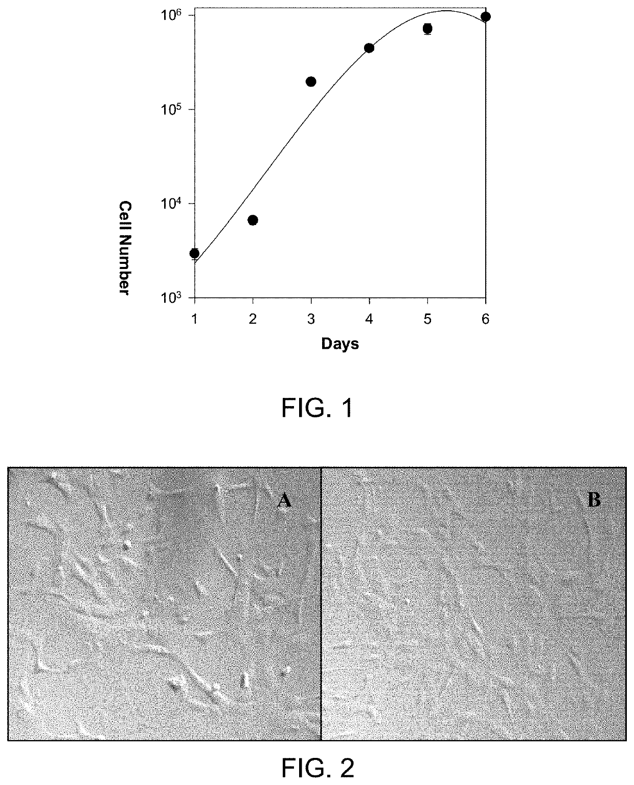
[0057]Obtaining First Trimester HUCPV cells

[0058]The following method describes obtaining first trimester human umbilical cord perivascular cells (HUPVC).

[0059]Materials—Reagents

[0060]A non-exhaustive list of possible reagents for use in the described method is provided below. Penicillin-streptomycin liquid containing 5000 U of penicillin and 5000 mg of streptomycin / mL (GIBCO; cat. no. 15070-63), aliquot and store at −20° C. Amphotericin B solution (250 μg / ml, Sigma; cat. no. A-2942), aliquot and store at −20° C. Dulbecco's phosphate-buffered saline (PBS) (+) (GIBCO™; cat. no. 14040-133), store at 4° C. Sterile water, tissue culture grade (GIBCO; cat. no. 15230-162), store at 4° C. Collagenase Type 1 (GIBCO; cat. no. 21985-023), store at 4° C. α-MEM (GIBCO; cat. no. 12571). Defined fetal bovine serum (HyClone, Logan, Utah; cat. no. 30070-03) aliquot and store at −20° C. Trypsin 0.25% / EDTA (GIBCO; cat. no. 25200-056), store at −20° C. DMSO (Sigma™, cat. no. D-5879), store at room tem...

example 2

[0084]Differentiation of First Trimester HUCPV Cells In Vitro and Expression of Tissue-Specific Markers.

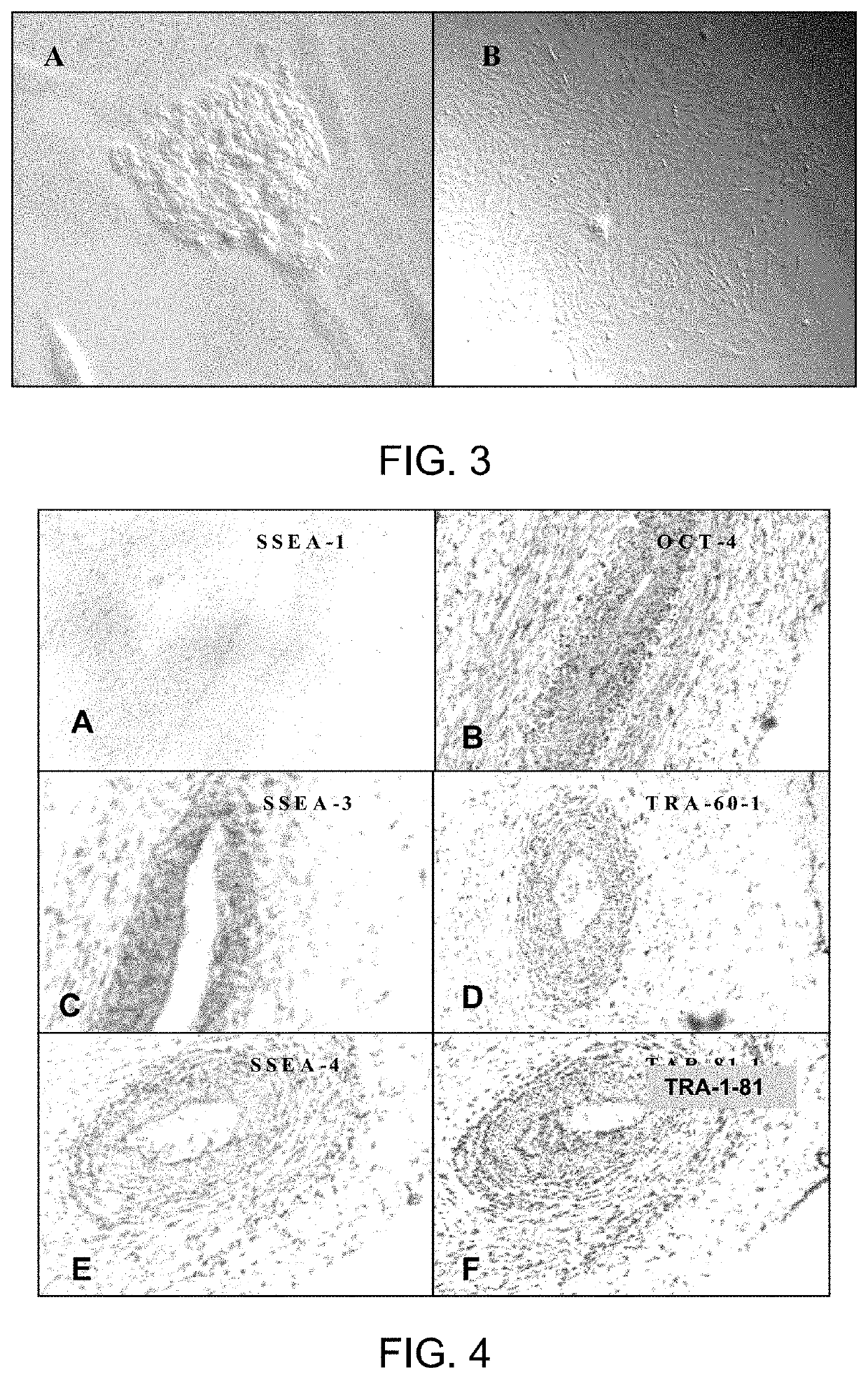
[0085]Stem cells were isolated and expanded from the first trimester human umbilical cord (HUCPV), demonstrating that the cells have embryonic stem cell characteristics. These cells can express embryonic stem cell markers from passage 0 to 16 and have the ability to self-renew.

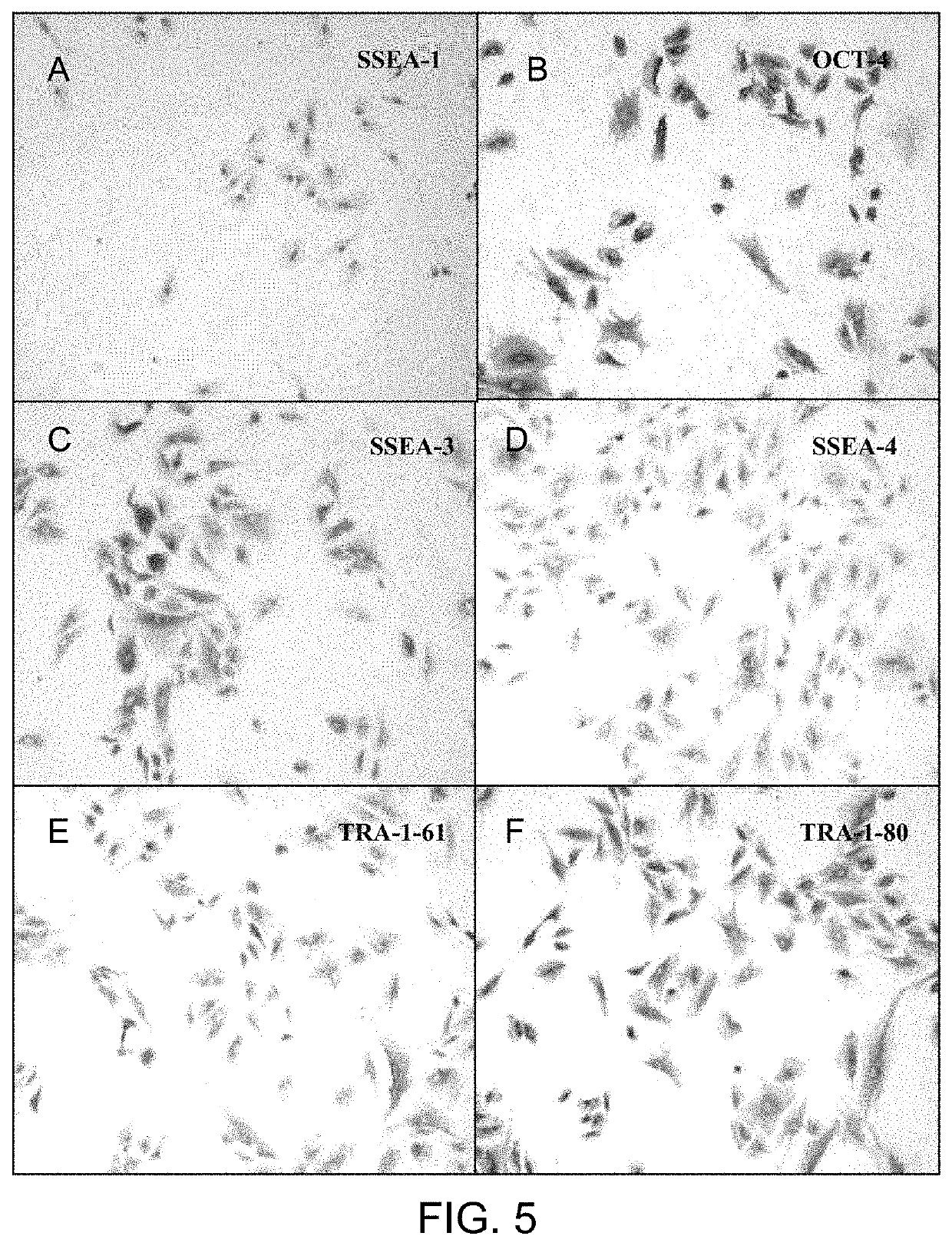
[0086]First trimester HUCPV cells were differentiated in vitro and examined for the expression of tissue-specific markers in the differentiated cells.

[0087]Methods

[0088]Perivascular cells were isolated from first trimester umbilical cords and were expanded in α-MEM containing 5-10% of FCS. These cells were grown in a suspension to induce their differentiation into EBs. EBs were transferred onto coated plates and cultured under appropriate condition. Morphology change was examined in these differentiation conditions. Dispersed EBs and differentiated cells were characterized using immunocytochemistry (ICC). RT...

example 3

[0103]Use of Early Intravenous First Trimester Human Umbilical Cord Cells in Spinal Cord Injury

[0104]Summary

[0105]This Example shows that early intravenous first trimester human umbilical cord-perivascular cell delivery after traumatic spinal cord injury significantly improves forelimb motor function and promotes weight gain. Localized vascular disruption as a result of spinal cord injury (SCI) triggers a cascade of secondary events, including inflammation, reactive gliosis and scarring that influence recovery. In addition to immunomodulatory and pathotropic properties, mesenchymal stromal cells (MSCs), including human umbilical cord matrix cells (HUCMCs), possess pericytic features. This makes MSCs ideally suited to targeting acute vascular disruption, which could reduce the severity of secondary injury, enhance tissue preservation and repair, and promote chronic functional recovery. Here, intravenous tail vein infusion of HUCMCs, first trimester human umbilical cord perivascular c...


PUM

PropertyMeasurementUnit
temperatureaaaaaaaaaa
timeaaaaaaaaaa
thickaaaaaaaaaa

Abstract

A method of isolating a pluripotent cell from human umbilical cord is described herein. The method involves collecting a sample of umbilical cord from fetal tissue obtained at less than 13 weeks of gestation, for example a first trimester umbilical cord. The sample is treated to obtain isolated umbilical cord cells, after which the isolated umbilical cord cells are incubated. Cells obtained in this way can be differentiated for use in treating conditions of cell damage, by supplanting the function of a damaged cell in a condition such as spinal cord injury, cardiovascular injury or heart disease.

Description

CROSS REFERENCE TO RELATED APPLICATIONS[0001]This application is a continuation-in-part of U.S. patent application Ser. No. 12 / 114,182 filed May 2, 2008; and claims the benefit of priority of U.S. Provisional Patent Application Ser. No. 60 / 972,022, filed Sep. 13, 2007 which is hereby incorporated by reference.FIELD[0002]The present disclosure relates generally to human stem cells. More particularly, the present disclosure relates to a method of isolating and expanding stem cells from first trimester umbilical cords, and uses therefor.BACKGROUND[0003]Stem cells are unspecialized human or animal cells that can produce mature specialized body cells and at the same time replicate themselves. This ability to differentiate into specialized cells has led to much research into their use for treating such fatal diseases and disorders as Parkinson's and Alzheimer's diseases, spinal cord injury, stroke, burns, heart disease, diabetes, osteoarthritis and rheumatoid arthritis. Stem cells are der...

Claims


Application Information

Patent Timeline
Patent Type & Authority Patents(United States)
IPC IPC(8): A61K35/51C12N5/073A01N1/02A61K35/44
CPCA61K35/51A01N1/0226A61K35/44C12N5/0605
Inventor LIBRACH, CLIFFORD L.YIE, SHANGMIANXIAO, RONG
Owner REPROBIOGEN INC
Learn More