Nervous system-specific transmembrane proteasome complex that modulates neuronal signaling through extracellular signaling via brain activity peptides
a neuronal system and transmembrane technology, applied in the direction of peptide/protein ingredients, instruments, boron compound active ingredients, etc., can solve the problems of undefined how proteasomes can rapidly alter neuronal function, and achieve the effects of reducing secretion, increasing detection signal, and increasing the amount of detection signal
- Summary
- Abstract
- Description
- Claims
- Application Information
AI Technical Summary
Benefits of technology
Problems solved by technology
Method used
Image

Examples
example 1
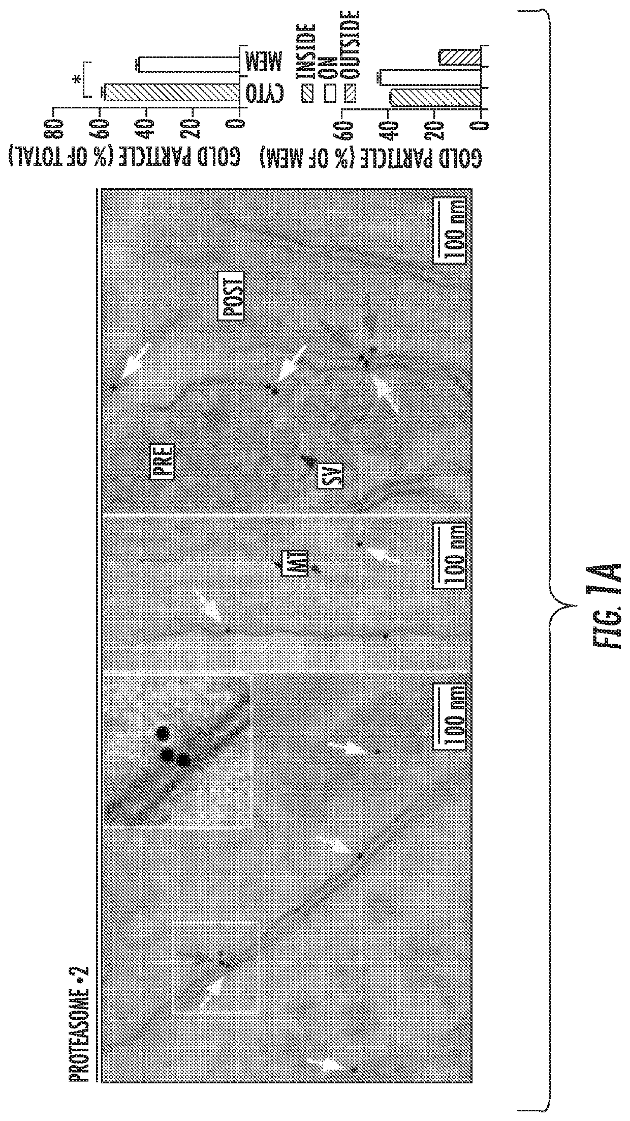
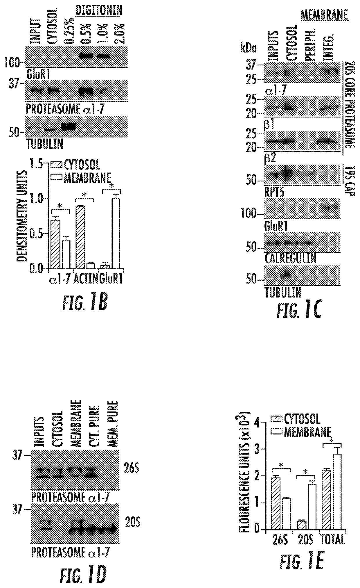
[0168]A catalytically active 20S proteasome is integral to neuronal plasma membranes.
[0169]Previous studies attempting to ascribe proteasomes with distinct function in the nervous system have identified localization as a key feature of determining proteasome function. However, all of these studies have focused on the 26S proteasome, either through the use of fluorescently-tagged cap subunits, or have used complex electron microscopy approaches to assess the distribution of 26S capped proteasomes in neurons. Taking a more unbiased approach to evaluate localization of all proteasomes in the nervous system, the inventors performed an immunogold electron microscopy (Immuno-EM) analysis of cortical neuronal cultures using an antibody that detects β2, a core proteasomal subunit common to all catalytically active proteasomes. Since core proteasome subunits have never been shown to be separated from the rest of the macromolecular proteasome complex, these data likely reflected the localizat...
example 2
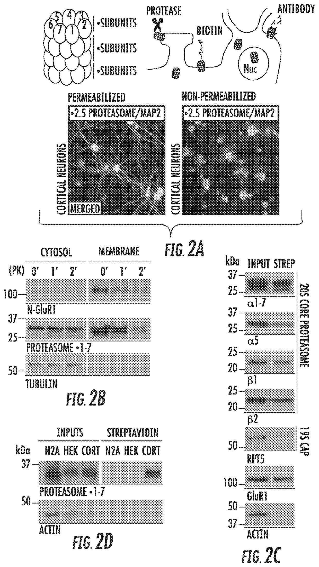
[0173]The NMP is exposed to the extracellular space, is specific to the nervous system, and is temporally regulated.
[0174]We postulated that a catalytically active proteasome with access to both the intracellular and extracellular space would be positioned in such a manner to rapidly modulate neuronal function through its proteolytic activity. Thus, to validate our Immuno-EM data, we proceeded with a series of classic approaches to assay whether proteins are exposed to the extracellular space (FIG. 2A). First, cortical neurons were fixed and stained using antibodies against catalytic proteasome subunits under both permeabilizing and non-permeabilizing conditions. Consistent with the NMP being surface exposed, we observed immunostaining against proteasomal subunits under both conditions, but only observed cytosolic MAP2 staining under permeabilizing conditions (FIG. 2A). To biochemically determine whether the NMP was surface-exposed, we found that proteasome subunits in neuronal memb...
example 3
[0177]The NMP Mediates Degradation of Intracellular Proteins into Extracellular Peptides
[0178]Given that it was catalytically active in vitro, we hypothesized that in intact cells, the NMP would promote proteasome-dependent degradation of intracellular proteins into the extracellular space. To test our hypothesis, we used 35S-radiolabelling approaches to trace the fate of newly synthesized intracellular proteins (Schubert et al., 2000). 35S-methionine / cysteine was quickly incorporated into neurons; addition of proteasome inhibitors had no effect on radiolabeling efficiency (FIG. 3A). After 10 minutes of radiolabeling, free radioactivity was washed away, and media was collected over a time course and analyzed by liquid scintillation. We observed rapid proteasome-dependent release of radioactivity into the culture medium. Of the released radioactive material, 90±3% was made up of Proteinase K-sensitive molecules that ranged between 500 and 3000 Daltons in size (FIG. 3B). From this, we...
PUM
| Property | Measurement | Unit |
|---|---|---|
| pH | aaaaa | aaaaa |
| physiological temperature | aaaaa | aaaaa |
| physiological temperature | aaaaa | aaaaa |
Abstract
Description
Claims
Application Information
 - R&D
- Intellectual Property
- Life Sciences
- Materials
- Tech Scout
- Unparalleled Data Quality
- Higher Quality Content
- 60% Fewer Hallucinations
Browse by: Latest US Patents, China's latest patents, Technical Efficacy Thesaurus, Application Domain, Technology Topic, Popular Technical Reports.
© 2025 PatSnap. All rights reserved.Legal|Privacy policy|Modern Slavery Act Transparency Statement|Sitemap|About US| Contact US: help@patsnap.com