Tissue grasping devices and related methods

a technology of tissue grasping and tissue, applied in the field of tissue grasping devices, can solve the problems of progressive heart failure, valve regurgitation, and affecting the patient's recovery, and achieve the effects of reducing the pumping efficiency of the heart, reducing the risk of severe heart failure, and reducing the risk of patients being rushed to hospital

Active Publication Date: 2020-06-02
EVALVE
View PDF658 Cites 3 Cited by
  • Summary
  • Abstract
  • Description
  • Claims
  • Application Information

AI Technical Summary

Benefits of technology

[0015]At least one embodiment of the present disclosure relates to a tissue fixation kit, the kit including: a tissue gripping system that includes an actuator rod, an actuator line, a first and second distal element, each including a first end pivotally coupled to the actuator rod and extending to a free second end and a tissue engagement surface between the first and second end, the first and second distal elements being positionable by the actuator rod, a tissue gripping device formed of a shape-memory material, the tissue gripping device including a base section, a first arm, and a second arm, each arm having a first end coupled to the base section and a free end extending from the base section, the tissue gripping device being positionable by the actuator line; a handle; and a delivery catheter having a proximal end and a distal end, the tissue gripping system being couplable to the distal end of the delivery catheter and the handle being couplable to the proximal end of the delivery catheter.

Problems solved by technology

Regurgitation of the valve can significantly decrease the pumping efficiency of the heart, placing the patient at risk of severe, progressive heart failure.
Mitral valve regurgitation can result from a number of different mechanical defects in the mitral valve or the left ventricular wall.
The valve leaflets, the valve chordae which connect the leaflets to the papillary muscles, the papillary muscles themselves, or the left ventricular wall may be damaged or otherwise dysfunctional.
Commonly, the valve annulus may be damaged, dilated, or weakened, limiting the ability of the mitral valve to close adequately against the high pressures of the left ventricle during systole.
The need to both open the chest and place the patient on bypass is traumatic and has associated high mortality and morbidity.
This moving and plastically deforming components of the fixation device during pre-deployment, positioning, and closure of the device can lead to the weakening and pre-mature degradation of the fixation device.
Additionally, some tissue fixation treatments require that the fixation device maintain a degree of flexibility and mobility to allow a range of physiological movement even after the device has been properly placed and the target tissue has been properly fixed into the desired position, This can increase the risk of pre-mature failure of the device as continued plastic deformation of the flexing components (e.g., from the continuous opening and closing of valve leaflets) leads to unfavorable degradation of the device.

Method used


Image

  • Tissue grasping devices and related methods
  • Tissue grasping devices and related methods
  • Tissue grasping devices and related methods

Examples


Embodiment Construction

I. Cardiac Physiology

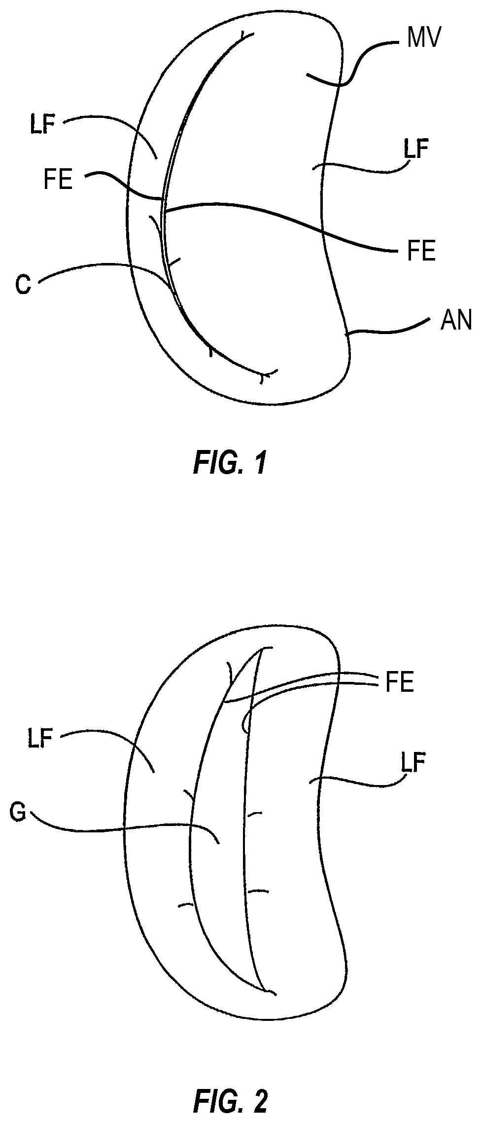
[0026]As shown in FIG. 1, the mitral valve (MV) consists of a pair of leaflets (LF) having free edges (FE) which, in patients with normal heart structure and function, meet evenly to close along a line of coaption (C). The leaflets (LF) attach to the surrounding heart structure along an annular region called the annulus (AN). The free edges (FE) of the leaflets (LF) are secured to the lower portions of the left ventricle LV through chordae tendinae (or “chordae”). As the left ventricle of a heart contracts (which is called “systole”), blood flow from the left ventricle to the left atrium through the mitral valve (MV) (called “mitral regurgitation”) is usually prevented by the mitral valve. Regurgitation occurs when the valve leaflets do not close properly and allow leakage from the left ventricle into the left atrium. A number of heart structural defects can cause mitral regurgitation. FIG. 2 shows a mitral valve with a defect causing regurgitation through a gap...


PUM


Abstract

A tissue gripping device is formed from a shape-memory material, and has a base section, a first arm, and a second arm disposed opposite the first arm, each arm having a first end coupled to the base section and a free end extending from the base section. The arms of the tissue gripping device are configured to resiliently flex toward a relaxed configuration in a distal direction as the tissue gripping device is moved from a pre-deployed configuration toward a deployed configuration. The tissue gripping device is usable in a method for gripping tissue. The method includes positioning the tissue gripping device near target tissue and moving the tissue gripping device from a pre-deployed configuration toward a deployed configuration in order to grip the target tissue.

Description

CROSS-REFERENCE TO RELATED APPLICATIONS[0001]N / ABACKGROUND[0002]The present disclosure relates generally to medical methods, devices, and systems. In particular, the present disclosure relates to methods, devices, and systems for the endovascular, percutaneous, or minimally invasive surgical treatment of bodily tissues, such as tissue approximation or valve repair. More particularly, the present disclosure relates to repair of valves of the heart and venous valves, and devices and methods for removing or disabling mitral valve repair components through minimally invasive procedures.[0003]Surgical repair of bodily tissues often involves tissue approximation and fastening of such tissues in the approximated arrangement. When repairing valves, tissue approximation often includes coapting the leaflets of the valves in a therapeutic arrangement which may then be maintained by fastening or fixing the leaflets. Such fixation of the leaflets can be used to treat regurgitation which most com...

Claims


Application Information

Patent Timeline
Patent Type & Authority Patents(United States)
IPC IPC(8): A61B17/08A61B17/00
CPCA61B17/08A61B17/00234A61B2017/00783A61B2017/00243A61B2017/00867A61B2017/00526A61F2/2445A61F2/246A61F2/2463A61F2/2466
Inventor KRONE, RYAN T.GREENBERG, JACOB L.BASUDE, RAGHUVEER
Owner EVALVE
Why Eureka
Try Eureka