
Medical imaging unit, medical imaging device with a medical imaging unit, and method for detecting a patient movement

a medical imaging and patient technology, applied in the field of medical imaging devices, can solve the problems of patient movement and artifacts in medical slice images, and achieve the effect of cost-effectiveness

Active Publication Date: 2020-01-28
SIEMENS HEALTHCARE GMBH
View PDF18 Cites 0 Cited by
  • Summary
  • Abstract
  • Description
  • Claims
  • Application Information

AI Technical Summary

Benefits of technology

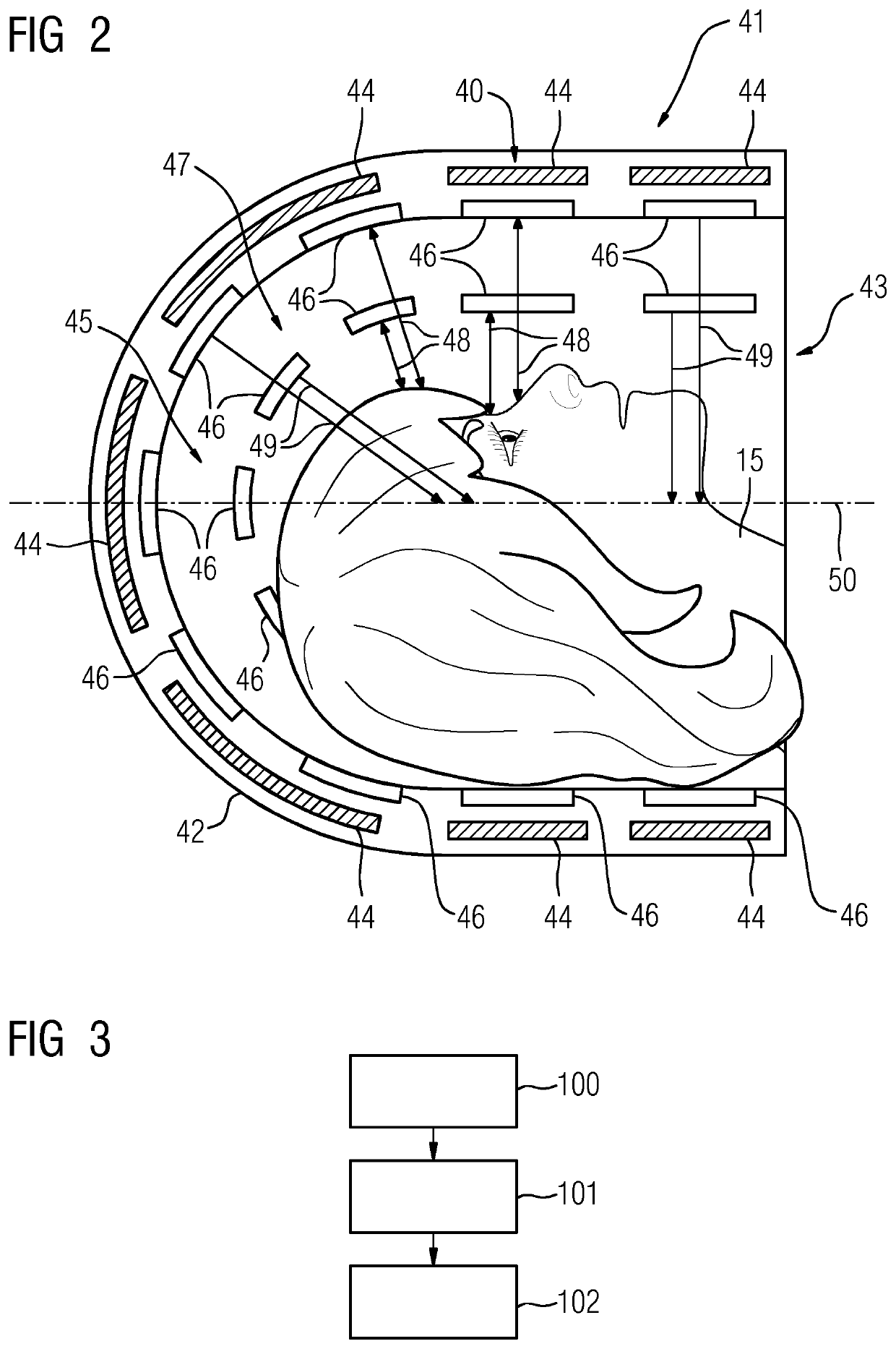
[0007]This enables a cost-effective movement detection that makes a reliable and rapid detection of a movement of the patient within the patient receiving area possible to be provided. In one embodiment, the two or more detection elements are read out with a high temporal resolution, so that an especially exact and always current position change and / or movement (e.g., a three-dimensional movement) of the patient may be detected for a medical imaging examination. The established patient movement enables the movement of the patient to be taken into account during an evaluation of medical image data of the medical imaging examination, and in this way, artifacts in medical slice images may be at least reduced and / or suppressed.
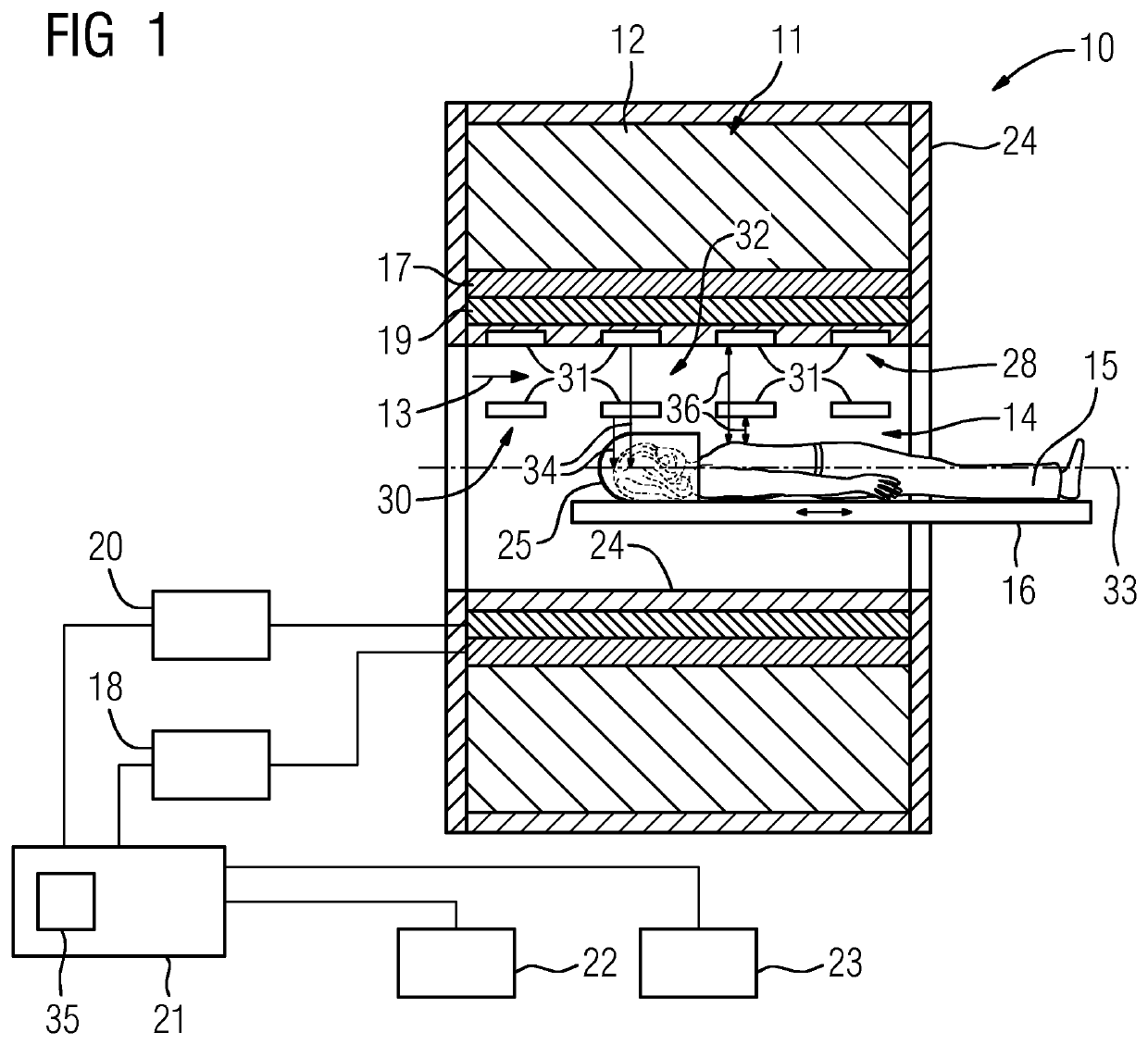
[0017]One or more of the present embodiments are based on a medical imaging device with a medical imaging unit. The medical imaging unit includes a patient receiving area, a housing unit at least partly surrounding a patient receiving area, a detector unit, and a movement detection unit. The movement detection unit includes two or more detection elements configured for detecting a distance between the detection elements and the patient, and including an evaluation unit. The evaluation unit is configured to establish a three-dimensional movement of the patient within the patient receiving area from the detected distance data of the individual detection elements. This configuration enables a cost-effective movement detection unit to be provided, which makes possible a reliable and rapid detection of a movement of the patient within the patient receiving area. Using the established patient movement, the movement of the patient may be taken into account in the evaluation of medical image data of the medical imaging examination. In this way, artifacts in medical slice images may be at least reduced and / or suppressed.

Problems solved by technology

In medical imaging examinations that use a long scanning time, such as, for example, magnetic resonance examinations, SPECT examinations, PET examinations, etc., a patient movement may lead to artifacts in the medical slice images.

Method used


Image

  • Medical imaging unit, medical imaging device with a medical imaging unit, and method for detecting a patient movement
  • Medical imaging unit, medical imaging device with a medical imaging unit, and method for detecting a patient movement

Examples


Embodiment Construction

[0028]FIG. 1 shows a schematic of one embodiment of a medical imaging device 10. In the present exemplary embodiment, the medical imaging device 10 is formed by a magnetic resonance device. The embodiment of the imaging device 10 is not restricted to a magnetic resonance device, however. Instead, the medical imaging device 10 may be formed by all medical imaging devices appearing sensible to the person skilled in the art, such as by a computed tomography device, a Positron Emission Tomography device, a SPECT device, etc., for example.

[0029]The magnetic resonance device includes a detector unit formed by a magnet unit 11 with a superconducting main magnet 12 for creating a strong and, for example, constant main magnetic field 13. In addition, the magnetic resonance device includes a patient receiving area 14 for receiving a patient 15. The patient receiving area 14 in the present exemplary embodiment is configured in a cylindrical shape and is surrounded in a circumferential directio...


PUM


Abstract

A medical imaging unit includes a patient receiving area, a housing unit at least partly surrounding the patient receiving area, a detector unit, and a movement detection unit. The movement detection unit includes two or more detection elements configured for detecting a distance from the detection elements to the patient, and including an evaluation unit. The evaluation unit is configured to establish from detected movement of the individual detection elements a three-dimensional movement of the patient within the patient receiving area.

Description

[0001]This application claims the benefit of DE 10 2014 215 651.3, filed on Aug. 7, 2015, which is hereby incorporated by reference in its entirety.BACKGROUND[0002]The present embodiments relate to a medical imaging device.[0003]In medical imaging examinations that use a long scanning time, such as, for example, magnetic resonance examinations, SPECT examinations, PET examinations, etc., a patient movement may lead to artifacts in the medical slice images. A correction of this patient movement in the detected medical data may at least reduce these artifacts.SUMMARY AND DESCRIPTION[0004]The scope of the present invention is defined solely by the appended claims and is not affected to any degree by the statements within this summary.[0005]The present embodiments may obviate one or more of the drawbacks or limitations in the related art. For example, a patient movement is accurately detected during a medical imaging.[0006]One or more of the present embodiments are based on a medical im...

Claims


Application Information

Patent Timeline
Patent Type & Authority Patents(United States)
IPC IPC(8): A61B5/055A61B5/11A61B5/00G16H40/63A61B6/03A61B6/00
CPCA61B5/6891G16H40/63A61B5/0059A61B5/721A61B5/055A61B5/6886A61B5/1113A61B5/1126A61B6/037A61B2562/04A61B5/7271A61B5/1128A61B5/1114A61B6/527
Inventor HEISMANN, BJÖRNNISZNANSKY, MARTINSCHRÖTER, STEFFENSUKKAU, JOHANN
Owner SIEMENS HEALTHCARE GMBH
Learn More